
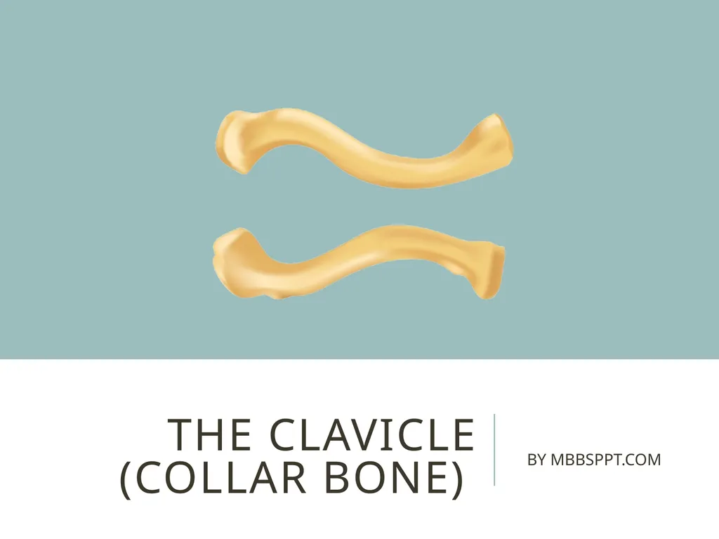
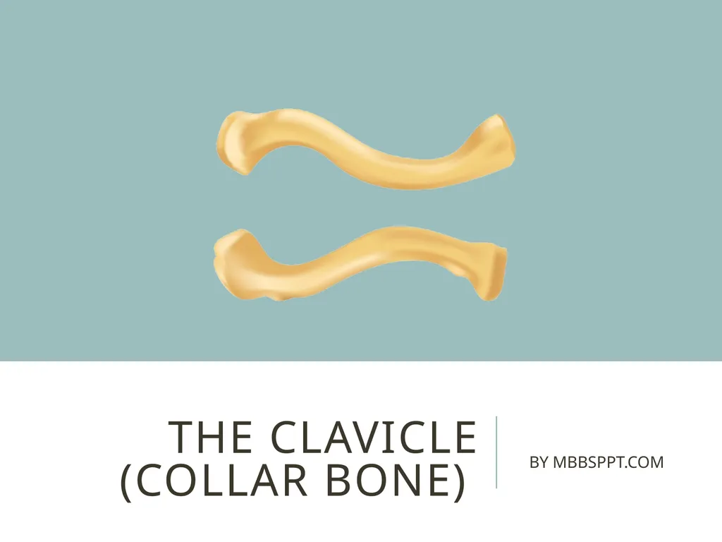
The clavicle (collar bone) BY MBBSPPT.COM The
CM
Published · 26 slides · 0 views
1 / 1

Description
The clavicle (collar bone) BY MBBSPPT.COM The clavicle (collarbone) extends between the manubrium of the sternum and the acromion of the scapula. It is classed as a long bone, and can be palpated along its length. In thin individuals, it is
Related Topics
Share
Embed code
Download this presentation From Below
"The clavicle (collar bone) BY MBBSPPT.COM The" is the property of its rightful owner. Permission is granted to download and print the materials on this website for personal, non-commercial use only, and to display it on your personal computer provided you do not modify the materials and that you retain all copyright notices contained in the materials. By downloading content from our website, you accept the terms of this agreement.
Presentation Transcript
01
The clavicle (collar bone) BY MBBSPPT.COM<br>
02
The clavicle (collarbone) extends between the manubrium of the sternum and the acromion of the scapula.
It is classed as a long bone, and can be palpated along its length.
In thin individuals, it is visible under the skin.
It has a slight S-shaped curve. The clavicle MBBSPPT.COM 2<br>
It is classed as a long bone, and can be palpated along its length.
In thin individuals, it is visible under the skin.
It has a slight S-shaped curve. The clavicle MBBSPPT.COM 2<br>
03
MBBSPPT.COM 3<br>
04
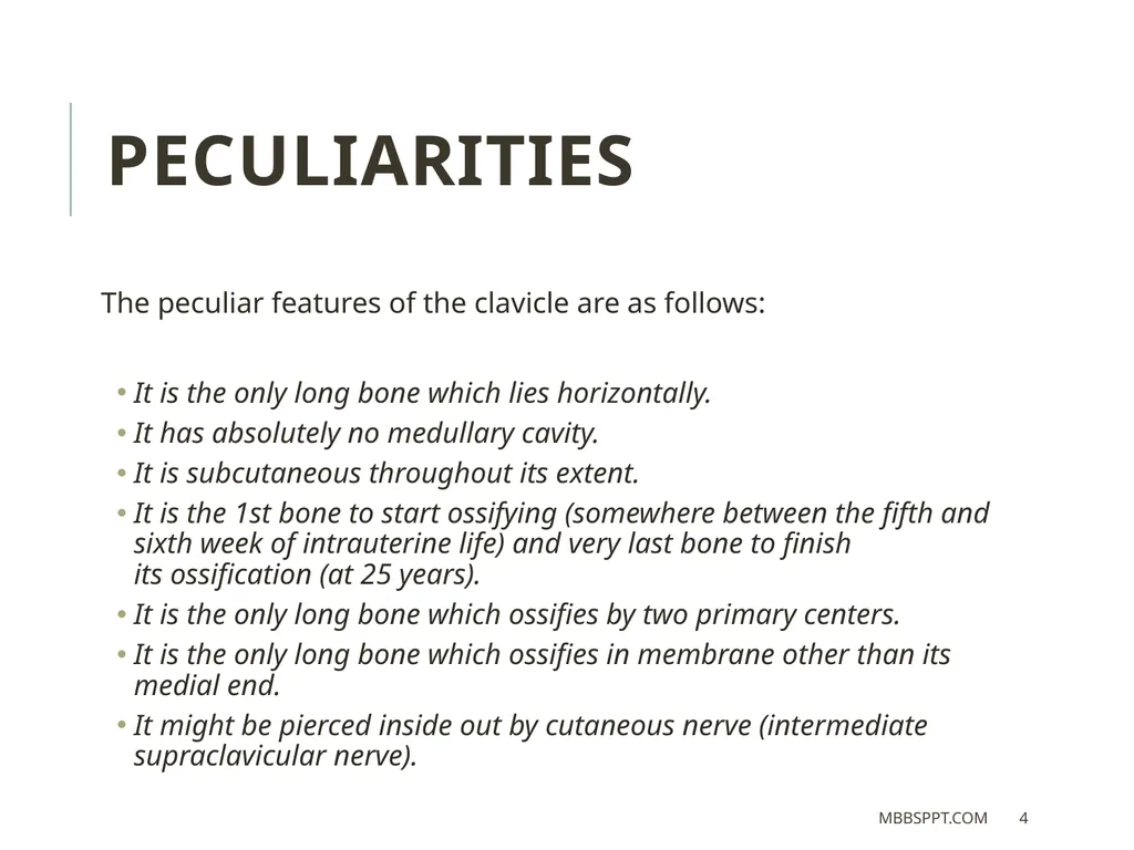
The peculiar features of the clavicle are as follows:
It is the only long bone which lies horizontally.
It has absolutely no medullary cavity.
It is subcutaneous throughout its extent.
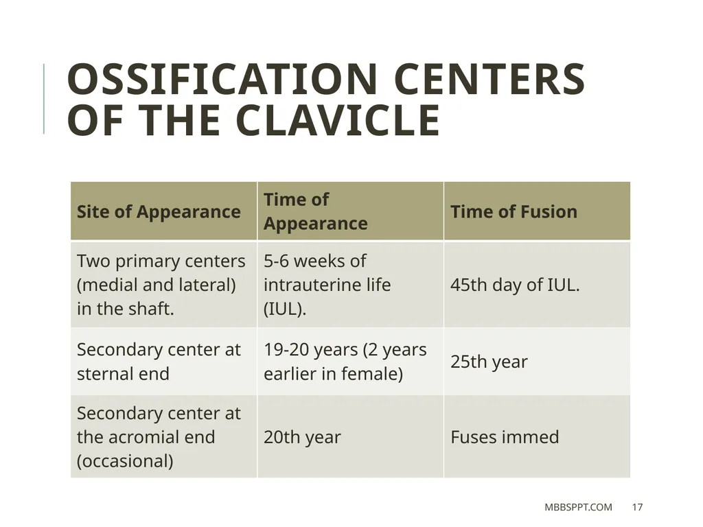
It is the 1st bone to start ossifying (somewhere between the fifth and sixth week of intrauterine life) and very last bone to finish its ossification (at 25 years).
It is the only long bone which ossifies by two primary centers.
It is the only long bone which ossifies in membrane other than its medial end.
It might be pierced inside out by cutaneous nerve (intermediate supraclavicular nerve). PECULIARITIES MBBSPPT.COM 4<br>
It is the only long bone which lies horizontally.
It has absolutely no medullary cavity.
It is subcutaneous throughout its extent.
It is the 1st bone to start ossifying (somewhere between the fifth and sixth week of intrauterine life) and very last bone to finish its ossification (at 25 years).
It is the only long bone which ossifies by two primary centers.
It is the only long bone which ossifies in membrane other than its medial end.
It might be pierced inside out by cutaneous nerve (intermediate supraclavicular nerve). PECULIARITIES MBBSPPT.COM 4<br>
05
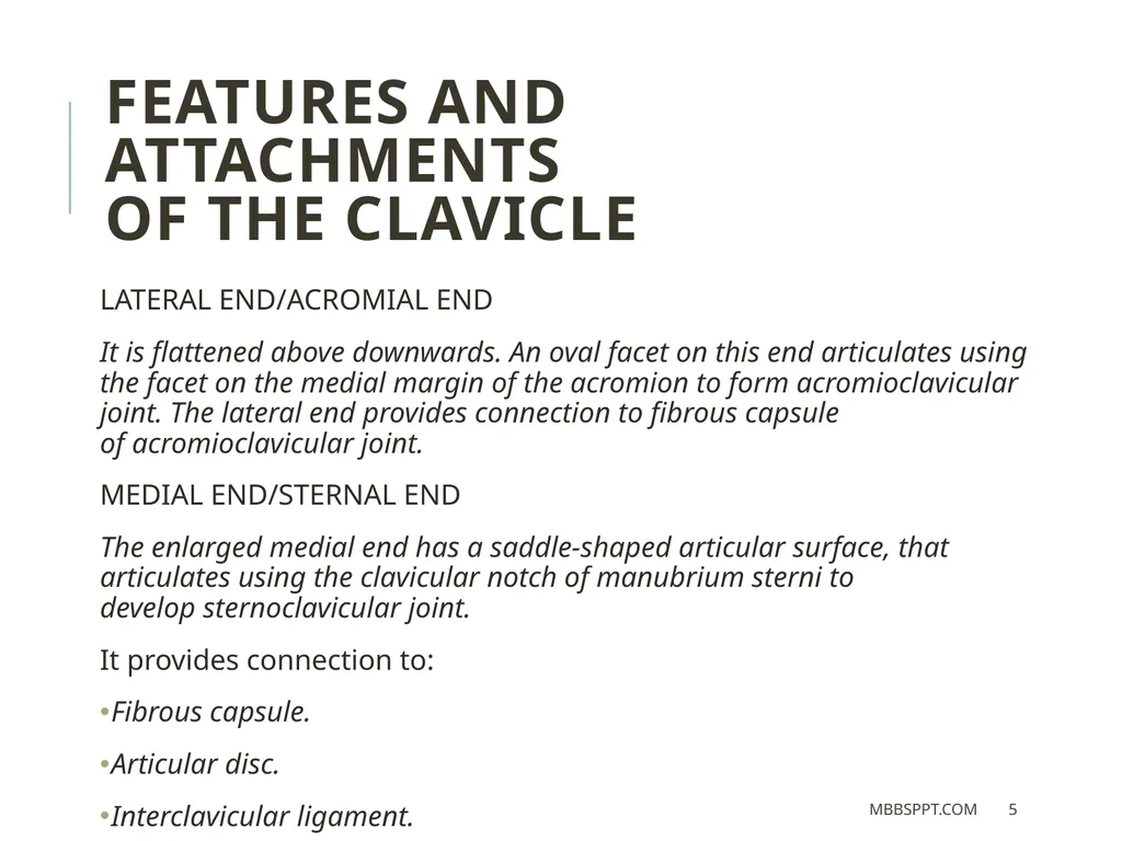
LATERAL END/ACROMIAL END
It is flattened above downwards. An oval facet on this end articulates using the facet on the medial margin of the acromion to form acromioclavicular joint. The lateral end provides connection to fibrous capsule of acromioclavicular joint.
MEDIAL END/STERNAL END
The enlarged medial end has a saddle-shaped articular surface, that articulates using the clavicular notch of manubrium sterni to develop sternoclavicular joint.
It provides connection to:
Fibrous capsule.
Articular disc.
Interclavicular ligament. Features and Attachments
of the clavicle MBBSPPT.COM 5<br>
It is flattened above downwards. An oval facet on this end articulates using the facet on the medial margin of the acromion to form acromioclavicular joint. The lateral end provides connection to fibrous capsule of acromioclavicular joint.
MEDIAL END/STERNAL END
The enlarged medial end has a saddle-shaped articular surface, that articulates using the clavicular notch of manubrium sterni to develop sternoclavicular joint.
It provides connection to:
Fibrous capsule.
Articular disc.
Interclavicular ligament. Features and Attachments
of the clavicle MBBSPPT.COM 5<br>
06
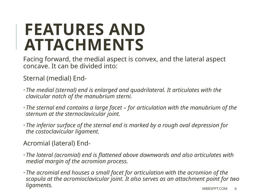
Facing forward, the medial aspect is convex, and the lateral aspect concave. It can be divided into:
Sternal (medial) End-
The medial (sternal) end is enlarged and quadrilateral. It articulates with the clavicular notch of the manubrium sterni.
The sternal end contains a large facet – for articulation with the manubrium of the sternum at the sternoclavicular joint.
The inferior surface of the sternal end is marked by a rough oval depression for the costoclavicular ligament.
Acromial (lateral) End-
The lateral (acromial) end is flattened above downwards and also articulates with medial margin of the acromion process.
The acromial end houses a small facet for articulation with the acromion of the scapula at the acromioclavicular joint. It also serves as an attachment point for two ligaments. Features and Attachments MBBSPPT.COM 6<br>
Sternal (medial) End-
The medial (sternal) end is enlarged and quadrilateral. It articulates with the clavicular notch of the manubrium sterni.
The sternal end contains a large facet – for articulation with the manubrium of the sternum at the sternoclavicular joint.
The inferior surface of the sternal end is marked by a rough oval depression for the costoclavicular ligament.
Acromial (lateral) End-
The lateral (acromial) end is flattened above downwards and also articulates with medial margin of the acromion process.
The acromial end houses a small facet for articulation with the acromion of the scapula at the acromioclavicular joint. It also serves as an attachment point for two ligaments. Features and Attachments MBBSPPT.COM 6<br>
07
Features and Attachments of
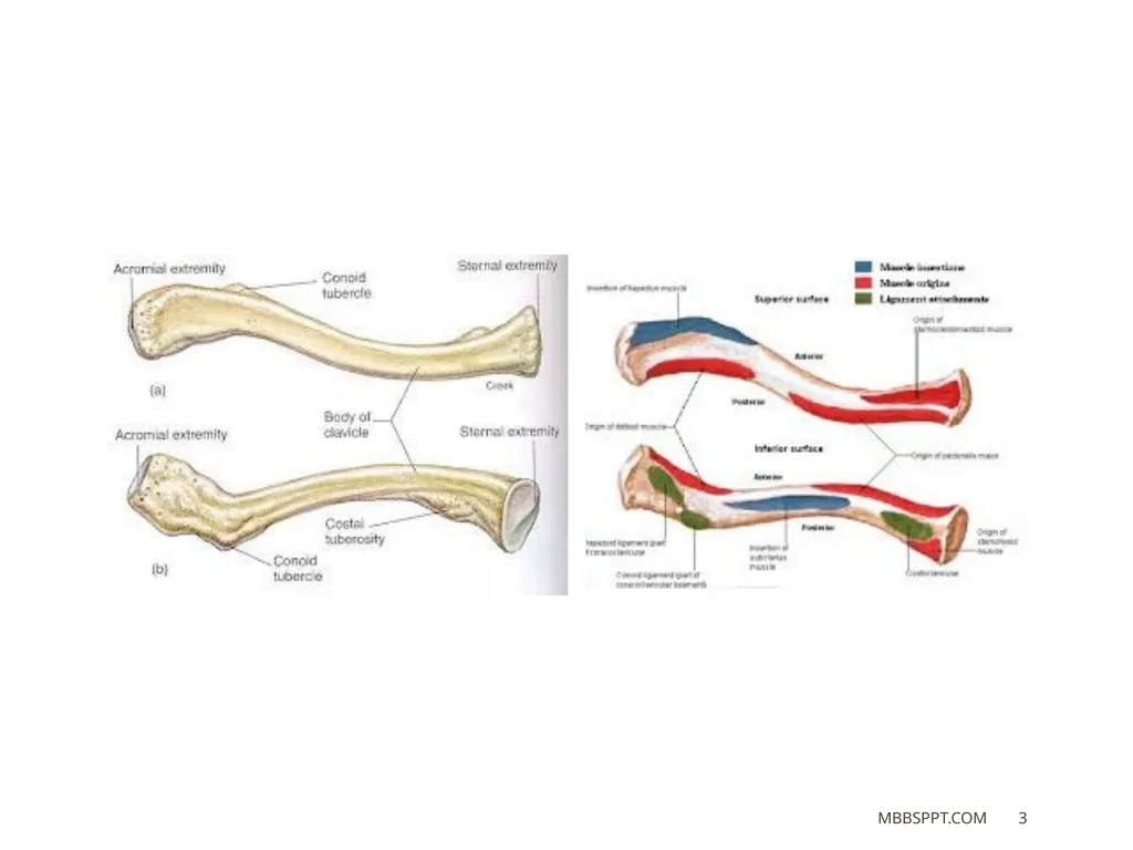
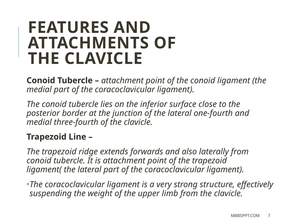
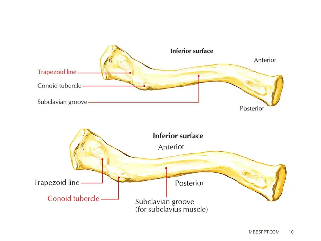
the clavicle Conoid Tubercle – attachment point of the conoid ligament (the medial part of the coracoclavicular ligament).
The conoid tubercle lies on the inferior surface close to the posterior border at the junction of the lateral one-fourth and medial three-fourth of the clavicle.
Trapezoid Line –
The trapezoid ridge extends forwards and also laterally from conoid tubercle. It is attachment point of the trapezoid ligament( the lateral part of the coracoclavicular ligament).
The coracoclavicular ligament is a very strong structure, effectively suspending the weight of the upper limb from the clavicle. MBBSPPT.COM 7<br>
The conoid tubercle lies on the inferior surface close to the posterior border at the junction of the lateral one-fourth and medial three-fourth of the clavicle.
Trapezoid Line –
The trapezoid ridge extends forwards and also laterally from conoid tubercle. It is attachment point of the trapezoid ligament( the lateral part of the coracoclavicular ligament).
The coracoclavicular ligament is a very strong structure, effectively suspending the weight of the upper limb from the clavicle. MBBSPPT.COM 7<br>
08
SHAFT The shaft of the clavicle is split into two parts: lateral one-third and medial two-third.
The medial two-third of shaft is convex forward but lateral one-third is concave forward.
Lateral One-third It is flattened from above downwards.
It has two surfaces, i.e., superior and inferior, and two borders, i.e., anterior and posterior.
The shaft of the clavicle acts a point of origin and attachment for several muscles –
Deltoid, trapezius, subclavius, pectoralis major, sternocleidomastoid and sternohyoid. MBBSPPT.COM 8<br>
The medial two-third of shaft is convex forward but lateral one-third is concave forward.
Lateral One-third It is flattened from above downwards.
It has two surfaces, i.e., superior and inferior, and two borders, i.e., anterior and posterior.
The shaft of the clavicle acts a point of origin and attachment for several muscles –
Deltoid, trapezius, subclavius, pectoralis major, sternocleidomastoid and sternohyoid. MBBSPPT.COM 8<br>
09
SURFACES Superior Surface:
The clavicular head of sternocleidomastoid muscle originates from medial half of this surface.
It is subcutaneous somewhere between the attachments of deltoid and trapezius.
Inferior Surface: It provides a conoid tubercle and trapezoid ridge, which provide attachments to conoid and trapezoid component of coracoclavicular ligament, respectively.
Costoclavicular ligament is attached to an oval impression at its medial end.
Subclavius muscle is inserted into the subclavian groove on this surface.
Clavipectoral fascia is attached to the margins of subclavian groove.
Nutrient foramen of clavicle is found on the lateral end of the subclavian groove. MBBSPPT.COM 9<br>
The clavicular head of sternocleidomastoid muscle originates from medial half of this surface.
It is subcutaneous somewhere between the attachments of deltoid and trapezius.
Inferior Surface: It provides a conoid tubercle and trapezoid ridge, which provide attachments to conoid and trapezoid component of coracoclavicular ligament, respectively.
Costoclavicular ligament is attached to an oval impression at its medial end.
Subclavius muscle is inserted into the subclavian groove on this surface.
Clavipectoral fascia is attached to the margins of subclavian groove.
Nutrient foramen of clavicle is found on the lateral end of the subclavian groove. MBBSPPT.COM 9<br>
10
MBBSPPT.COM 10<br>
11
SURFACES Anterior surface: It is convex forwards and provides origin to clavicular head of pectoralis major.
Posterior surface: It is concave backwards and provides origin to sternohyoid muscle close to its medial end. The lateral component of this surface forms the anterior boundary of cervico-axillary canal and relates to the following structures:
Trunks of brachial plexus.
Third part of subclavian artery. MBBSPPT.COM 11<br>
Posterior surface: It is concave backwards and provides origin to sternohyoid muscle close to its medial end. The lateral component of this surface forms the anterior boundary of cervico-axillary canal and relates to the following structures:
Trunks of brachial plexus.
Third part of subclavian artery. MBBSPPT.COM 11<br>
12
BORDERS Anterior border: It is concave forwards and provides origin to deltoid muscle. A small tubercle know as deltoid tubercle might exist on this border.
Posterior border: It is convex backwards and supplies insertion to the trapezius muscle.
Medial Two-third It is cylindrical in shape and provides four surfaces: anterior, posterior, superior, and inferior. MBBSPPT.COM 12<br>
Posterior border: It is convex backwards and supplies insertion to the trapezius muscle.
Medial Two-third It is cylindrical in shape and provides four surfaces: anterior, posterior, superior, and inferior. MBBSPPT.COM 12<br>
13
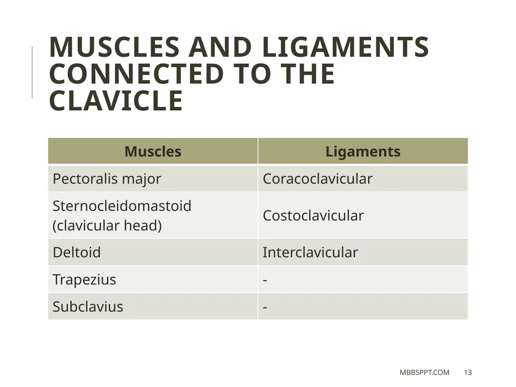
MUSCLES AND LIGAMENTS CONNECTED TO THE CLAVICLE MBBSPPT.COM 13<br>
14
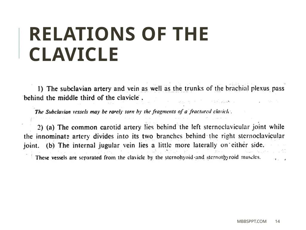
Relations of the clavicle MBBSPPT.COM 14<br>
15
ANATOMICAL POSITION
AND SIDE DETERMINATION The side of clavicle can be identified by holding the bone horizontally as though its flattened end lies on the lateral side and its enlarged quadrilateral end lies on the medial side.
The convexity of its medial two-third and concavity of its lateral one-third face forwards having longitudinal groove in the middle third of shaft facing inferiorly. MBBSPPT.COM 15<br>
The convexity of its medial two-third and concavity of its lateral one-third face forwards having longitudinal groove in the middle third of shaft facing inferiorly. MBBSPPT.COM 15<br>
16
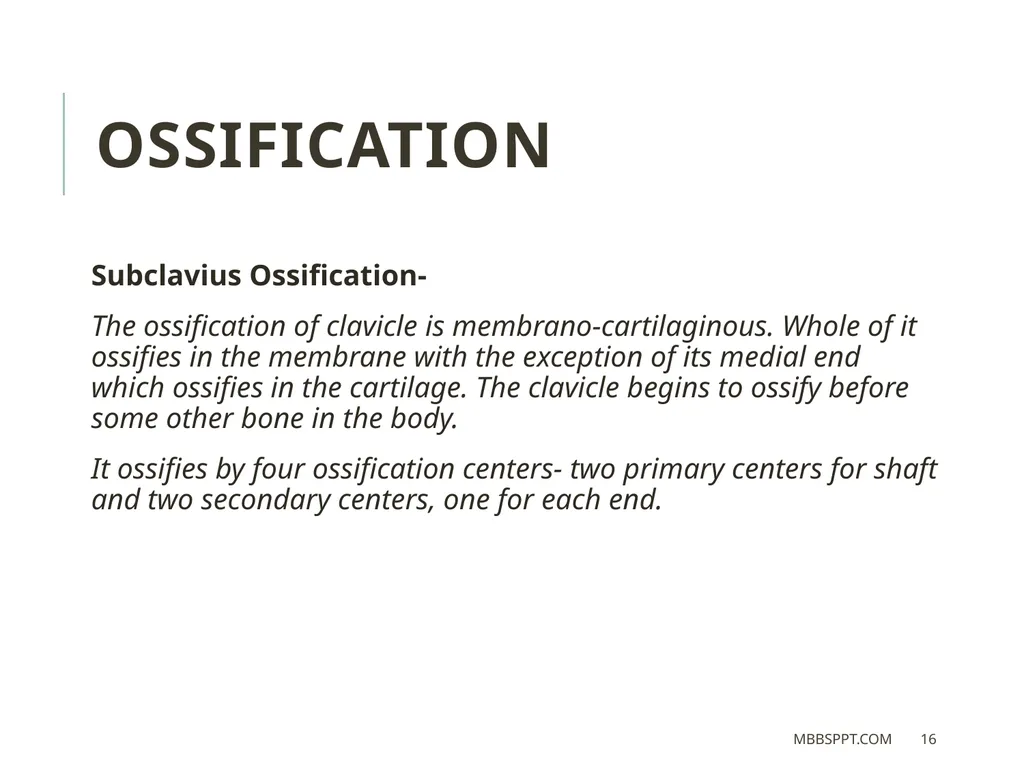
OSSIFICATION Subclavius Ossification-
The ossification of clavicle is membrano-cartilaginous. Whole of it ossifies in the membrane with the exception of its medial end which ossifies in the cartilage. The clavicle begins to ossify before some other bone in the body.
It ossifies by four ossification centers- two primary centers for shaft and two secondary centers, one for each end. MBBSPPT.COM 16<br>
The ossification of clavicle is membrano-cartilaginous. Whole of it ossifies in the membrane with the exception of its medial end which ossifies in the cartilage. The clavicle begins to ossify before some other bone in the body.
It ossifies by four ossification centers- two primary centers for shaft and two secondary centers, one for each end. MBBSPPT.COM 16<br>
17
Ossification centers
of the Clavicle MBBSPPT.COM 17<br>
of the Clavicle MBBSPPT.COM 17<br>
18
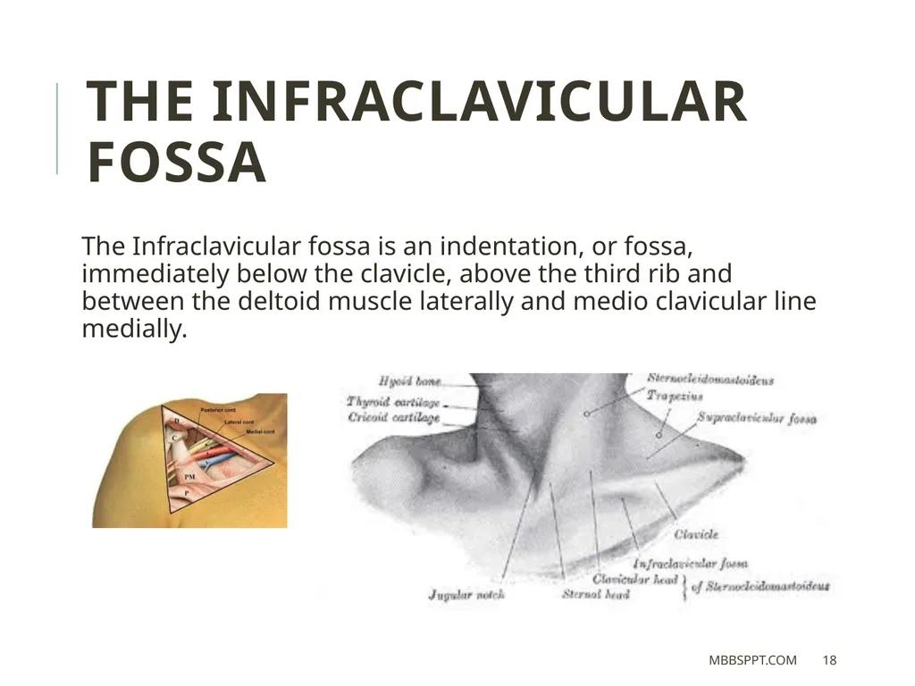
The Infraclavicular fossa The Infraclavicular fossa is an indentation, or fossa, immediately below the clavicle, above the third rib and between the deltoid muscle laterally and medio clavicular line medially. MBBSPPT.COM 18<br>
19
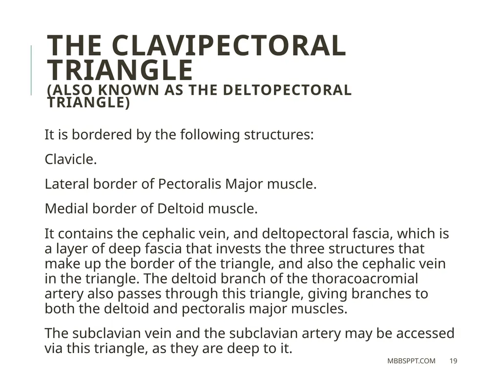
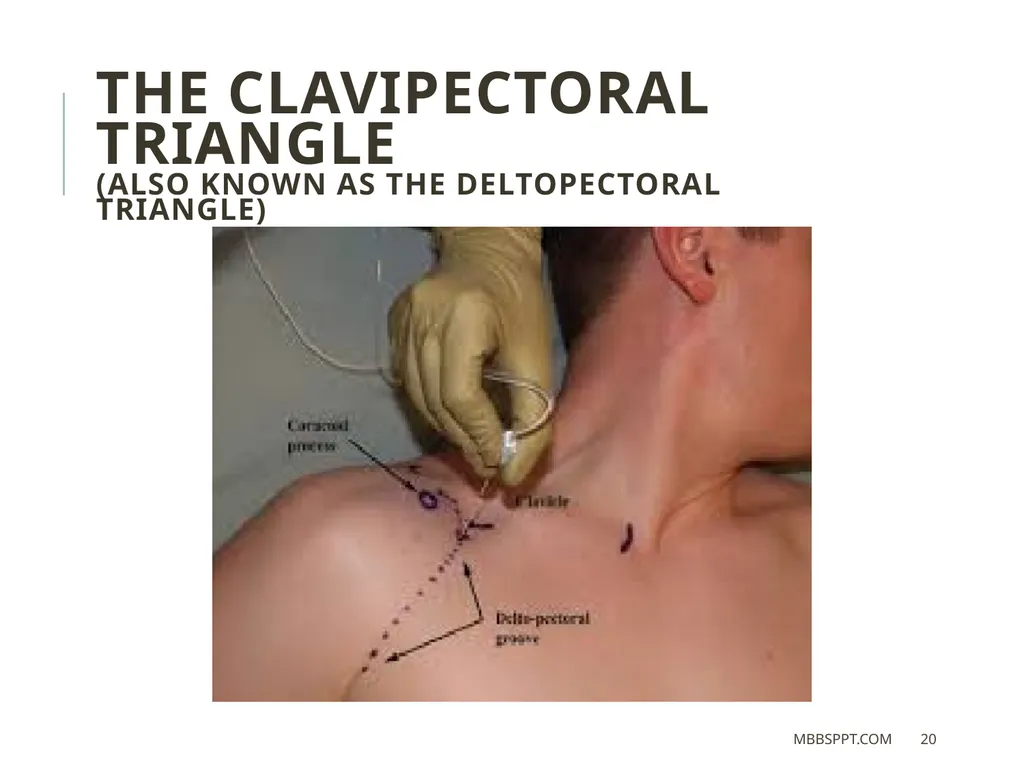
The clavipectoral triangle
(also known as the deltopectoral triangle) It is bordered by the following structures:
Clavicle.
Lateral border of Pectoralis Major muscle.
Medial border of Deltoid muscle.
It contains the cephalic vein, and deltopectoral fascia, which is a layer of deep fascia that invests the three structures that make up the border of the triangle, and also the cephalic vein in the triangle. The deltoid branch of the thoracoacromial artery also passes through this triangle, giving branches to both the deltoid and pectoralis major muscles.
The subclavian vein and the subclavian artery may be accessed via this triangle, as they are deep to it. MBBSPPT.COM 19<br>
(also known as the deltopectoral triangle) It is bordered by the following structures:
Clavicle.
Lateral border of Pectoralis Major muscle.
Medial border of Deltoid muscle.
It contains the cephalic vein, and deltopectoral fascia, which is a layer of deep fascia that invests the three structures that make up the border of the triangle, and also the cephalic vein in the triangle. The deltoid branch of the thoracoacromial artery also passes through this triangle, giving branches to both the deltoid and pectoralis major muscles.
The subclavian vein and the subclavian artery may be accessed via this triangle, as they are deep to it. MBBSPPT.COM 19<br>
20
The clavipectoral triangle
(also known as the deltopectoral triangle) MBBSPPT.COM 20<br>
(also known as the deltopectoral triangle) MBBSPPT.COM 20<br>
21
Fracture of clavicle The clavicle is the most commonly fractured bone in the body. It commonly fractures at the junction of its lateral one-third and medial two-third due to blows to the shoulder or indirect forces, usually as a result of strong impact on the hand or shoulder, when person falls on the outstretched hand or the shoulder.
When fracture occurs, the lateral fragment is displaced downward by the weight of the upper limb because trapezius alone is unable to support the weight of the upper limb.
In addition, the lateral fragment is drawn medially by shoulder adductors viz. teres major, etc. The medial fragment is slightly elevated by the sternocleidomastoid muscle.
The characteristic clinical picture of the patient with fractured clavicle is that of a man/woman supporting his sagging upper limb with the opposite hand. MBBSPPT.COM 21<br>
When fracture occurs, the lateral fragment is displaced downward by the weight of the upper limb because trapezius alone is unable to support the weight of the upper limb.
In addition, the lateral fragment is drawn medially by shoulder adductors viz. teres major, etc. The medial fragment is slightly elevated by the sternocleidomastoid muscle.
The characteristic clinical picture of the patient with fractured clavicle is that of a man/woman supporting his sagging upper limb with the opposite hand. MBBSPPT.COM 21<br>
22
Clinical Relevance
Fracture of the Clavicle The clavicle acts to transmit forces from the upper limb to the axial skeleton. Given its relative size, this leaves it particularly susceptible to fracture. The most common mechanism of injury is a fall onto the shoulder, or onto an outstretched hand.
With the clavicle arbitrarily divided into thirds:
15% of fractures occur in the lateral third.
80% occur in the middle third
5% occur in the medial third.
After a fracture, the lateral end of the clavicle is displaced inferiorly by the weight of the arm, and displaced medially by the pectoralis major. The medial end is pulled superiorly by the sternocleidomastoid muscle.
Management of a clavicular fracture can be conservative (e.g. sling immobilisation) or operative (e.g. open reduction and internal fixation, ORIF). In ORIF, the supraclavicular nerves are often sacrificed – resulting in a post-operative numb patch over the shoulder. MBBSPPT.COM 22<br>
With the clavicle arbitrarily divided into thirds:
15% of fractures occur in the lateral third.
80% occur in the middle third
5% occur in the medial third.
After a fracture, the lateral end of the clavicle is displaced inferiorly by the weight of the arm, and displaced medially by the pectoralis major. The medial end is pulled superiorly by the sternocleidomastoid muscle.
Management of a clavicular fracture can be conservative (e.g. sling immobilisation) or operative (e.g. open reduction and internal fixation, ORIF). In ORIF, the supraclavicular nerves are often sacrificed – resulting in a post-operative numb patch over the shoulder. MBBSPPT.COM 22<br>
23
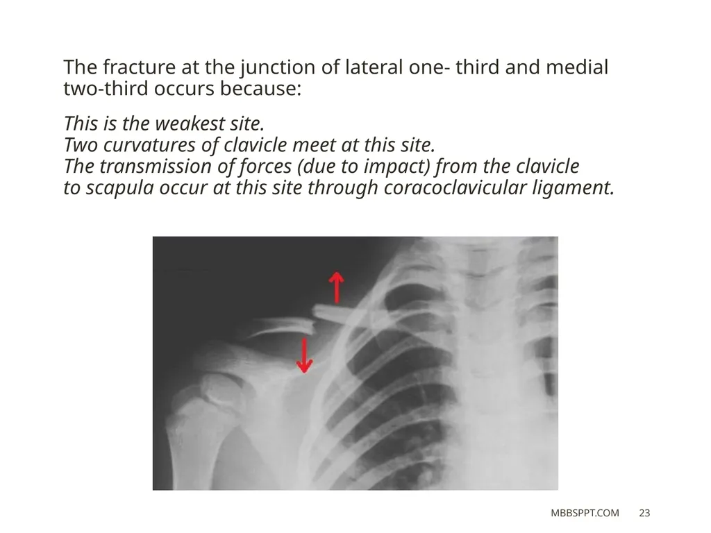
The fracture at the junction of lateral one- third and medial two-third occurs because:
This is the weakest site. Two curvatures of clavicle meet at this site. The transmission of forces (due to impact) from the clavicle to scapula occur at this site through coracoclavicular ligament. MBBSPPT.COM 23<br>
This is the weakest site. Two curvatures of clavicle meet at this site. The transmission of forces (due to impact) from the clavicle to scapula occur at this site through coracoclavicular ligament. MBBSPPT.COM 23<br>
24
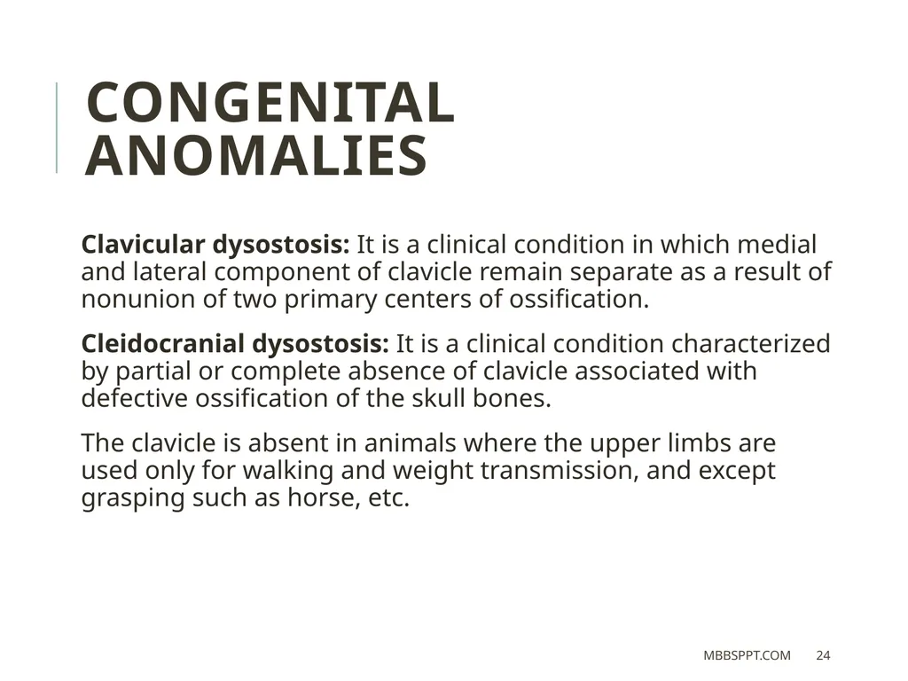
CONGENITAL ANOMALIES Clavicular dysostosis: It is a clinical condition in which medial and lateral component of clavicle remain separate as a result of nonunion of two primary centers of ossification.
Cleidocranial dysostosis: It is a clinical condition characterized by partial or complete absence of clavicle associated with defective ossification of the skull bones.
The clavicle is absent in animals where the upper limbs are used only for walking and weight transmission, and except grasping such as horse, etc. MBBSPPT.COM 24<br>
Cleidocranial dysostosis: It is a clinical condition characterized by partial or complete absence of clavicle associated with defective ossification of the skull bones.
The clavicle is absent in animals where the upper limbs are used only for walking and weight transmission, and except grasping such as horse, etc. MBBSPPT.COM 24<br>
25
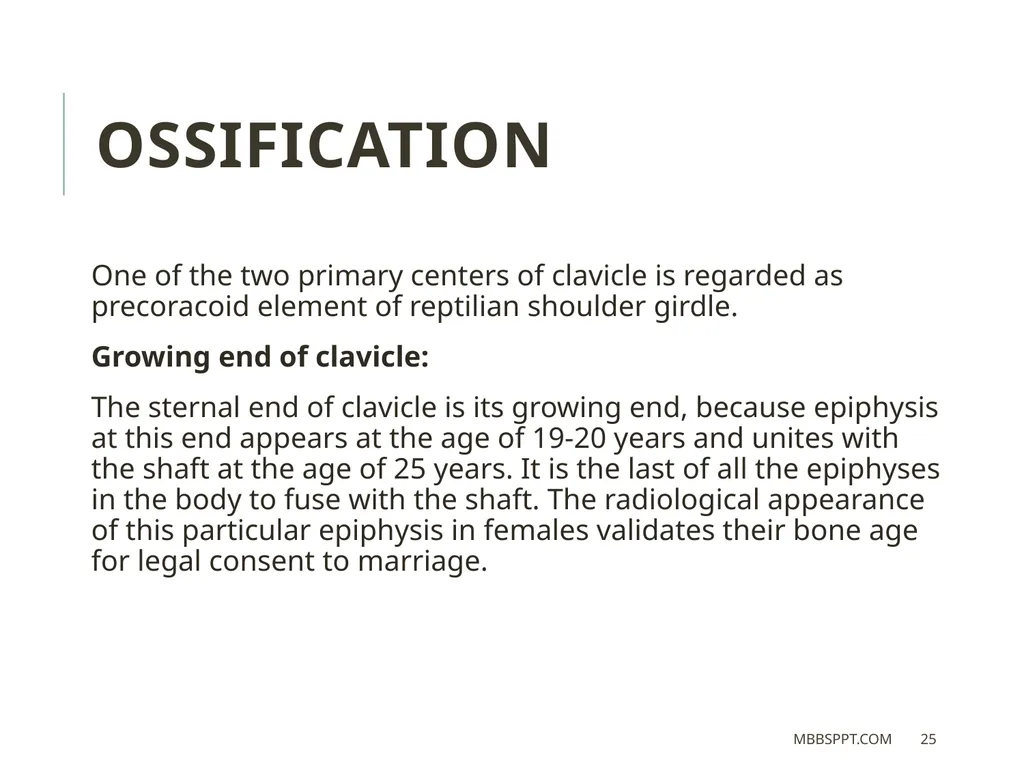
OSSIFICATION One of the two primary centers of clavicle is regarded as precoracoid element of reptilian shoulder girdle.
Growing end of clavicle:
The sternal end of clavicle is its growing end, because epiphysis at this end appears at the age of 19-20 years and unites with the shaft at the age of 25 years. It is the last of all the epiphyses in the body to fuse with the shaft. The radiological appearance of this particular epiphysis in females validates their bone age for legal consent to marriage. MBBSPPT.COM 25<br>
Growing end of clavicle:
The sternal end of clavicle is its growing end, because epiphysis at this end appears at the age of 19-20 years and unites with the shaft at the age of 25 years. It is the last of all the epiphyses in the body to fuse with the shaft. The radiological appearance of this particular epiphysis in females validates their bone age for legal consent to marriage. MBBSPPT.COM 25<br>
26
Thank You MBBSPPT.COM 26<br>