
The kidneys 54 Negative pressure on the dialysate
EM
Published · 3 slides · 0 views

1 / 1
Description
The kidneys 54 Negative pressure on the dialysate side of the membrane - a high rate of excretion of the metabolic can be varied to adjust the amount of water removed. product forming the stone, due either to In intermittent and continuous
Related Topics
Share
Embed code
Download this presentation From Below
"The kidneys 54 Negative pressure on the dialysate" is the property of its rightful owner. Permission is granted to download and print the materials on this website for personal, non-commercial use only, and to display it on your personal computer provided you do not modify the materials and that you retain all copyright notices contained in the materials. By downloading content from our website, you accept the terms of this agreement.
Presentation Transcript
01
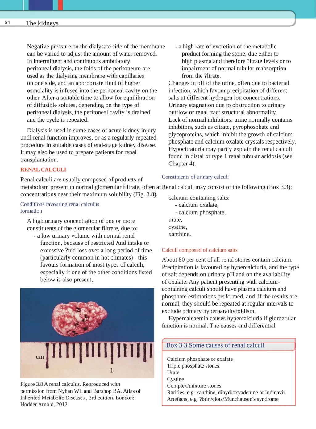
The kidneys 54 Negative pressure on the dialysate side of the membrane - a high rate of excretion of the metabolic can be varied to adjust the amount of water removed. product forming the stone, due either to In intermittent and continuous ambulatory high plasma and therefore ?ltrate levels or to peritoneal dialysis, the folds of the peritoneum are impairment of normal tubular reabsorption used as the dialysing membrane with capillaries from the ?ltrate. on one side, and an appropriate fluid of higher Changes in pH of the urine, often due to bacterial osmolality is infused into the peritoneal cavity on the infection, which favour precipitation of different other. After a suitable time to allow for equilibration salts at different hydrogen ion concentrations. of diffusible solutes, depending on the type of Urinary stagnation due to obstruction to urinary peritoneal dialysis, the peritoneal cavity is drained outflow or renal tract structural abnormality. and the cycle is repeated. Lack of normal inhibitors: urine normally contains inhibitors, such as citrate, pyrophosphate and Dialysis is used in some cases of acute kidney injury glycoproteins, which inhibit the growth of calcium until renal function improves, or as a regularly repeated phosphate and calcium oxalate crystals respectively. procedure in suitable cases of end-stage kidney disease. Hypocitraturia may partly explain the renal calculi It may also be used to prepare patients for renal found in distal or type 1 renal tubular acidosis (see transplantation. Chapter 4). RENAL CALCULI Constituents of urinary calculi Renal calculi are usually composed of products of metabolism present in normal glomerular filtrate, often at Renal calculi may consist of the following (Box 3.3): concentrations near their maximum solubility (Fig. 3.8). calcium-containing salts: Conditions favouring renal calculus - calcium oxalate, formation - calcium phosphate, urate, A high urinary concentration of one or more cystine, constituents of the glomerular filtrate, due to: xanthine. - a low urinary volume with normal renal function, because of restricted ?uid intake or excessive ?uid loss over a long period of time Calculi composed of calcium salts (particularly common in hot climates) - this About 80 per cent of all renal stones contain calcium. favours formation of most types of calculi, Precipitation is favoured by hypercalciuria, and the type especially if one of the other conditions listed of salt depends on urinary pH and on the availability below is also present, of oxalate. Any patient presenting with calcium- containing calculi should have plasma calcium and phosphate estimations performed, and, if the results are normal, they should be repeated at regular intervals to exclude primary hyperparathyroidism. Hypercalcaemia causes hypercalciuria if glomerular function is normal. The causes and differential Box 3.3 Some causes of renal calculi cm Calcium phosphate or oxalate Triple phosphate stones 1 Urate Cystine Figure 3.8 A renal calculus. Reproduced with Complex/mixture stones permission from Nyhan WL and Barshop BA. Atlas of Rarities, e.g. xanthine, dihydroxyadenine or indinavir Inherited Metabolic Diseases , 3rd edition. London: Artefacts, e.g. ?brin/clots/Munchausen's syndrome Hodder Arnold, 2012.<br>
02
Renal calculi 55 diagnosis of hypercalcaemia are discussed in Chapter by treating hypercalcaemia if present, 6. In many subjects with calcium-containing renal if this is not possible, by reducing dietary calcium calculi the plasma calcium concentration is normal. (although this alone may exacerbate hyperoxaluria) Any increased release of calcium from bone (as and oxalate intake, in actively progressing osteoporosis, in which loss by maintaining a high fluid intake, unless there is of matrix causes secondary decalci?cation, or in glomerular failure. prolonged acidosis, in which ionization of calcium Thiazide diuretics reduce urinary calcium excretion, is increased) causes hypercalciuria; hypercalcaemia and treatment of urinary tract infection may reduce the is unusual in such cases. In distal renal tubular risk of calculi formation. acidosis there is an increased calcium load and, because of the relative alkalinity of the urine, calcium Struvite (magnesium ammonium phosphate) precipitation in the kidney and renal tract may occur These stones (about 10 per cent of all renal calculi) - nephrocalcinosis. are associated with chronic urinary tract infections by Hypercalciuria has been de?ned as a daily urinary organisms such as Proteus species capable of splitting calcium excretion of more than 6.2 mmol in adult ammonium. The urinary pH is usually greater than 7. females and 7.5 mmol in adult males. These urease-containing bacteria convert urea to A signi?cant proportion of cases remain in which ammonia and bicarbonate. there is no apparent cause for calcium precipitation. A common cause is hypercalciuria despite Uric acid stones normocalcaemia (see Chapter 6). About 8 per cent of renal calculi contain uric acid; these Hyperoxaluria favours the formation of the very are sometimes associated with hyperuricaemia, with or poorly soluble calcium oxalate, even if calcium without clinical gout. In most cases, no predisposing excretion is normal. The source of the oxalate may be cause can be found. Precipitation is favoured in an acid derived exogenously from the diet. Oxalate absorption urine. Uric acid stones are usually small, friable and is increased by fat malabsorption: calcium in the bowel yellowish brown, but can occasionally be large enough is bound to fat instead of precipitating with oxalate, to form `staghorn' calculi. They are radiolucent but which is then free to be absorbed. Foods rich in oxalate may be visualized by ultrasound or by an intravenous include rhubarb, chocolate, beetroot, spinach, nuts pyelogram. and tea. The treatment of hyperuricaemia is discussed Primary hyperoxaluria, a rare inborn error, should in Chapter 20. If the plasma urate concentration is be considered if renal calculi occur in childhood. normal, ?uid intake should be kept high and the urine There are two main types, 1 and 2, the former being alkalinized. A low-purine diet may help to reduce urate more common. Type 1 is due to de?ciency of alanine production and excretion. glyoxylate aminotransferase, and type 2 is due to de?cient D -glycerate dehydrogenase. Hyperoxaluria Cystine stones (urinary oxalate greater than 400 æmol/24 h) is a more Cystine stones are rare. In normal subjects the important risk factor for formation of renal stones than concentration of cystine in urine is soluble, but in is hypercalciuria. homozygous cystinuria this may be exceeded and the Calcium-containing calculi are usually hard, white patient may present with radio-opaque renal calculi. and radio-opaque. Calcium phosphate may form Like urate, cystine is more soluble in alkaline than in `staghorn' calculi in the renal pelvis, while calcium acidic urine; the principles of treatment are the same as oxalate stones tend to be smaller and to lodge in the for uric acid stones. Penicillamine can also be used to ureters, where they are compressed into a fusiform treat the condition (see Chapter 27). shape. Alkaline conditions favouring calcium phosphate precipitation and stone formation are particularly Miscellaneous stones common in patients with chronic renal infection. Xanthine stones The treatment of calcium-containing calculi depends Xanthine stones are very uncommon and may be the on the cause. Urinary calcium concentration should be result of the rare inborn error xanthinuria. reduced:<br>
03
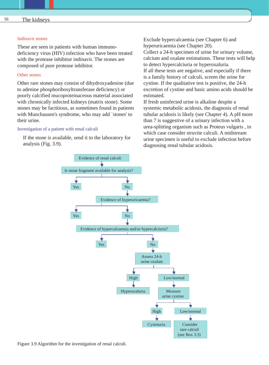
The kidneys 56 Indinavir stones Exclude hypercalcaemia (see Chapter 6) and hyperuricaemia (see Chapter 20). These are seen in patients with human immuno- Collect a 24-h specimen of urine for urinary volume, deficiency virus (HIV) infection who have been treated calcium and oxalate estimations. These tests will help with the protease inhibitor indinavir. The stones are to detect hypercalciuria or hyperoxaluria. composed of pure protease inhibitor. If all these tests are negative, and especially if there Other stones is a family history of calculi, screen the urine for cystine. If the qualitative test is positive, the 24-h Other rare stones may consist of dihydroxyadenine (due excretion of cystine and basic amino acids should be to adenine phosphoribosyltransferase deficiency) or estimated. poorly calcified mucoproteinaceous material associated If fresh uninfected urine is alkaline despite a with chronically infected kidneys (matrix stone). Some systemic metabolic acidosis, the diagnosis of renal stones may be factitious, as sometimes found in patients tubular acidosis is likely (see Chapter 4). A pH more with Munchausen's syndrome, who may add `stones' to than 7 is suggestive of a urinary infection with a their urine. urea-splitting organism such as Proteus vulgaris , in Investigation of a patient with renal calculi which case consider struvite calculi. A midstream If the stone is available, send it to the laboratory for urine specimen is useful to exclude infection before analysis (Fig. 3.9). diagnosing renal tubular acidosis. Evidence of renal calculi Is stone fragment available for analysis? No Yes Evidence of hyperuricaemia? No Yes Evidence of hypercalcaemia and/or hypercalciuria? No Yes Assess 24-h urine oxalate Low/normal High Hyperoxaluria Measure urine cystine Low/normal High Cystinuria Consider rare calculi (see Box 3.3) Figure 3.9 Algorithm for the investigation of renal calculi.<br>