
Tishk International University Dentistry Faculty
FD
Published · 69 slides · 0 views

1 / 1







Description
Tishk International University Dentistry Faculty Periodontics Department Periodontology Furcation Involvement and Its Management Instructor: Dr. Jafar Naghshbandi D.D.S;M.S Diplomate of American Board of Periodontology Furcation Involvement
Related Topics
Share
Embed code
Download this presentation From Below
"Tishk International University Dentistry Faculty" is the property of its rightful owner. Permission is granted to download and print the materials on this website for personal, non-commercial use only, and to display it on your personal computer provided you do not modify the materials and that you retain all copyright notices contained in the materials. By downloading content from our website, you accept the terms of this agreement.
Presentation Transcript
01
Tishk International University
Dentistry Faculty
Periodontics Department Periodontology Furcation Involvement and Its Management Instructor: Dr. Jafar Naghshbandi D.D.S;M.S
Diplomate of American Board of Periodontology<br>
Dentistry Faculty
Periodontics Department Periodontology Furcation Involvement and Its Management Instructor: Dr. Jafar Naghshbandi D.D.S;M.S
Diplomate of American Board of Periodontology<br>
02
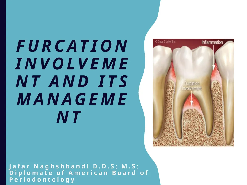
Furcation Involvement and Its Management Jafar Naghshbandi D.D.S; M.S;
Diplomate of American Board of Periodontology<br>
03
Objevtives Introduction
Anatomical considerations
Classification
Etiopathogenesis
Diagnosis
Management
Factors affecting clinical outcome
Key Notes<br>
Anatomical considerations
Classification
Etiopathogenesis
Diagnosis
Management
Factors affecting clinical outcome
Key Notes<br>
04
‘’whoever taught me a word made me his servant’’ Dr.Raul Caffesse Dr. Jim Simon ‘’whoever taught me a word made me his servant’’ Dr.Raul Caffesse Dr. Jim Simon<br>
05
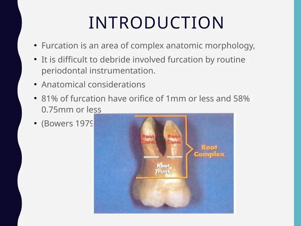
Introduction Furcation is an area of complex anatomic morphology,
It is difficult to debride involved furcation by routine periodontal instrumentation.
Anatomical considerations
81% of furcation have orifice of 1mm or less and 58% 0.75mm or less
(Bowers 1979)<br>
It is difficult to debride involved furcation by routine periodontal instrumentation.
Anatomical considerations
81% of furcation have orifice of 1mm or less and 58% 0.75mm or less
(Bowers 1979)<br>
06
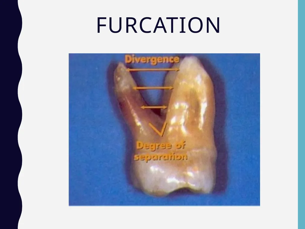
Furcation<br>
07
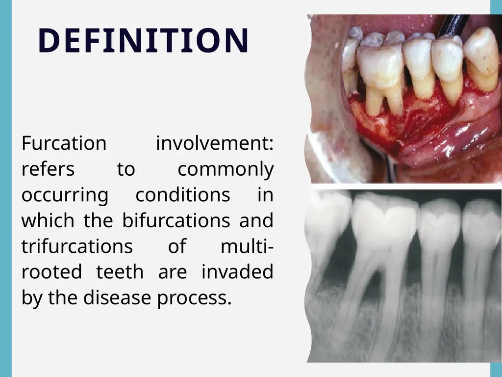
DEFINITION Furcation involvement: refers to commonly occurring conditions in which the bifurcations and trifurcations of multi-rooted teeth are invaded by the disease process.<br>
08
Furcation Involvements • The term furcation involvement refers to the invasion of the bifurcation and trifurcation of multi-rooted teeth by periodontal disease
• The prevalence of furcation involved molars is not clear
Some studies indicate that the mandibular first molars are the most common sites and the maxillary premolars are the least common, others have found higher prevalence in upper molars.
• The number of furcation involvements increases with age<br>
• The prevalence of furcation involved molars is not clear
Some studies indicate that the mandibular first molars are the most common sites and the maxillary premolars are the least common, others have found higher prevalence in upper molars.
• The number of furcation involvements increases with age<br>
09
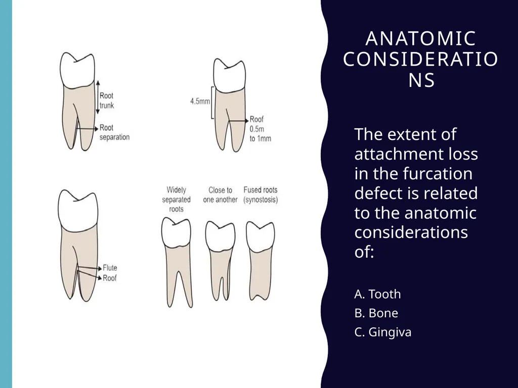
Anatomic considerations The extent of attachment loss in the furcation defect is related to the anatomic considerations of:
A. Tooth
B. Bone
C. Gingiva<br>
A. Tooth
B. Bone
C. Gingiva<br>
10
Tooth The following features should be considered:
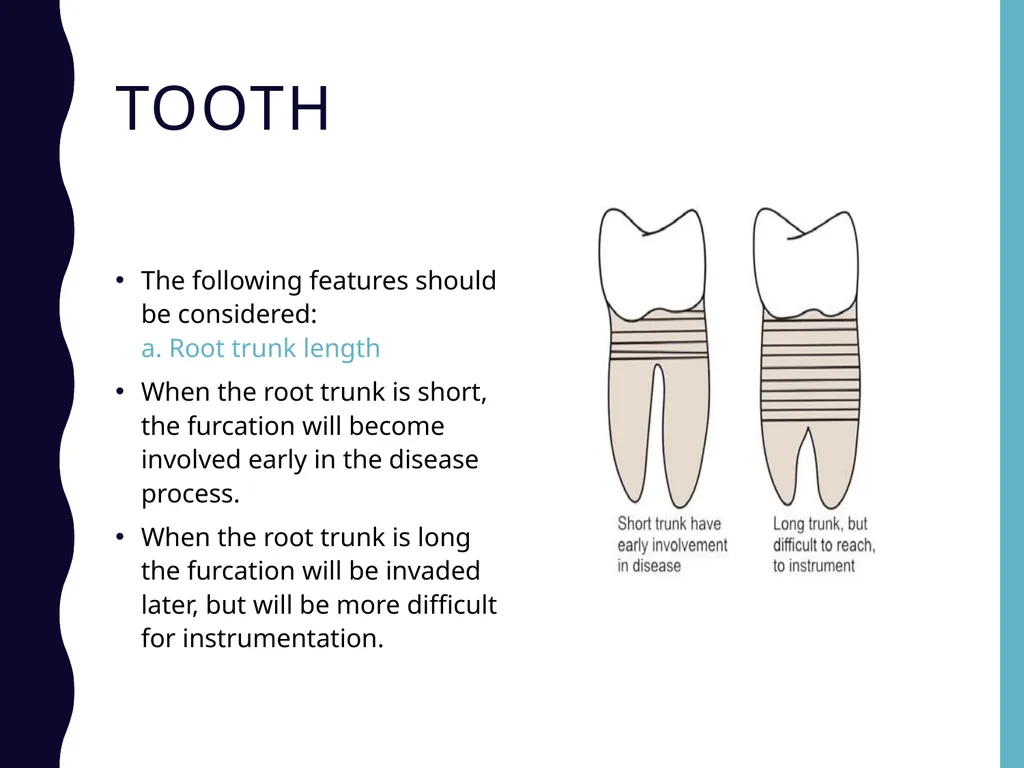
a. Root trunk length
When the root trunk is short, the furcation will become involved early in the disease process.
When the root trunk is long the furcation will be invaded later, but will be more difficult for instrumentation.<br>
When the root trunk is short, the furcation will become involved early in the disease process.
When the root trunk is long the furcation will be invaded later, but will be more difficult for instrumentation.<br>
11
Tooth b. Concavity of the inner surface of exposed roots
All the root surfaces facing the furcation exhibit some degree of concavity or depression in an occluso-apical direction.
This may make instrumentation for plaque removal and root planing almost impossible.
But these concavities increases the attachment area of a tooth and produce a root shape that is resistant to torque.
It is common in mesiobuccal root of maxillary first molar and mesial root of mandibular first molar.<br>
All the root surfaces facing the furcation exhibit some degree of concavity or depression in an occluso-apical direction.
This may make instrumentation for plaque removal and root planing almost impossible.
But these concavities increases the attachment area of a tooth and produce a root shape that is resistant to torque.
It is common in mesiobuccal root of maxillary first molar and mesial root of mandibular first molar.<br>
12
Tooth c. Degree of separation of roots/inter-radicular dimension
Wide separation of roots improves access, thereby facilitating instrumentation.
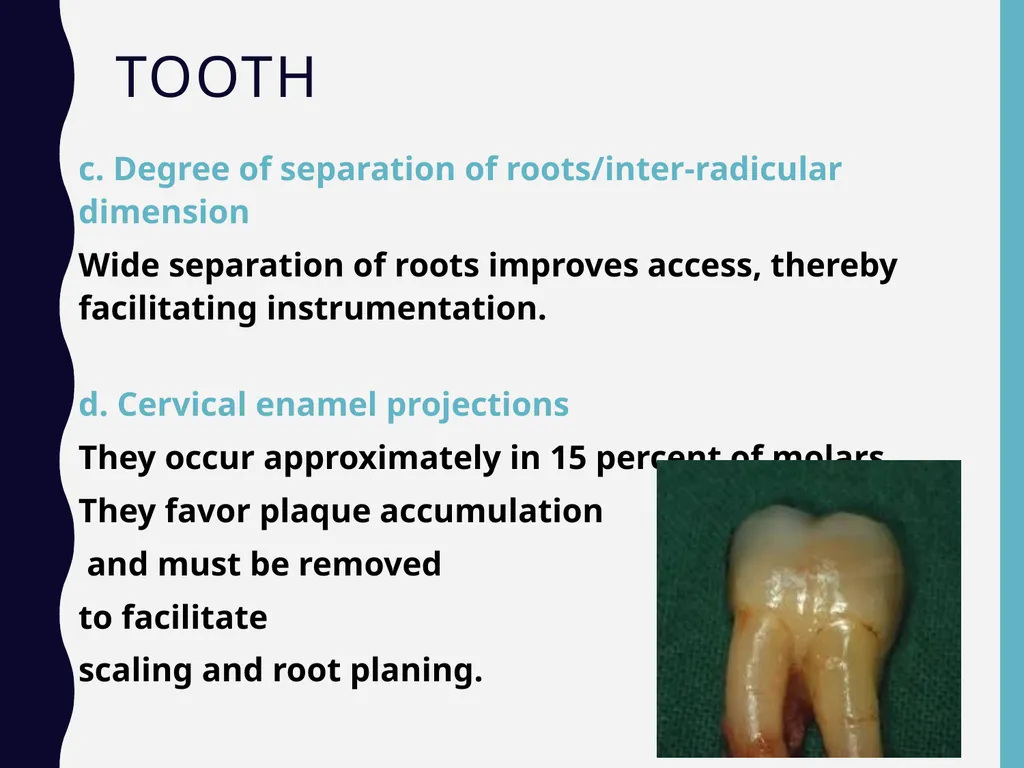
d. Cervical enamel projections
They occur approximately in 15 percent of molars.
They favor plaque accumulation
and must be removed
to facilitate
scaling and root planing.<br>
Wide separation of roots improves access, thereby facilitating instrumentation.
d. Cervical enamel projections
They occur approximately in 15 percent of molars.
They favor plaque accumulation
and must be removed
to facilitate
scaling and root planing.<br>
13
Tooth e. Anatomy of the furcation
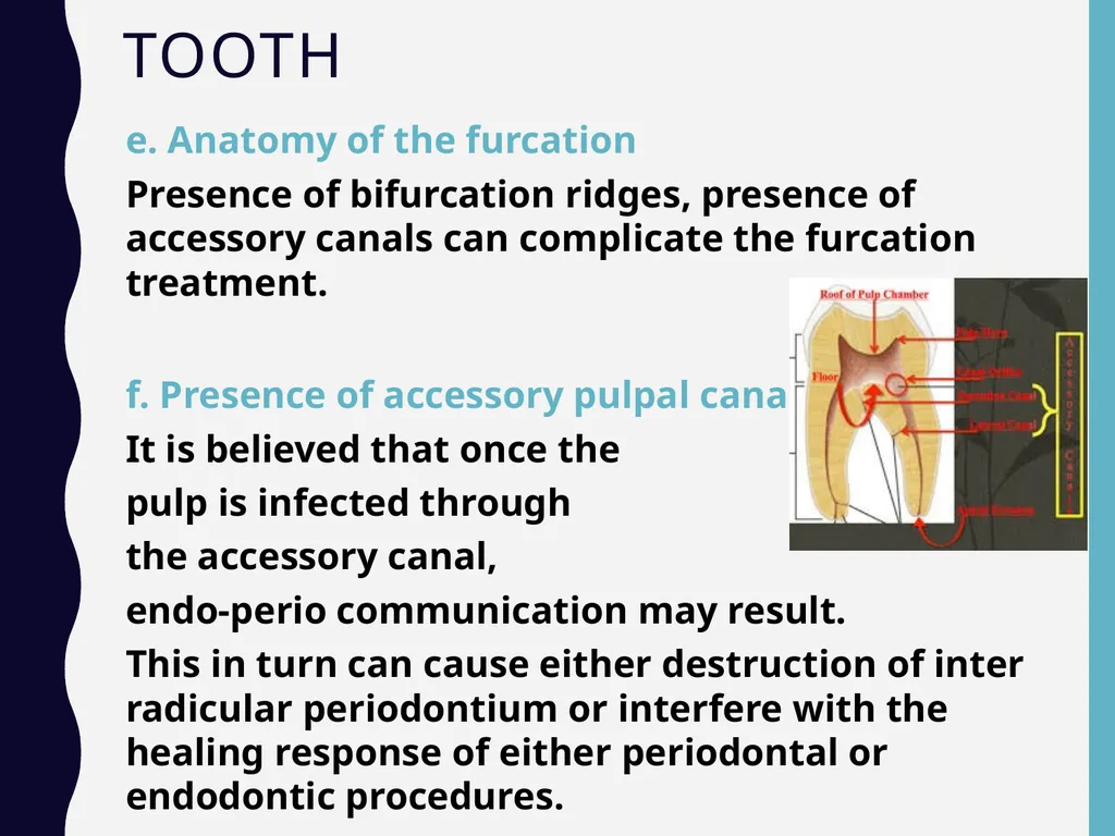
Presence of bifurcation ridges, presence of accessory canals can complicate the furcation treatment.
f. Presence of accessory pulpal canals
It is believed that once the
pulp is infected through
the accessory canal,
endo-perio communication may result.
This in turn can cause either destruction of inter radicular periodontium or interfere with the healing response of either periodontal or endodontic procedures.<br>
Presence of bifurcation ridges, presence of accessory canals can complicate the furcation treatment.
f. Presence of accessory pulpal canals
It is believed that once the
pulp is infected through
the accessory canal,
endo-perio communication may result.
This in turn can cause either destruction of inter radicular periodontium or interfere with the healing response of either periodontal or endodontic procedures.<br>
14
Bone Bone shape in the exposed furcation area has a horizontal component that determines the grade (I, II and III) of the involvement and a vertical component that most often creates a depression in the center of the remaining bone similar to a crater in an interdental area. The vertical component can also appear as a vertical or angular loss towards one of the roots.
The vertical defect can have one, two or three osseous walls or can be funnel-shaped around one root.<br>
The vertical defect can have one, two or three osseous walls or can be funnel-shaped around one root.<br>
15
Gingiva The presence of sufficient attached keratinized gingival tissue and adequate vestibular depth will facilitate the gingival management of the furcation area.<br>
16
Furcation Involvements Classification 1. Based on horizontal attachment loss
Glickman’s classification (1953)
Hamp’s classification (1975)
2. Based on Horizontal and vertical componenets
Tarnow and Fletcher’s classification (1984)
3. Based on Combination of these findings and morphology of bone deformity
Easley and Drennan’s classification (1969)<br>
Glickman’s classification (1953)
Hamp’s classification (1975)
2. Based on Horizontal and vertical componenets
Tarnow and Fletcher’s classification (1984)
3. Based on Combination of these findings and morphology of bone deformity
Easley and Drennan’s classification (1969)<br>
17
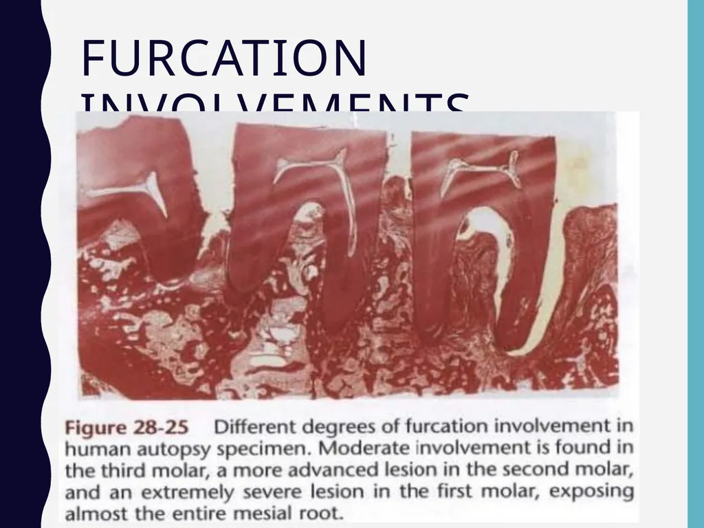
Furcation Involvements<br>
18
Furcation Involvements Classification One of the most widely used furcation classification systems was developed by Glickman (1958 )
In this system, furcation involvement is divided into four categories, primarily on the basis of the horizontal component of destruction<br>
In this system, furcation involvement is divided into four categories, primarily on the basis of the horizontal component of destruction<br>
19
Furcation Involvements
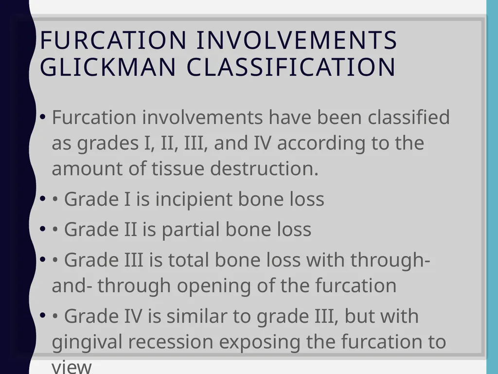
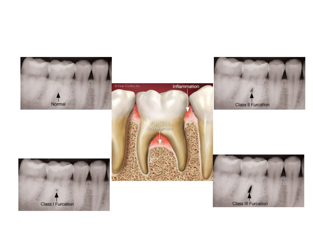
Glickman Classification Furcation involvements have been classified as grades I, II, III, and IV according to the amount of tissue destruction.
• Grade I is incipient bone loss
• Grade II is partial bone loss
• Grade III is total bone loss with through-and- through opening of the furcation
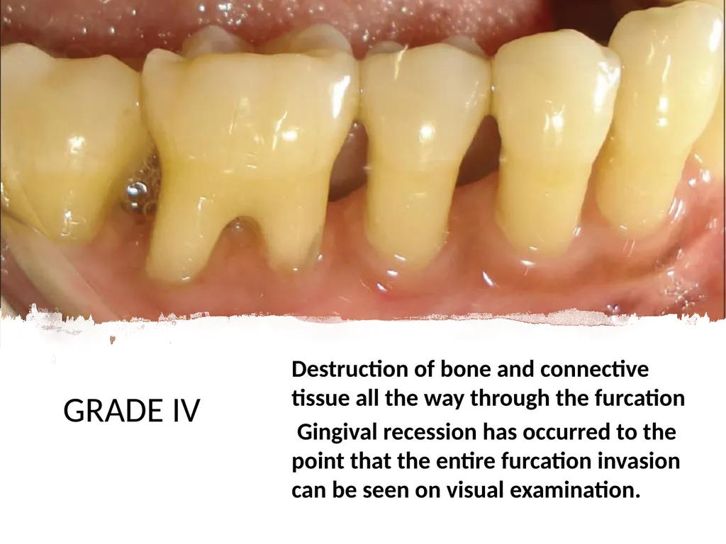
• Grade IV is similar to grade III, but with gingival recession exposing the furcation to view<br>
• Grade I is incipient bone loss
• Grade II is partial bone loss
• Grade III is total bone loss with through-and- through opening of the furcation
• Grade IV is similar to grade III, but with gingival recession exposing the furcation to view<br>
20
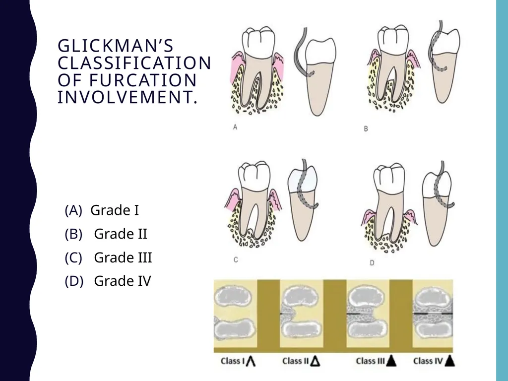
Glickman’s classification of furcation involvement. Grade I
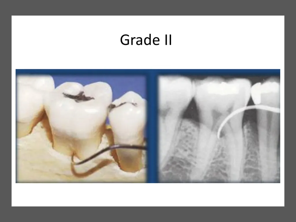
Grade II
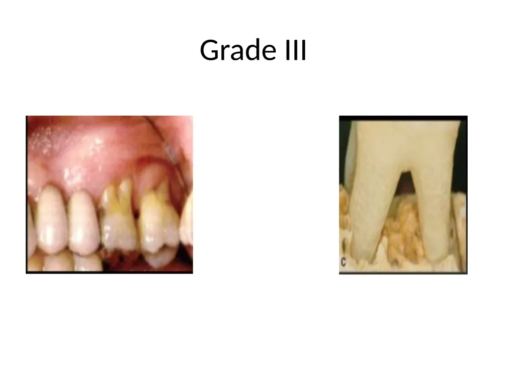
Grade III
Grade IV<br>
Grade II
Grade III
Grade IV<br>
21
Furcation Involvements
Glickman Classification
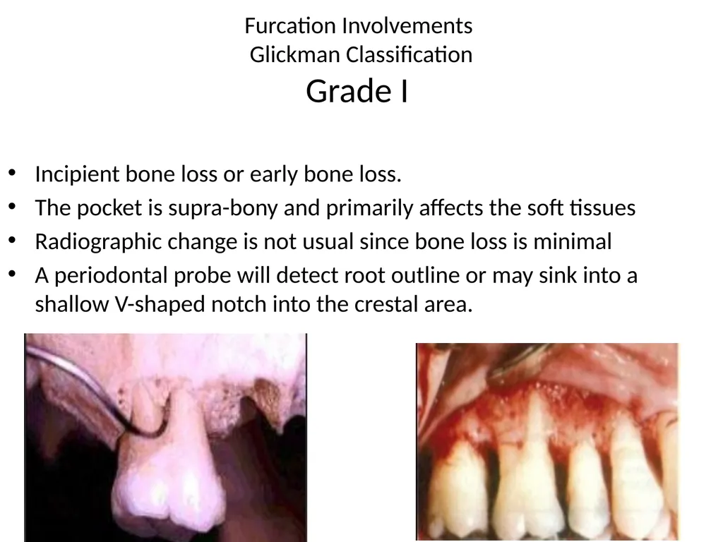
Grade I Incipient bone loss or early bone loss.
The pocket is supra-bony and primarily affects the soft tissues
Radiographic change is not usual since bone loss is minimal
A periodontal probe will detect root outline or may sink into a shallow V-shaped notch into the crestal area.<br>
The pocket is supra-bony and primarily affects the soft tissues
Radiographic change is not usual since bone loss is minimal
A periodontal probe will detect root outline or may sink into a shallow V-shaped notch into the crestal area.<br>
22
Furcation Involvements
Glickman Classification
Grade II Partial bone loss
Distinct horizontal destruction of the furcation area is present
This lesion has been called a "cul de sac" because destruction may extend to any depth within the furcation, but does not extend all the way through the furcation to other side
The extent of horizontal probing determines whether the Grade II furcation is shallow or deep.
Vertical bone loss may or may not be present
It can affect one or more furcation's of the same tooth<br>
Distinct horizontal destruction of the furcation area is present
This lesion has been called a "cul de sac" because destruction may extend to any depth within the furcation, but does not extend all the way through the furcation to other side
The extent of horizontal probing determines whether the Grade II furcation is shallow or deep.
Vertical bone loss may or may not be present
It can affect one or more furcation's of the same tooth<br>
23
Furcation Involvements
Glickman Classification
Grade III Total bone loss with through and through opening
Bone is not attached to dome of furcation
In early lesion, opening may be filled with soft tissue and may not be visible
Destruction of bone and connective tissue all the way through the furcation such that an instrument can be passed from its opening to its exit
The furcation defect is not visible to the eye because the gingival tissues cover the furcation entrance.<br>
Bone is not attached to dome of furcation
In early lesion, opening may be filled with soft tissue and may not be visible
Destruction of bone and connective tissue all the way through the furcation such that an instrument can be passed from its opening to its exit
The furcation defect is not visible to the eye because the gingival tissues cover the furcation entrance.<br>
24
Grade III<br>
25
GRADE IV Destruction of bone and connective tissue all the way through the furcation
Gingival recession has occurred to the point that the entire furcation invasion can be seen on visual examination.<br>
Gingival recession has occurred to the point that the entire furcation invasion can be seen on visual examination.<br>
26
Based on vertical component
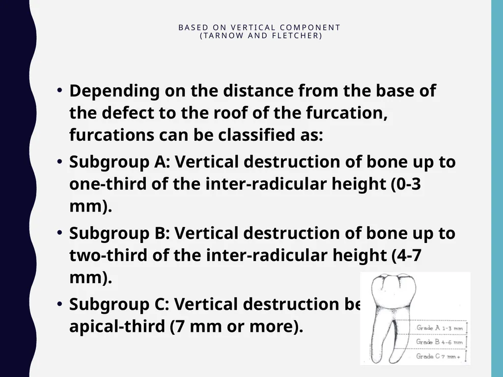
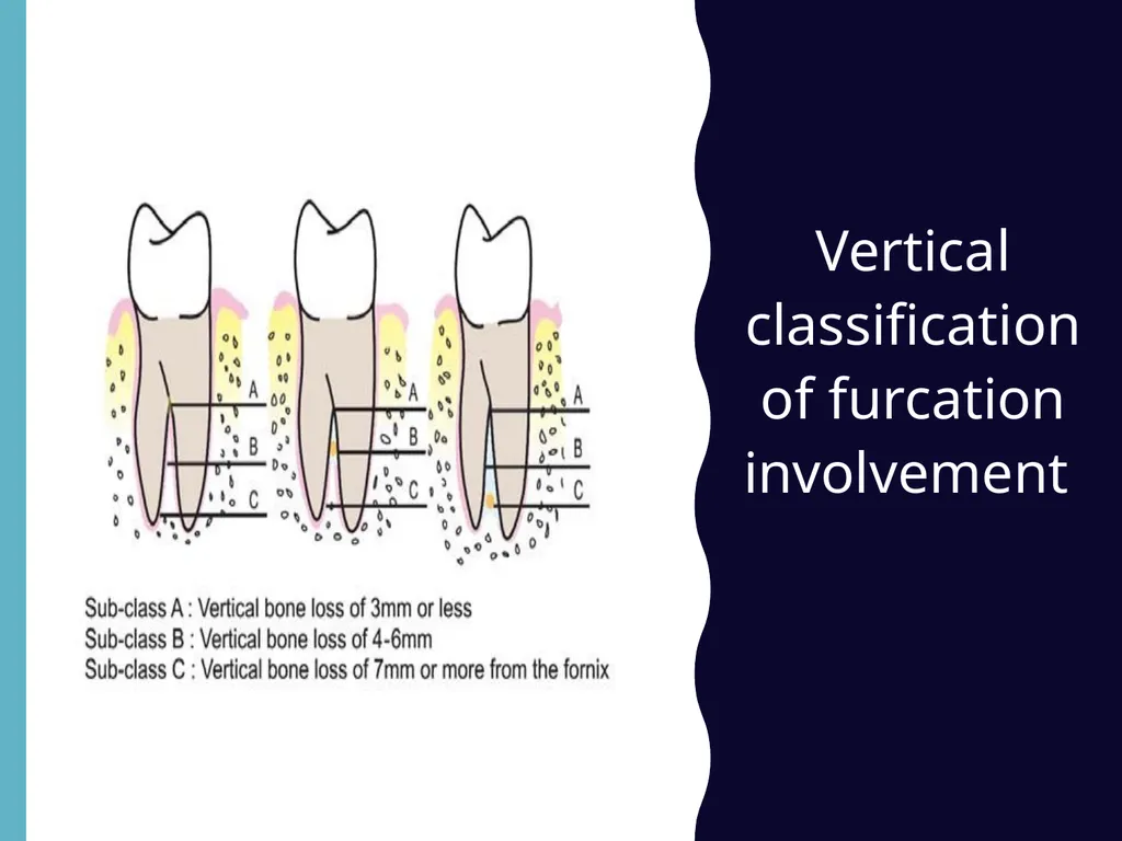
(Tarnow and Fletcher) Depending on the distance from the base of the defect to the roof of the furcation, furcations can be classified as:
Subgroup A: Vertical destruction of bone up to one-third of the inter-radicular height (0-3 mm).
Subgroup B: Vertical destruction of bone up to two-third of the inter-radicular height (4-7 mm).
Subgroup C: Vertical destruction beyond the apical-third (7 mm or more).<br>
Subgroup A: Vertical destruction of bone up to one-third of the inter-radicular height (0-3 mm).
Subgroup B: Vertical destruction of bone up to two-third of the inter-radicular height (4-7 mm).
Subgroup C: Vertical destruction beyond the apical-third (7 mm or more).<br>
27
Vertical classification of furcation involvement<br>
28
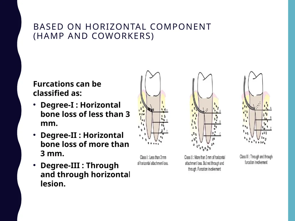
Based on horizontal component (Hamp and Coworkers) Furcations can be classified as:
Degree-I : Horizontal bone loss of less than 3 mm.
Degree-II : Horizontal bone loss of more than 3 mm.
Degree-III : Through and through horizontal lesion.<br>
Degree-I : Horizontal bone loss of less than 3 mm.
Degree-II : Horizontal bone loss of more than 3 mm.
Degree-III : Through and through horizontal lesion.<br>
29
Primary Etiologic Factor :
It is bacterial plaque and long-standing inflammation of periodontal tissues.
Anatomic and clinical characteristics of tooth, bone and gingiva are of importance for the clinical management of furcation lesions.<br>
It is bacterial plaque and long-standing inflammation of periodontal tissues.
Anatomic and clinical characteristics of tooth, bone and gingiva are of importance for the clinical management of furcation lesions.<br>
30
Diagnosis
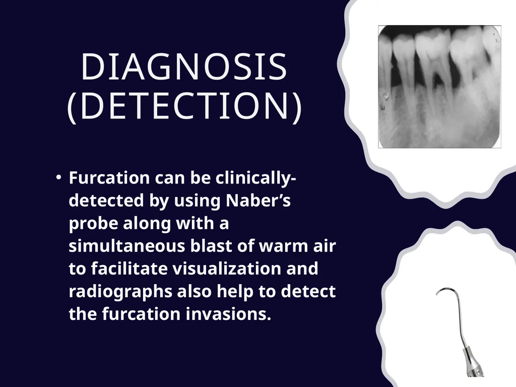
(Detection) Furcation can be clinically-detected by using Naber’s probe along with a simultaneous blast of warm air to facilitate visualization and radiographs also help to detect the furcation invasions.<br>
31
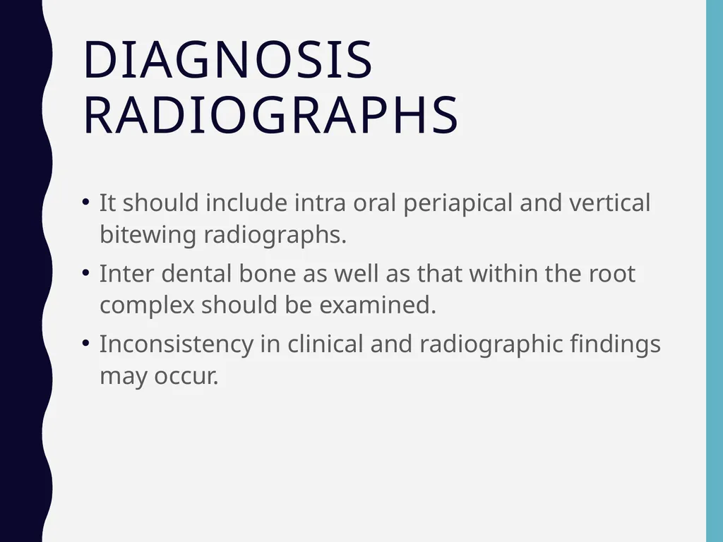
Diagnosis Radiographs It should include intra oral periapical and vertical bitewing radiographs.
Inter dental bone as well as that within the root complex should be examined.
Inconsistency in clinical and radiographic findings may occur.<br>
Inter dental bone as well as that within the root complex should be examined.
Inconsistency in clinical and radiographic findings may occur.<br>
32
CLINICAL FEATURES Clinically
The mandibular first molars are the most common sites and maxillary premolars are the least common.
The denuded furcation may be visible clinically or covered by the wall of the pocket.
Associated with suprabony and infrabony pockets.
Periodontal abscess.
Root caries and tooth mobility are common.<br>
The mandibular first molars are the most common sites and maxillary premolars are the least common.
The denuded furcation may be visible clinically or covered by the wall of the pocket.
Associated with suprabony and infrabony pockets.
Periodontal abscess.
Root caries and tooth mobility are common.<br>
33
CLINICAL FEATURES Microscopically
It is simply a phase in the rootward extension of the periodontal pocket.
In its early stages, there is a widening of the periodontal space with cellular and inflammatory fluid exudation, followed by epithelial proliferation into the furcation area from an adjoining periodontal pocket.<br>
It is simply a phase in the rootward extension of the periodontal pocket.
In its early stages, there is a widening of the periodontal space with cellular and inflammatory fluid exudation, followed by epithelial proliferation into the furcation area from an adjoining periodontal pocket.<br>
34
CLINICAL FEATURES Extension of the inflammation into the bone leads to resorption and reduction in bone height.
The bone destructive pattern may produce horizontal loss, or there may be angular osseous defects associated with infrabony pockets. Plaque, calculus and bacterial debris occupy the denuded furcation space.<br>
The bone destructive pattern may produce horizontal loss, or there may be angular osseous defects associated with infrabony pockets. Plaque, calculus and bacterial debris occupy the denuded furcation space.<br>
35
PROGNOSIS Maxillary first premolars and maxillary molars as well as mandibular molars are the teeth with multiple roots.
Furcation involvement will be likely to occur in these multirooted teeth.
Maxillary first premolar often shows fusion of the roots and the furcation area may be located very much apically and also the roots of the maxillary first premolars are placed buccally and palatally with furcation opening in a mesiodistal direction. For these reasons, furcation involvement in maxillary first premolar has poor prognosis.<br>
Furcation involvement will be likely to occur in these multirooted teeth.
Maxillary first premolar often shows fusion of the roots and the furcation area may be located very much apically and also the roots of the maxillary first premolars are placed buccally and palatally with furcation opening in a mesiodistal direction. For these reasons, furcation involvement in maxillary first premolar has poor prognosis.<br>
36
PROGNOSIS In the case of maxillary molars furcations may open bucally, mesially and distally because of the presence of the three roots. Since access from proximal areas is difficult for plaque control, prognosis of furcation involvement in maxillary molars is not good.
Mandibular molars have two roots, placed mesially and distally and the furcation opens buccolingually. The roots are usually divergent especially in mandibular first molars. As a result prognosis of furcation involvement in mandibular molar (especially the first molar) is considered good.<br>
Mandibular molars have two roots, placed mesially and distally and the furcation opens buccolingually. The roots are usually divergent especially in mandibular first molars. As a result prognosis of furcation involvement in mandibular molar (especially the first molar) is considered good.<br>
37
TREATMENT It is aimed to prevent further attachment loss and improve the maintenance of furcation area.
Two treatment modalities have been proposed:
Traditional treatment procedures.
Reconstructive or regenerative treatment.<br>
Two treatment modalities have been proposed:
Traditional treatment procedures.
Reconstructive or regenerative treatment.<br>
38
Factors to be considered when deciding on a mode
of therapy are as follows: 1. Degree of involvement.
2. Crown root ratio.
3. Length of roots.
4. Degree of root separation.
5. Strategic value of the tooth or teeth in question. 6. Root anatomy of the involved tooth.
7. Residual tooth mobility.
8. Endodontic therapy and complications.
9. Ability to eliminate the defect.
10. Periodontal condition of the adjacent teeth.<br>
10. Periodontal condition of the adjacent teeth.<br>
39
Traditional Treatment Procedures They are those which are directed to maintain the state of the health but do not attempt to regenerate the lost periodontium.
The goal is to prevent the further progression of the disease and provide an environment which will help in adequate plaque control.<br>
The goal is to prevent the further progression of the disease and provide an environment which will help in adequate plaque control.<br>
40
Traditional Treatment Procedures The procedures are:
Grade I: They are usually associated with suprabony pockets, hence,
a. Initial preparation or scaling and root planing.
b. Curettage or gingivectomy to expose the furcation area.
c. Odontoplasty—to reshape the facial groove in order to prevent plaque accumulation.
Grade II: In shallow grade-II invasions, Osteoplasty with limited ostectomy may be helpful. Odontoplasty can be performed.<br>
Grade II: In shallow grade-II invasions, Osteoplasty with limited ostectomy may be helpful. Odontoplasty can be performed.<br>
41
Traditional Treatment Procedures In severe grade-II to IV invasions elimination of furcation by:
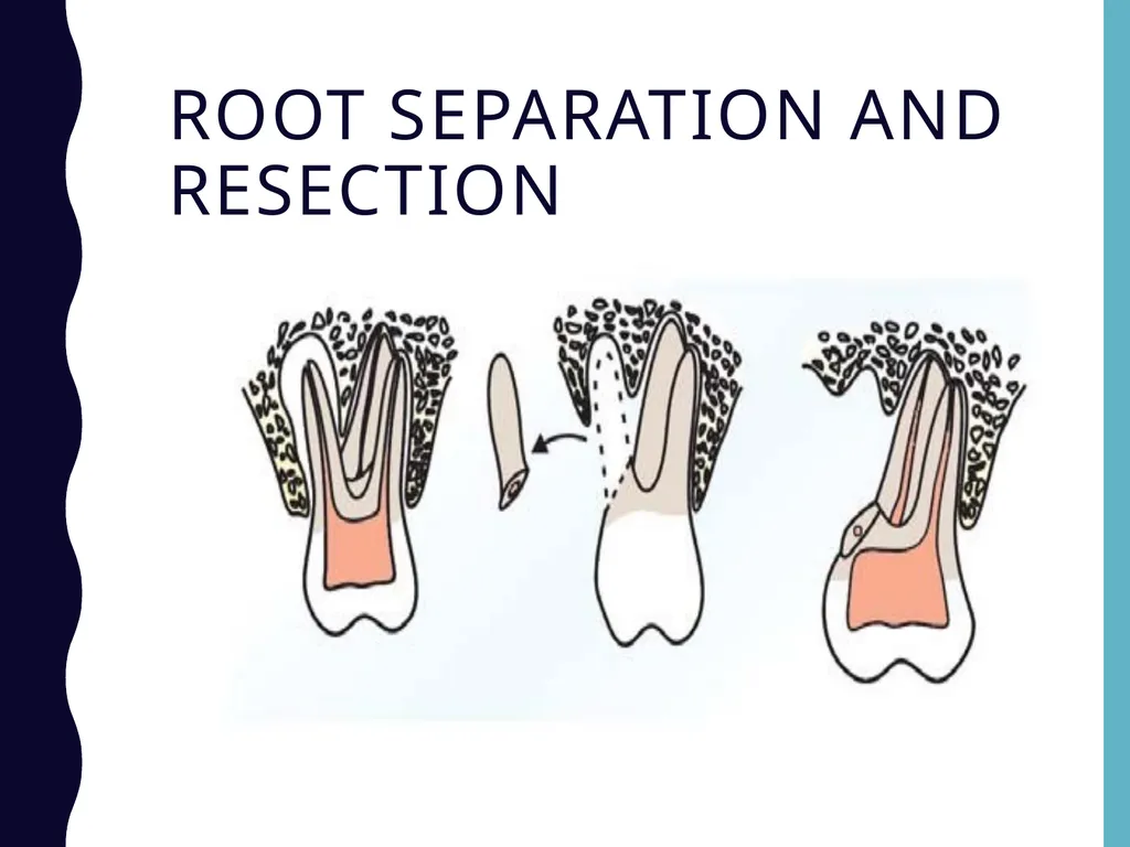
a. Root resection or amputation: After periodontal flap reflection, surgical removal of the root portion of the affected tooth is most commonly performed in maxillary first molars.
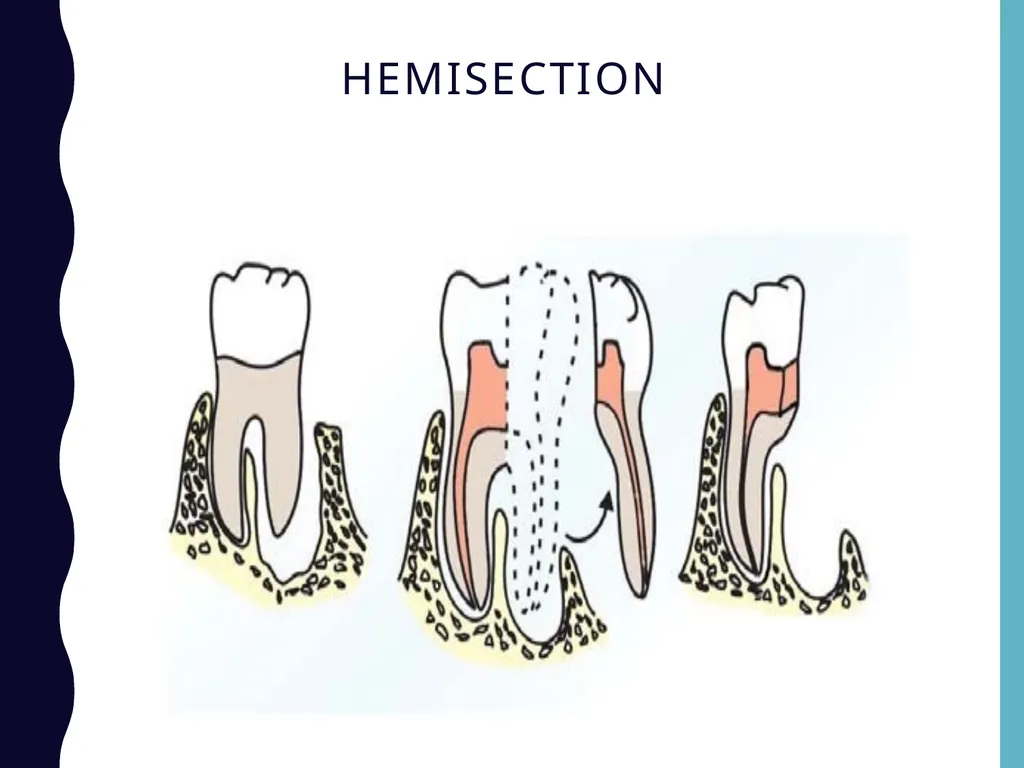
b. Hemisection or root separation: It is the surgical removal of the root along with the crown. Most commonly done in mandibular molars.
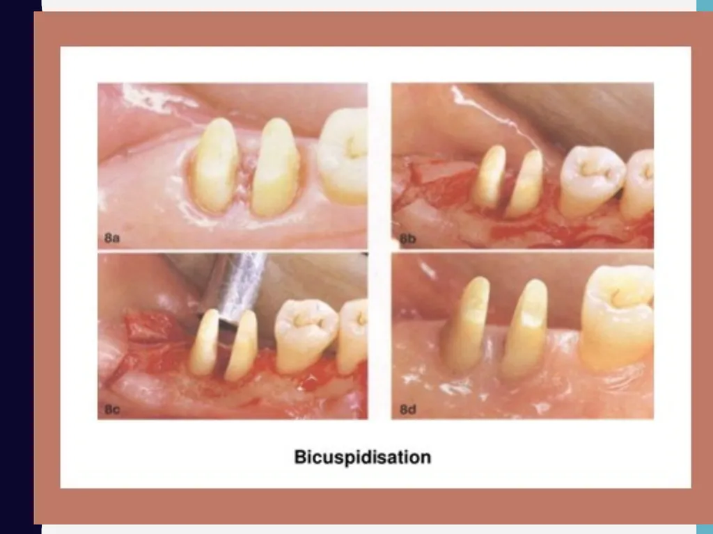
c. Bicuspidization/root separation: Splitting of a two- rooted tooth into two separate portions. Frequently performed in mandibular molars.
d. Tunnel preparation: It is by transforming the grade II lesion to grade III and IV for better access, but it is not performed anymore because of increased incidence of root caries.
Grade III and grade IV can be treated with root resection and root separation.<br>
a. Root resection or amputation: After periodontal flap reflection, surgical removal of the root portion of the affected tooth is most commonly performed in maxillary first molars.
b. Hemisection or root separation: It is the surgical removal of the root along with the crown. Most commonly done in mandibular molars.
c. Bicuspidization/root separation: Splitting of a two- rooted tooth into two separate portions. Frequently performed in mandibular molars.
d. Tunnel preparation: It is by transforming the grade II lesion to grade III and IV for better access, but it is not performed anymore because of increased incidence of root caries.
Grade III and grade IV can be treated with root resection and root separation.<br>
42
Treatment options Maintain the furcation
Increase the access to furcation
Removal of furcation
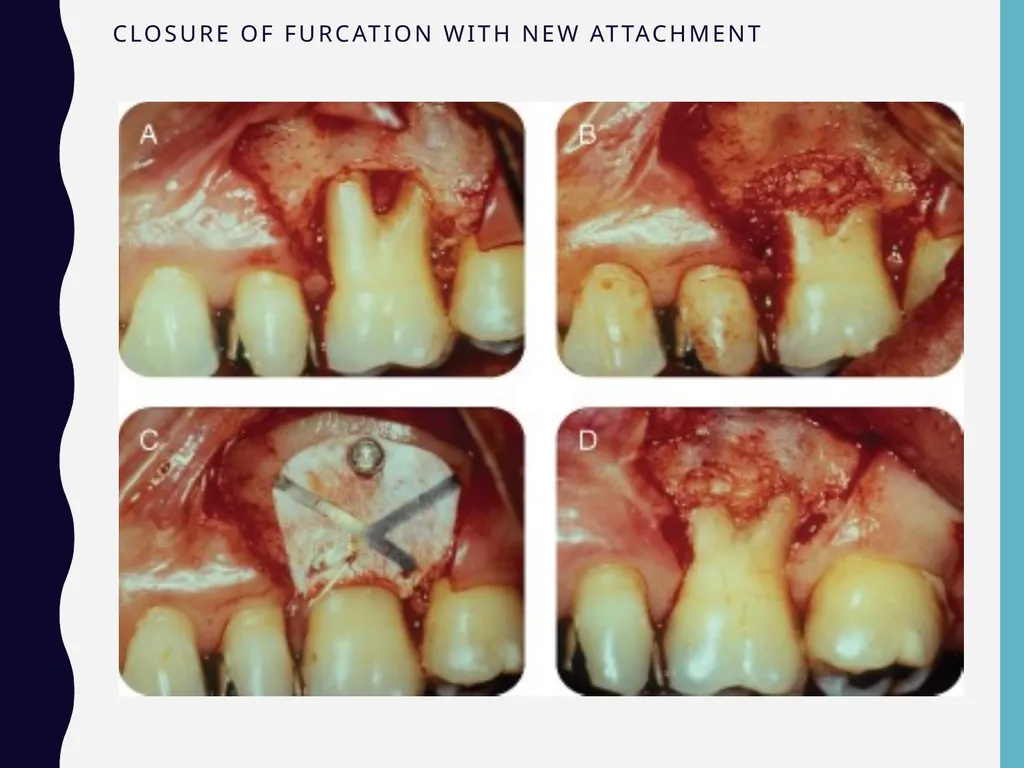
Closure of furcation with new attachment
Kalkwarf and Reinhardt (1988)<br>
Increase the access to furcation
Removal of furcation
Closure of furcation with new attachment
Kalkwarf and Reinhardt (1988)<br>
43
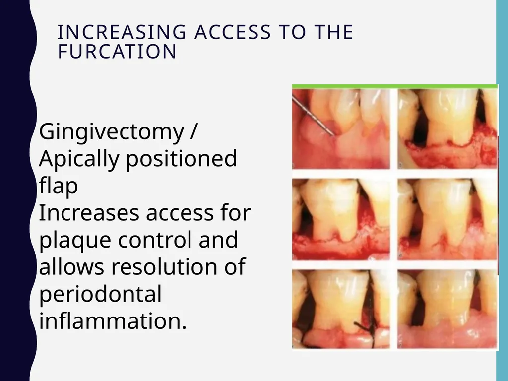
Increasing access to the furcation Gingivectomy / Apically positioned flap
Increases access for plaque control and allows resolution of periodontal inflammation.<br>
Increases access for plaque control and allows resolution of periodontal inflammation.<br>
44
Odontoplasty :
It is the reshaping of the tooth coronal to the furcation to improve access for plaque control
It increases entrance to the furcation and reduces its horizontal depth
Mainly used for Grade I and Grade II furcation defects treatment
Possible complications:
Hypersensitivity
Pulpal irritation leading to permanent damage
Pulp exposure
Increase risk of root caries.<br>
It is the reshaping of the tooth coronal to the furcation to improve access for plaque control
It increases entrance to the furcation and reduces its horizontal depth
Mainly used for Grade I and Grade II furcation defects treatment
Possible complications:
Hypersensitivity
Pulpal irritation leading to permanent damage
Pulp exposure
Increase risk of root caries.<br>
45
b. Osteoplasty and Ostectomy:
Osteoplasty: Reshaping surfaces of bone without removing tooth supporting bone
Ostectomy: Reshaping and removal of tooth supporting bone
Improved plaque control through osteoplasty is reported to be accomplished by:
Creating bony ramps into the furcation area allowing the gingiva to tuck into tooth concavities
Removing lip of the bony defect to decrease horizontal depth of the involvement
Reducing pocket depth by allowing apical adaptation of the flap
Recommended for Grade I and II furcation involvements
In advanced cases of Grade II and Grade III furcations ostectomy may be extended into creating a tunnel to expose the entire furcation area.<br>
Osteoplasty: Reshaping surfaces of bone without removing tooth supporting bone
Ostectomy: Reshaping and removal of tooth supporting bone
Improved plaque control through osteoplasty is reported to be accomplished by:
Creating bony ramps into the furcation area allowing the gingiva to tuck into tooth concavities
Removing lip of the bony defect to decrease horizontal depth of the involvement
Reducing pocket depth by allowing apical adaptation of the flap
Recommended for Grade I and II furcation involvements
In advanced cases of Grade II and Grade III furcations ostectomy may be extended into creating a tunnel to expose the entire furcation area.<br>
46
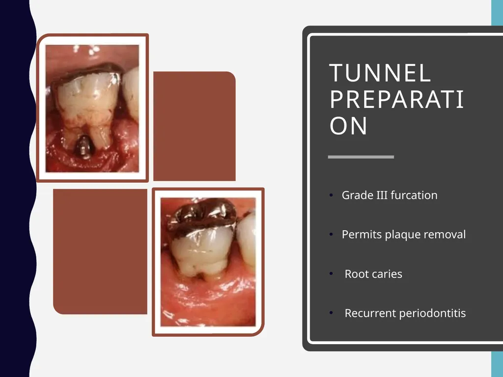
Tunnel preparation Grade III furcation
Permits plaque removal
Root caries
Recurrent periodontitis<br>
Permits plaque removal
Root caries
Recurrent periodontitis<br>
47
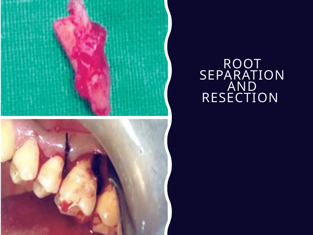
Root separation and resection<br>
48
INDICATIONS AND CONTRAINDICATIONS FOR ROOT RESECTION AND SEPARATION Indications
Severe bone loss affecting one or more roots untreatable with regenerative procedures.
Class II or III furcation invasions or involvement.
Severe recession or dehiscence of a root.
Contraindications
a. General contraindications like systemic diseases and poor oral hygiene.
b. Fused roots, unfavorable tissue architecture.
c. Roots that are endodontically-untreatable.<br>
Severe bone loss affecting one or more roots untreatable with regenerative procedures.
Class II or III furcation invasions or involvement.
Severe recession or dehiscence of a root.
Contraindications
a. General contraindications like systemic diseases and poor oral hygiene.
b. Fused roots, unfavorable tissue architecture.
c. Roots that are endodontically-untreatable.<br>
49
Root separation and resection<br>
50
Hemisection<br>
51
Reconstructive and Regenerative Treatment Procedures Grade I: Traditional treatment will do.
Grade II: Various regenerative techniques include
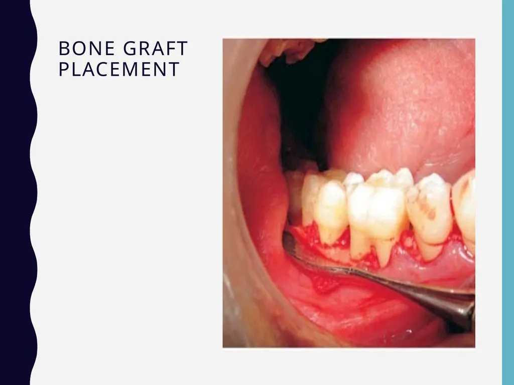
a. Autogenous bone grafting, e.g. osseous coagulum,
bone blend.
b. Allografts, e.g. freeze, dried bone allografts, demineralized freeze-dried bone allografts (FDBA, DFDBA).<br>
Grade II: Various regenerative techniques include
a. Autogenous bone grafting, e.g. osseous coagulum,
bone blend.
b. Allografts, e.g. freeze, dried bone allografts, demineralized freeze-dried bone allografts (FDBA, DFDBA).<br>
52
Reconstructive and Regenerative Treatment Procedures c. Alloplasts—hydroxyapatite, tricalcium phosphate.
d. Citric acid root conditioning with coronally positioned flap.
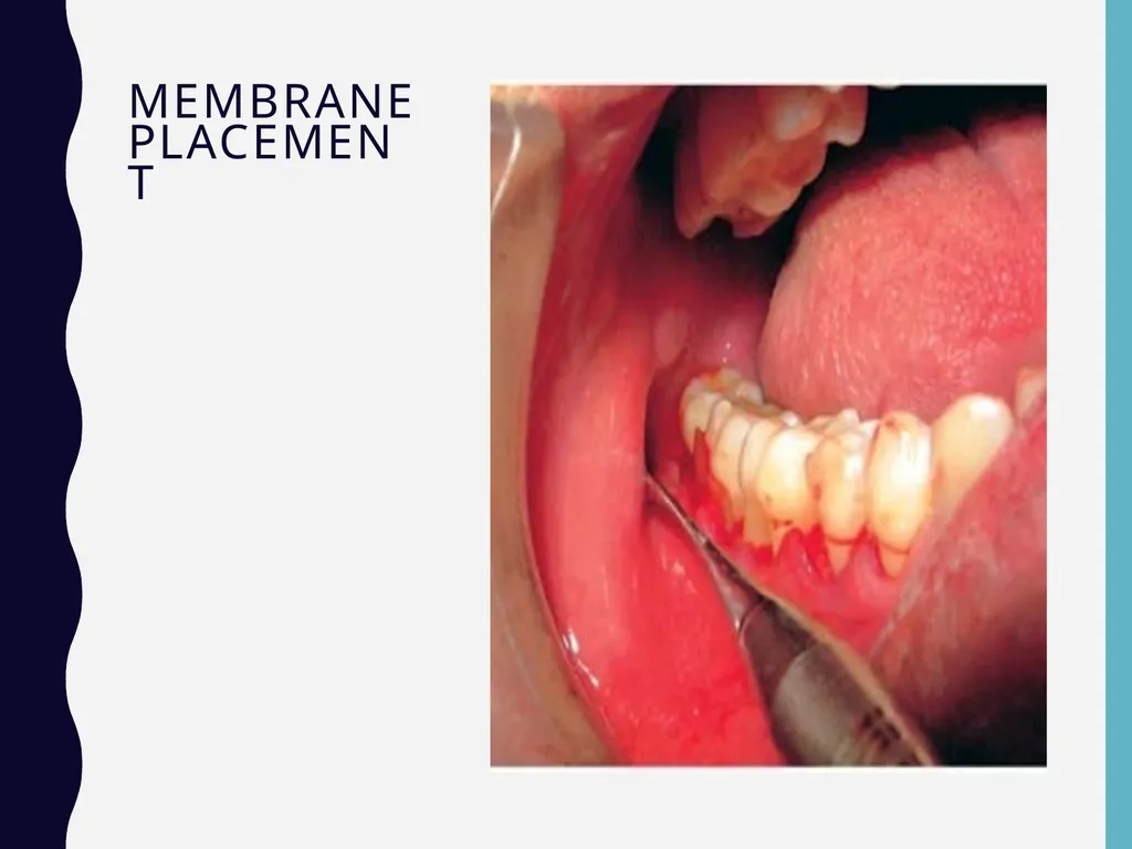
e. Guided tissue regeneration and combination techniques.
For grade III and grade IV furcation involvements the success rate is limited.<br>
d. Citric acid root conditioning with coronally positioned flap.
e. Guided tissue regeneration and combination techniques.
For grade III and grade IV furcation involvements the success rate is limited.<br>
53
Closure of furcation with new attachment<br>
54
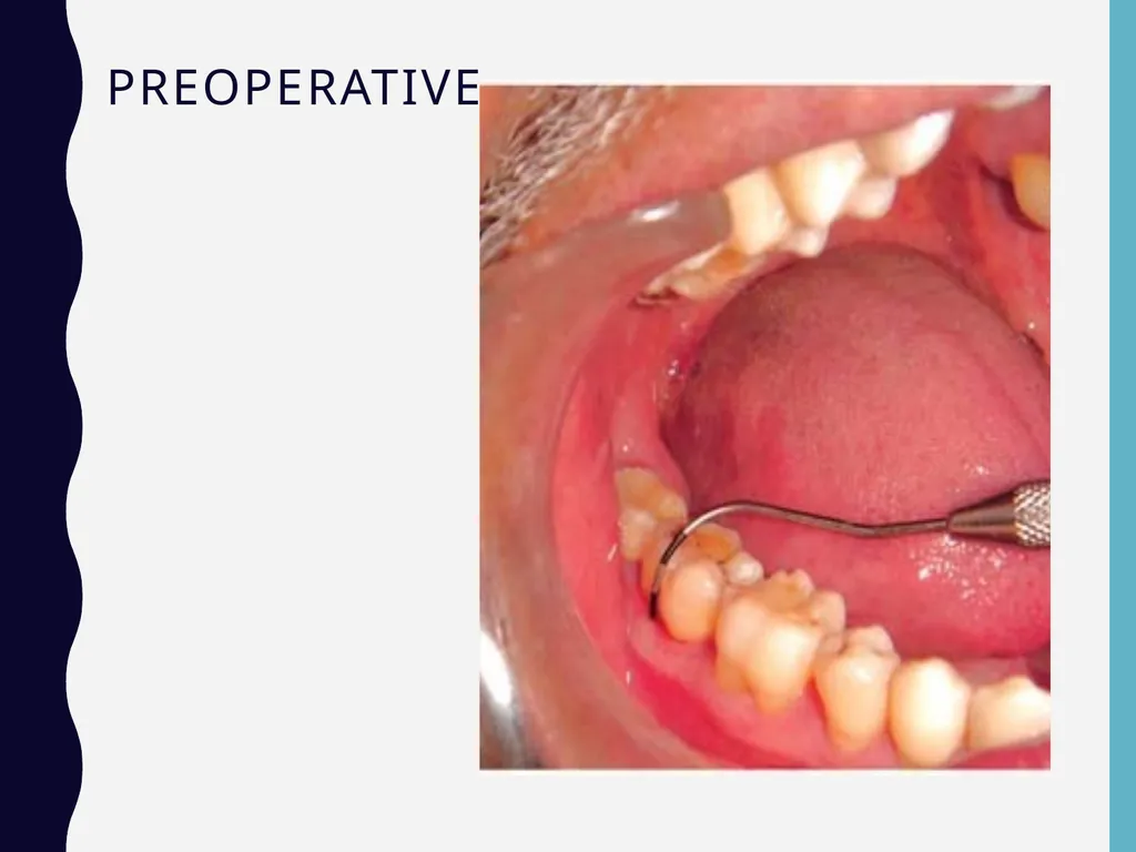
Preoperative<br>
55
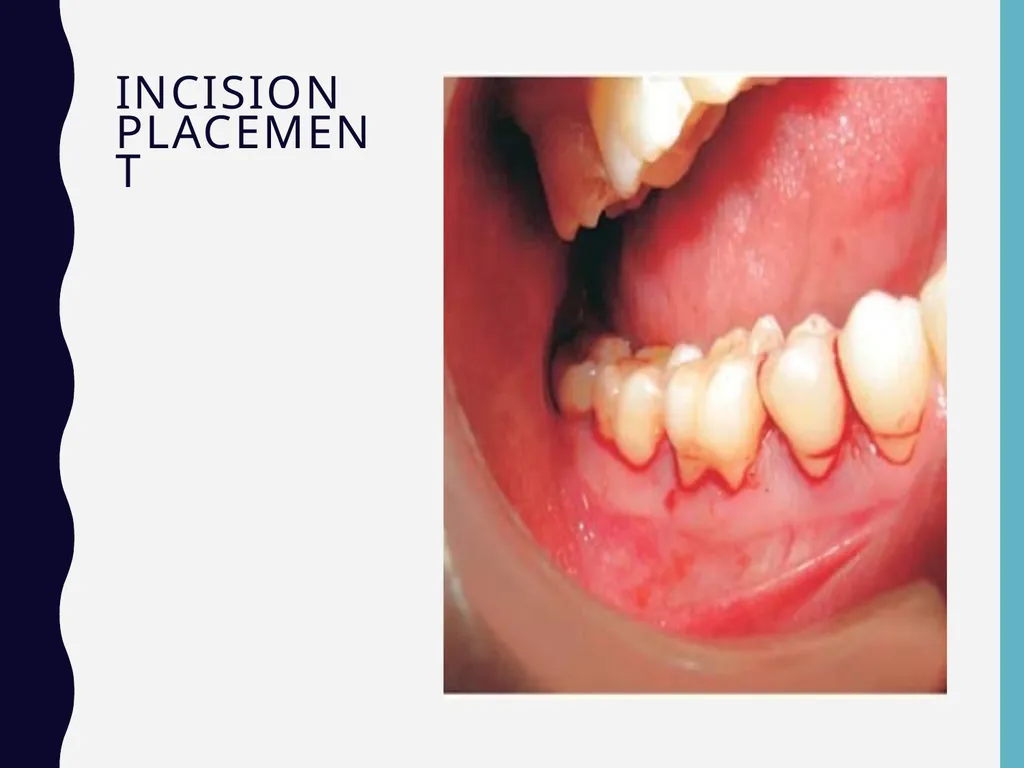
Incision placement<br>
56
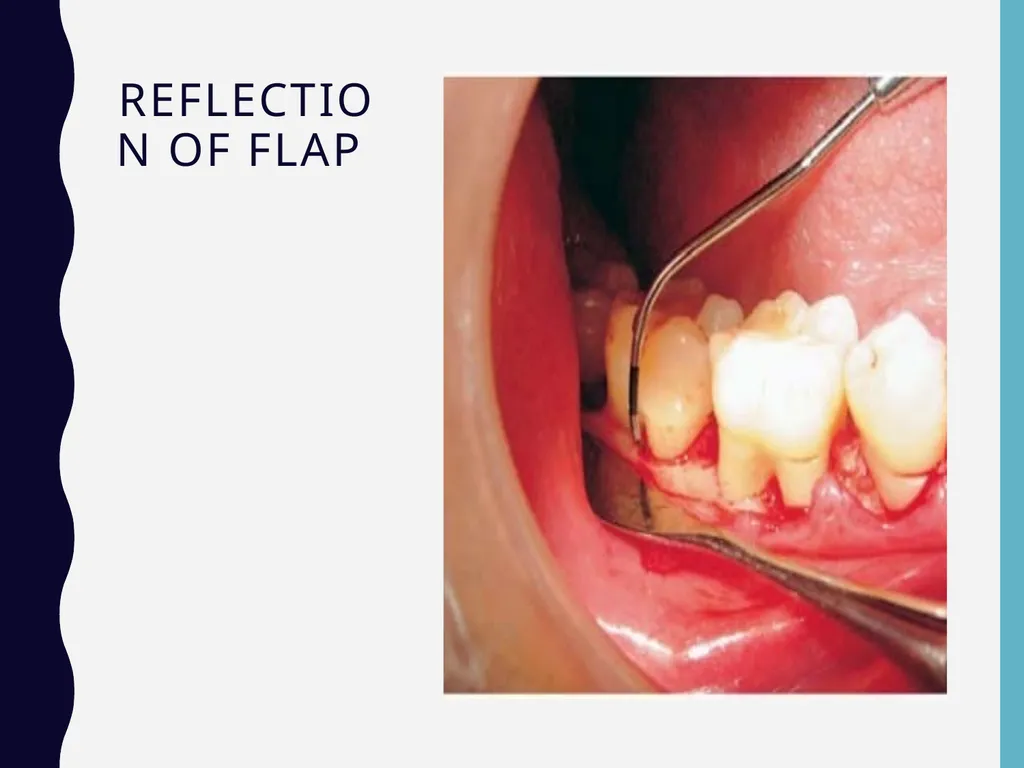
Reflection of flap<br>
57
Bone graft placement<br>
58
Membrane placement<br>
59
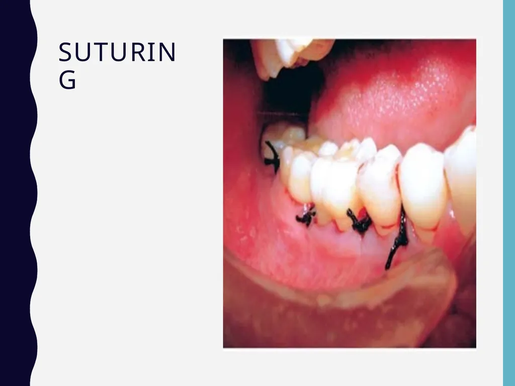
Suturing<br>
60
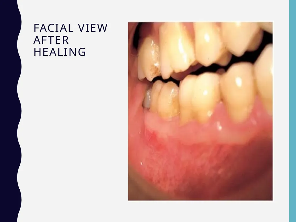
Facial view after healing<br>
61
KEY POINTS TO NOTE 1. Furcation involvement refers to commonly occurring conditions in which the bifurcation and trifurcation of multi- rooted teeth are invaded by the disease process.
2. Primary etiologic factor is bacterial plaque, predisposing or anatomic factors include, aberrant root morphology, cervical enamel projections or pearls and presence of accessory canals.
3. Furcation involvement can be classified according to Glickman as Grade I, Grade II, Grade III and Grade IV.
4. Depending on the distance from the base of the defect to the roof of the furcation, (vertical component) Tarnow and Fletcher classified furcation involvement into—subgroup A, subgroup B, subgroup C.
5. Furcation involvement can be diagnosed by Naber’s probe and radiographs.
6. Treatment of furcation involvement is divided into: a. Traditional treatment procedures. b. Reconstructive or regenerative treatment<br>
2. Primary etiologic factor is bacterial plaque, predisposing or anatomic factors include, aberrant root morphology, cervical enamel projections or pearls and presence of accessory canals.
3. Furcation involvement can be classified according to Glickman as Grade I, Grade II, Grade III and Grade IV.
4. Depending on the distance from the base of the defect to the roof of the furcation, (vertical component) Tarnow and Fletcher classified furcation involvement into—subgroup A, subgroup B, subgroup C.
5. Furcation involvement can be diagnosed by Naber’s probe and radiographs.
6. Treatment of furcation involvement is divided into: a. Traditional treatment procedures. b. Reconstructive or regenerative treatment<br>
62
READING ASSIGNMENT Chapter 64, Furcation: The problem and its management
Newman and Carranza’s Clinical Periodontology
Thirteenth Edition
Besides the lectures in slides presentations , contents of the reading assignment are included in the exams and quizzes<br>
Newman and Carranza’s Clinical Periodontology
Thirteenth Edition
Besides the lectures in slides presentations , contents of the reading assignment are included in the exams and quizzes<br>
63
Words Are Weapons<br>