
Topic : MEDICAL EMERGENCIES IN DENTAL PRACTICE



Description: Topic : MEDICAL EMERGENCIES IN DENTAL PRACTICE Subject Code : 008 Subject Name : Pedodontics Delivered by : Dr Joyson Moses Department : Pedodontics Topic :Medical Emergencies Staff name : Dr Joyson Moses MEDICAL EMERGENCIES IN DENTAL
Related Topics
Download Presentation
"Topic : MEDICAL EMERGENCIES IN DENTAL PRACTICE" is the property of its rightful owner. Permission is granted to download and print the materials on this website for personal, non-commercial use only, and to display it on your personal computer provided you do not modify the materials and that you retain all copyright notices contained in the materials. By downloading content from our website, you accept the terms of this agreement.
Presentation Transcript
slide1. Topic : MEDICAL EMERGENCIES IN DENTAL PRACTICE
Subject Code : 008
Subject Name : Pedodontics
Delivered by : Dr Joyson Moses<br>
slide2. Department : Pedodontics Topic :Medical Emergencies Staff name : Dr Joyson Moses MEDICAL EMERGENCIES IN DENTAL PRACTICE MEDICAL EMERGENCIES IN DENTAL PRACTICE MEDICAL EMERGENCIES IN DENTAL PRACTICE<br>
slide3. MEDICAL EMERGENCIES IN DENTAL PRACTICE<br>
slide4. INTRODUCTION Emergency- a condition that warrants or immediate attention by the doctor.
Many emergency problems can be definitely avoided by simple preventive measures like a careful medical history, general physical examination regarding patient health status & proper preoperative preparation of the patient.
Emergency according to Dorland’s Medical Dictionary is defined as a sudden, urgent, usually unforeseen occurrence requiring immediate action.<br>
slide5. SHOCK It is a phenomenon marked by circulatory deficiency which is either cardiac or vasomotor in origin exhibiting marked hypotension.
Signs & Symptoms:
Patient –unconscious with ashen grey face & cold, clammy skin.
Mucous membrane –pale whereas lips,nails, finger tips & lobules of the ear are grayish blue. Face is expression less with sunken eyes.
Pupils are dilated but react feebly to light.
Pulse is weak & thread.
Shallow & irregular respiration
Temperature is subnormal
Treatment :
Position: Put the patients head at the lower level than feet 15 degree- Trendelenburg position<br>
slide6. Maintain the body heat by covering the patient with blanket & keep a hot water bottle between the thighs.
Check for any airway obstruction & patency of airway be maintained.
Control the loss of blood in haemorrhage shock by pressure packs.
Restore the lost body fluids. Infusion with plasma expanders or Ringer’s lactate solution should be carried out to maintain the intravenous line & restore the volume loss
Administer 100 percent oxygen
Blood pressure,pulse rate & respiratory rate should be constantly monitored to assess the vitals.
Injection hydrocortisone sodium hemisuccinate 100mg in 5mL of water intravenously as stress bearing factor of he body
Injection mephentermine to raise BP
Injection atropine is given for bradycardia.
Broad spectrum antibiotics.
Narcotic analgesic to relieve pain<br>
slide7. ALLERGIC REACTION It is an unwanted response of the body to a complete dose of the drug. It is the result of immunological response of /in the individual
Gell & Coombs classification:
Type I ( IgE- mediated hypersensitivity )- most life- theatening few minutes.
Type 2 ( Cytotoxic / cytolytic antibdy-mediated) IgM or Ig G soluble metabolite.
Type 3 ( Immune complex mediated ) 1-4 weeks, IgM or IgG soluble metabolite.
Type 4 ( Delayed hypersensitivity )- sensitized T-cell lymphocytes.
Signs & symptoms:
Cutaneous reactions are the most common occurrence & include urticarial,exanthematous & eczematoid reactions. Itching is common & can also find exfoliative dermatitis & bullous dermatosis.<br>
slide8. Angioedema ( swelling) this varies from localized slight swelling of the lips, eyelids, & face to more uncomfortable swelling of the mouth, throat & extremities.
Respiratory ( tightness in chest,sneezing, bronchospasm) bronchospasm is a generalized contraction of bronchial smooth muscles resulting in the restriction of airflow. This may also be accompanied by edema of the bronchiolar mucosa.
Bronchospasm is more common with preexisting pulmonary disease such as asthma or infection but can also be caused by the nhalation of a foreign substance.
Ocular reactions include conjunctivitis & watering of eyes.
Hypotension can occur with any allergic reaction.<br>
slide9. ANAPLYLAXIS This is a severe systemic type allergic reaction & is a medical emergency.
Signs & symptoms :
Cardiovascular shock including : Pallor, syncope, palpitations,tachycardia,hypotension,arrhythmias & convulsions.
Respiratory symptoms include: Sneezing , cough,wheezing,tightness in chest, bronchospasm,laryngospasm.
Skin is warm & flushed with itching,urticaria & angioedema.
Nausea, vomiting , abdominal cramps & diarrhea are also possible.
Treatment :
General treatment :
ABC’s ( is expansion required or removes the bullet from the next point)<br>
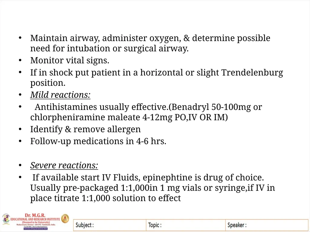
slide10. Maintain airway, administer oxygen, & determine possible need for intubation or surgical airway.
Monitor vital signs.
If in shock put patient in a horizontal or slight Trendelenburg position.
Mild reactions:
Antihistamines usually effective.(Benadryl 50-100mg or chlorpheniramine maleate 4-12mg PO,IV OR IM)
Identify & remove allergen
Follow-up medications in 4-6 hrs.
Severe reactions:
If available start IV Fluids, epinephtine is drug of choice. Usually pre-packaged 1:1,000in 1 mg vials or syringe,if IV in place titrate 1:1,000 solution to effect<br>
slide11. If drop in blood pressure is minimal,start with 0.5mL (0.5mg)
If drop in blood pressure is severe start with 2mL (2mg), repeat after 2 minute if needed.
If no IV use 1;1,000(1mg/cc),IM 0.3-0.5mg ( 0.3-0.5cc), for an adult repeat this dose in 10-20mins.
If the patient is intubated can give epinephrine endotracheally.
If asthma,edema or pruritus ( itching) is present, can use corticosteroids.However,these drugs are too slow-acting to be used for an emergency situation
Hydrocortisone sodium succinate (solu-cortef) 100-500mg IV or IM. Dexamethasone ( Decadron) 4-12mg IV or IM
Repeat dose at 1,3,6 & 10 hrs as indicated by severity of symptoms.<br>
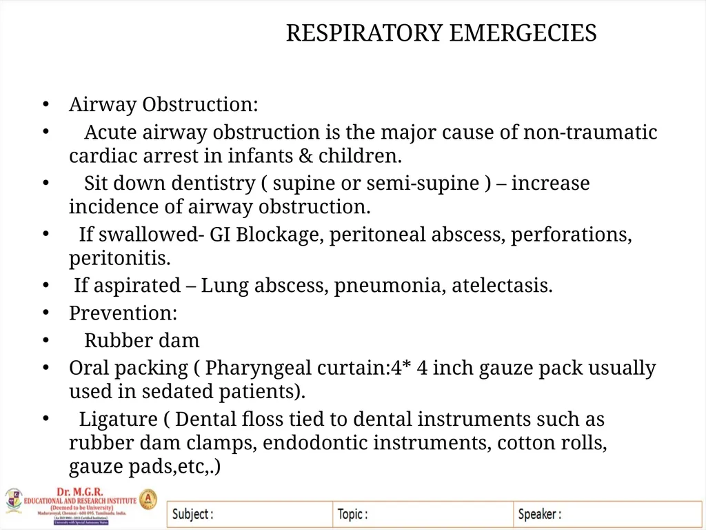
slide12. RESPIRATORY EMERGECIES Airway Obstruction:
Acute airway obstruction is the major cause of non-traumatic cardiac arrest in infants & children.
Sit down dentistry ( supine or semi-supine ) – increase incidence of airway obstruction.
If swallowed- GI Blockage, peritoneal abscess, perforations, peritonitis.
If aspirated – Lung abscess, pneumonia, atelectasis.
Prevention:
Rubber dam
Oral packing ( Pharyngeal curtain:4* 4 inch gauze pack usually used in sedated patients).
Ligature ( Dental floss tied to dental instruments such as rubber dam clamps, endodontic instruments, cotton rolls, gauze pads,etc,.)<br>
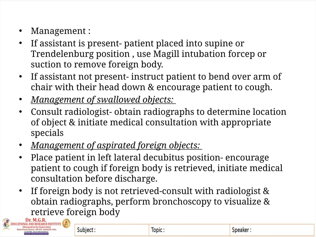
slide13. Management :
If assistant is present- patient placed into supine or Trendelenburg position , use Magill intubation forcep or suction to remove foreign body.
If assistant not present- instruct patient to bend over arm of chair with their head down & encourage patient to cough.
Management of swallowed objects:
Consult radiologist- obtain radiographs to determine location of object & initiate medical consultation with appropriate specials
Management of aspirated foreign objects:
Place patient in left lateral decubitus position- encourage patient to cough if foreign body is retrieved, initiate medical consultation before discharge.
If foreign body is not retrieved-consult with radiologist & obtain radiographs, perform bronchoscopy to visualize & retrieve foreign body<br>
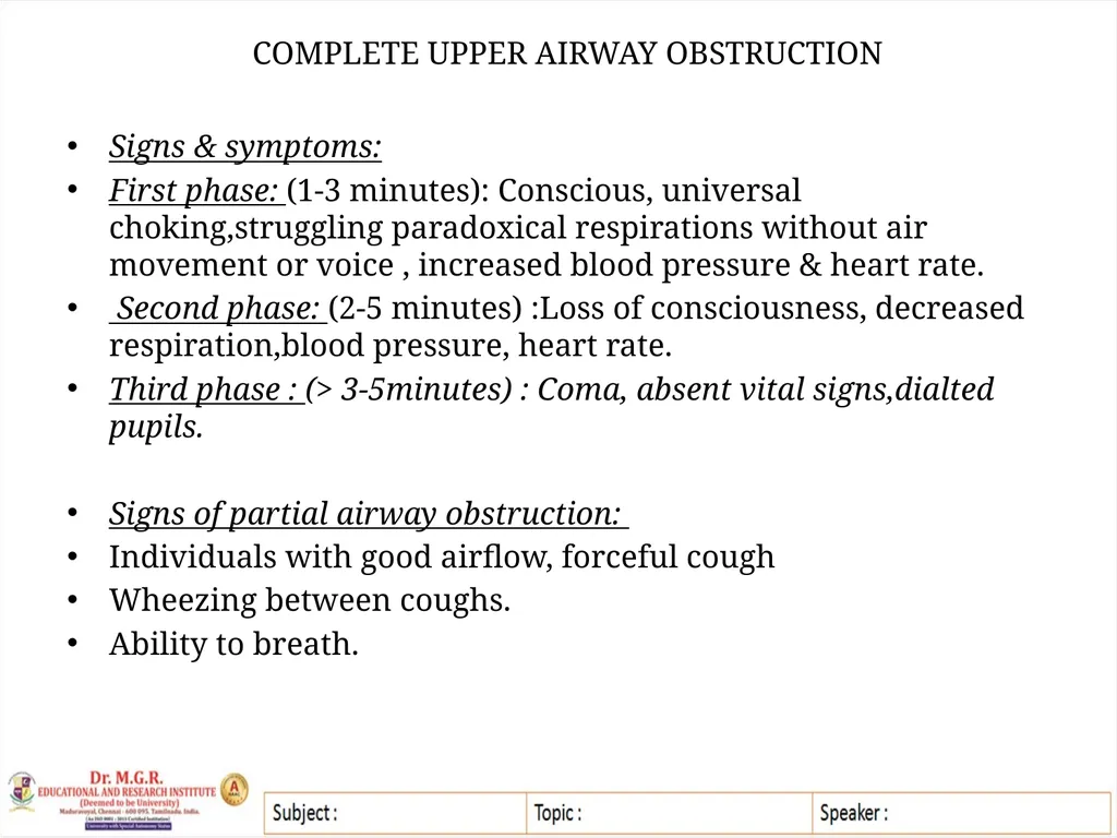
slide14. COMPLETE UPPER AIRWAY OBSTRUCTION Signs & symptoms:
First phase: (1-3 minutes): Conscious, universal choking,struggling paradoxical respirations without air movement or voice , increased blood pressure & heart rate.
Second phase: (2-5 minutes) :Loss of consciousness, decreased respiration,blood pressure, heart rate.
Third phase : (> 3-5minutes) : Coma, absent vital signs,dialted pupils.
Signs of partial airway obstruction:
Individuals with good airflow, forceful cough
Wheezing between coughs.
Ability to breath.<br>
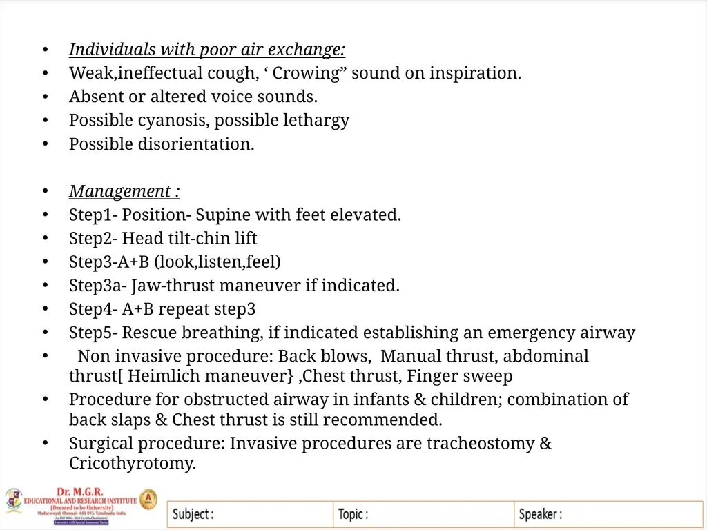
slide15. Individuals with poor air exchange:
Weak,ineffectual cough, ‘ Crowing” sound on inspiration.
Absent or altered voice sounds.
Possible cyanosis, possible lethargy
Possible disorientation.
Management :
Step1- Position- Supine with feet elevated.
Step2- Head tilt-chin lift
Step3-A+B (look,listen,feel)
Step3a- Jaw-thrust maneuver if indicated.
Step4- A+B repeat step3
Step5- Rescue breathing, if indicated establishing an emergency airway
Non invasive procedure: Back blows, Manual thrust, abdominal thrust[ Heimlich maneuver} ,Chest thrust, Finger sweep
Procedure for obstructed airway in infants & children; combination of back slaps & Chest thrust is still recommended.
Surgical procedure: Invasive procedures are tracheostomy & Cricothyrotomy.<br>
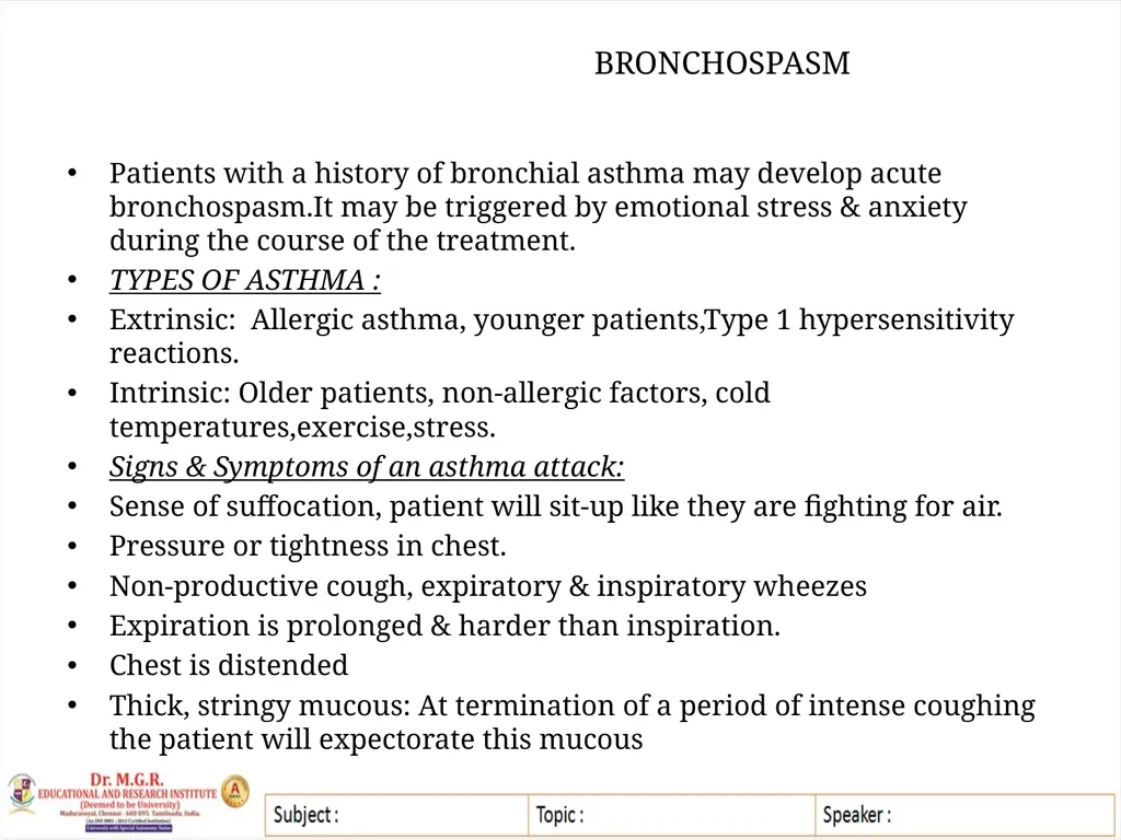
slide16. BRONCHOSPASM Patients with a history of bronchial asthma may develop acute bronchospasm.It may be triggered by emotional stress & anxiety during the course of the treatment.
TYPES OF ASTHMA :
Extrinsic: Allergic asthma, younger patients,Type 1 hypersensitivity reactions.
Intrinsic: Older patients, non-allergic factors, cold temperatures,exercise,stress.
Signs & Symptoms of an asthma attack:
Sense of suffocation, patient will sit-up like they are fighting for air.
Pressure or tightness in chest.
Non-productive cough, expiratory & inspiratory wheezes
Expiration is prolonged & harder than inspiration.
Chest is distended
Thick, stringy mucous: At termination of a period of intense coughing the patient will expectorate this mucous<br>
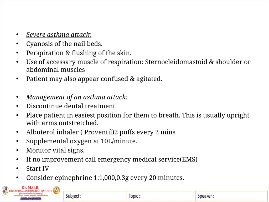
slide17. Severe asthma attack:
Cyanosis of the nail beds.
Perspiration & flushing of the skin.
Use of accessary muscle of respiration: Sternocleidomastoid & shoulder or abdominal muscles
Patient may also appear confused & agitated.
Management of an asthma attack:
Discontinue dental treatment
Place patient in easiest position for them to breath. This is usually upright with arms outstretched.
Albuterol inhaler ( Proventil)2 puffs every 2 mins
Supplemental oxygen at 10L/minute.
Monitor vital signs.
If no improvement call emergency medical service(EMS)
Start IV
Consider epinephrine 1:1,000,0.3g every 20 minutes.<br>
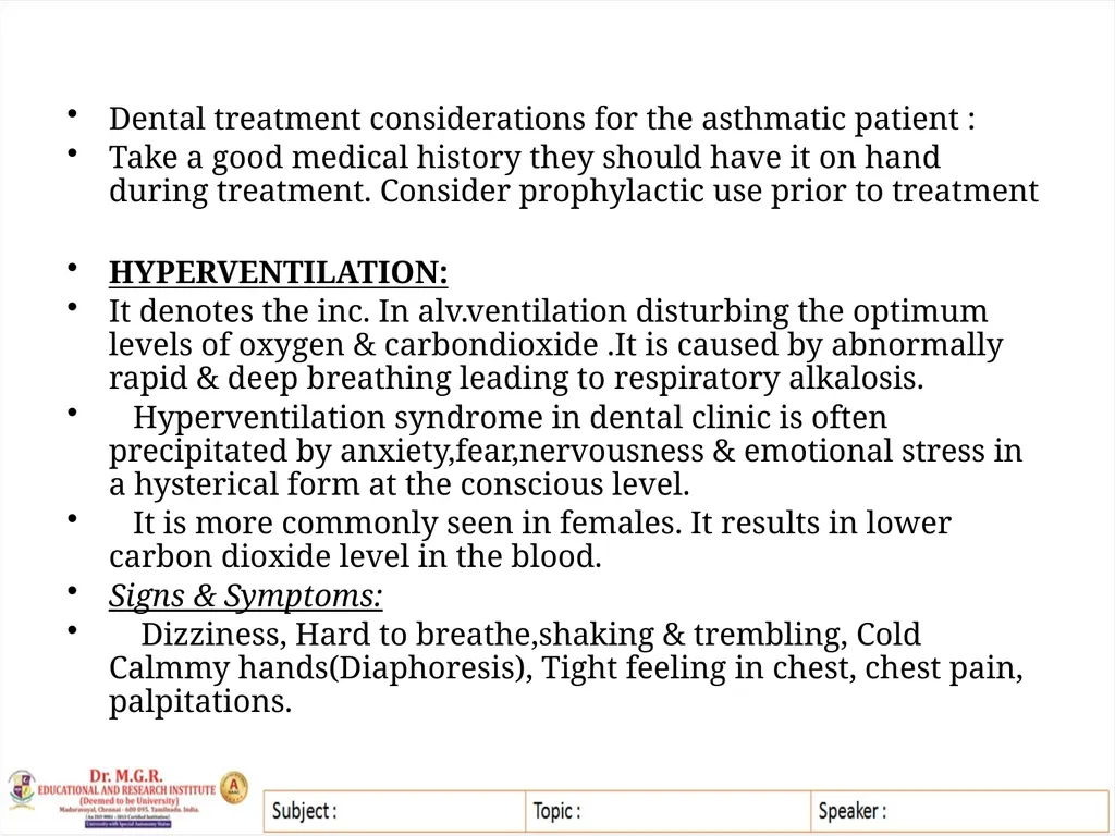
slide18. Dental treatment considerations for the asthmatic patient :
Take a good medical history they should have it on hand during treatment. Consider prophylactic use prior to treatment
HYPERVENTILATION:
It denotes the inc. In alv.ventilation disturbing the optimum levels of oxygen & carbondioxide .It is caused by abnormally rapid & deep breathing leading to respiratory alkalosis.
Hyperventilation syndrome in dental clinic is often precipitated by anxiety,fear,nervousness & emotional stress in a hysterical form at the conscious level.
It is more commonly seen in females. It results in lower carbon dioxide level in the blood.
Signs & Symptoms:
Dizziness, Hard to breathe,shaking & trembling, Cold Calmmy hands(Diaphoresis), Tight feeling in chest, chest pain, palpitations.<br>
slide19. Lightheaded,giddy,impaired consciousness.
Uncontrolled over breathing.Respiration rate increase to 25-30/minute.
Globus Hystericus: Feeling of lump in throat & suffocating.
Tingling in hands, feet & perioral areas.
Inc. In blood pressure & increase heart rate
Treatment :
Discontinue treatment & remove any foreign objects from the patient’s mouth.
Position patient upright, assess airway , reassure patient & try to calm them
Have patient breath slowly & shallowly into a paper bag or mask 6-10 times / minute.
Monitor vital signs
If available it can use versed IV 1mg / minute upto 4-6mg or IM 5mg to calm the patient
Determine what precipitated attack<br>
slide20. HYPERTENSION When a patient exhibits blood pressure above 160/100mm Hg is in the preoperative phase, he is labeled as hypertensive.
Patient complains of headache, dizziness, nausea & even vomiting.
Fundus examination reveals hemorrhages or exhibits blood spots over the retina with increased intracranial tension.
Treatment :
This can be avoided by proper premedication
Should this emergency arise intraoperatively? The patient is allowed to take rest in a semi-sitting position.
Oxygen may be administered to bring down the blood pressure.
Injection diazepam may be administered. This normally settles the stress-related hypertension.
Injection furosemide IV & is maintained as a preparation to any serious emergency.
10mg capsule nifedipine sublingually to bring the blood pressure down.<br>
slide21. HYPOTENSION A fall in blood pressure or hypotension during oral surgical procedures can be due to a simple common fainting attack.
Signs & symptoms:
Associated weak pulse, bradycardia.
Confusion, restlessness, nausea, stupor.
Treatment:
Put the patient in supine position with legs raised, 100% oxygen should be administered, IV line maintained with Ringer’s lactate solution, Atropine 0.6mg in 5mL of sterile water is given IV slowly if the pulse is less than 6 per minute.
Atropine should be stopped when a good volume radial pulse with rate of 72 per minute appears.
Inj .mephentermine 15mg can be administered intramuscularly.
Inj hydrocortisone succinate 100mg should be administered or combating stress.<br>
slide22. ISCHEMIC HEART DISEASE It denotes ischemia of the myocardium leading to arrythmias,angina pectoris, myocardial infarction & sudden death.
Condition is characterized by tightness in the chest, sensation of choking & a referred to jaw & neck. The attack may be precipitated on exertion by the stress during the dental treatment
Treatment :
In case of an acute attack of angina pectoris occurs in dental chair, dental treatment should immediately be stopped,adjust the chair in semi-reclining position.
A tablet of nitroglycerin is placed sublingually & can be repeated after 5 mins.
Oxygen administration, patient physician should be immediately called
In case of MI pain may be controlled with small amounts of morphine.
Inj atropine should be given if there is bradycardia.
In case of recent history of MI, any elective surgery should not be undertaken for a period of 6 months.<br>
slide23. TO BE CONTINUED......<br>
slide2. Department : Pedodontics Topic :Medical Emergencies Staff name : Dr Joyson Moses MEDICAL EMERGENCIES IN DENTAL PRACTICE MEDICAL EMERGENCIES IN DENTAL PRACTICE MEDICAL EMERGENCIES IN DENTAL PRACTICE<br>
slide3. MEDICAL EMERGENCIES IN DENTAL PRACTICE<br>
slide4. INTRODUCTION Emergency- a condition that warrants or immediate attention by the doctor.
Many emergency problems can be definitely avoided by simple preventive measures like a careful medical history, general physical examination regarding patient health status & proper preoperative preparation of the patient.
Emergency according to Dorland’s Medical Dictionary is defined as a sudden, urgent, usually unforeseen occurrence requiring immediate action.<br>
slide5. SHOCK It is a phenomenon marked by circulatory deficiency which is either cardiac or vasomotor in origin exhibiting marked hypotension.
Signs & Symptoms:
Patient –unconscious with ashen grey face & cold, clammy skin.
Mucous membrane –pale whereas lips,nails, finger tips & lobules of the ear are grayish blue. Face is expression less with sunken eyes.
Pupils are dilated but react feebly to light.
Pulse is weak & thread.
Shallow & irregular respiration
Temperature is subnormal
Treatment :
Position: Put the patients head at the lower level than feet 15 degree- Trendelenburg position<br>
slide6. Maintain the body heat by covering the patient with blanket & keep a hot water bottle between the thighs.
Check for any airway obstruction & patency of airway be maintained.
Control the loss of blood in haemorrhage shock by pressure packs.
Restore the lost body fluids. Infusion with plasma expanders or Ringer’s lactate solution should be carried out to maintain the intravenous line & restore the volume loss
Administer 100 percent oxygen
Blood pressure,pulse rate & respiratory rate should be constantly monitored to assess the vitals.
Injection hydrocortisone sodium hemisuccinate 100mg in 5mL of water intravenously as stress bearing factor of he body
Injection mephentermine to raise BP
Injection atropine is given for bradycardia.
Broad spectrum antibiotics.
Narcotic analgesic to relieve pain<br>
slide7. ALLERGIC REACTION It is an unwanted response of the body to a complete dose of the drug. It is the result of immunological response of /in the individual
Gell & Coombs classification:
Type I ( IgE- mediated hypersensitivity )- most life- theatening few minutes.
Type 2 ( Cytotoxic / cytolytic antibdy-mediated) IgM or Ig G soluble metabolite.
Type 3 ( Immune complex mediated ) 1-4 weeks, IgM or IgG soluble metabolite.
Type 4 ( Delayed hypersensitivity )- sensitized T-cell lymphocytes.
Signs & symptoms:
Cutaneous reactions are the most common occurrence & include urticarial,exanthematous & eczematoid reactions. Itching is common & can also find exfoliative dermatitis & bullous dermatosis.<br>
slide8. Angioedema ( swelling) this varies from localized slight swelling of the lips, eyelids, & face to more uncomfortable swelling of the mouth, throat & extremities.
Respiratory ( tightness in chest,sneezing, bronchospasm) bronchospasm is a generalized contraction of bronchial smooth muscles resulting in the restriction of airflow. This may also be accompanied by edema of the bronchiolar mucosa.
Bronchospasm is more common with preexisting pulmonary disease such as asthma or infection but can also be caused by the nhalation of a foreign substance.
Ocular reactions include conjunctivitis & watering of eyes.
Hypotension can occur with any allergic reaction.<br>
slide9. ANAPLYLAXIS This is a severe systemic type allergic reaction & is a medical emergency.
Signs & symptoms :
Cardiovascular shock including : Pallor, syncope, palpitations,tachycardia,hypotension,arrhythmias & convulsions.
Respiratory symptoms include: Sneezing , cough,wheezing,tightness in chest, bronchospasm,laryngospasm.
Skin is warm & flushed with itching,urticaria & angioedema.
Nausea, vomiting , abdominal cramps & diarrhea are also possible.
Treatment :
General treatment :
ABC’s ( is expansion required or removes the bullet from the next point)<br>
slide10. Maintain airway, administer oxygen, & determine possible need for intubation or surgical airway.
Monitor vital signs.
If in shock put patient in a horizontal or slight Trendelenburg position.
Mild reactions:
Antihistamines usually effective.(Benadryl 50-100mg or chlorpheniramine maleate 4-12mg PO,IV OR IM)
Identify & remove allergen
Follow-up medications in 4-6 hrs.
Severe reactions:
If available start IV Fluids, epinephtine is drug of choice. Usually pre-packaged 1:1,000in 1 mg vials or syringe,if IV in place titrate 1:1,000 solution to effect<br>
slide11. If drop in blood pressure is minimal,start with 0.5mL (0.5mg)
If drop in blood pressure is severe start with 2mL (2mg), repeat after 2 minute if needed.
If no IV use 1;1,000(1mg/cc),IM 0.3-0.5mg ( 0.3-0.5cc), for an adult repeat this dose in 10-20mins.
If the patient is intubated can give epinephrine endotracheally.
If asthma,edema or pruritus ( itching) is present, can use corticosteroids.However,these drugs are too slow-acting to be used for an emergency situation
Hydrocortisone sodium succinate (solu-cortef) 100-500mg IV or IM. Dexamethasone ( Decadron) 4-12mg IV or IM
Repeat dose at 1,3,6 & 10 hrs as indicated by severity of symptoms.<br>
slide12. RESPIRATORY EMERGECIES Airway Obstruction:
Acute airway obstruction is the major cause of non-traumatic cardiac arrest in infants & children.
Sit down dentistry ( supine or semi-supine ) – increase incidence of airway obstruction.
If swallowed- GI Blockage, peritoneal abscess, perforations, peritonitis.
If aspirated – Lung abscess, pneumonia, atelectasis.
Prevention:
Rubber dam
Oral packing ( Pharyngeal curtain:4* 4 inch gauze pack usually used in sedated patients).
Ligature ( Dental floss tied to dental instruments such as rubber dam clamps, endodontic instruments, cotton rolls, gauze pads,etc,.)<br>
slide13. Management :
If assistant is present- patient placed into supine or Trendelenburg position , use Magill intubation forcep or suction to remove foreign body.
If assistant not present- instruct patient to bend over arm of chair with their head down & encourage patient to cough.
Management of swallowed objects:
Consult radiologist- obtain radiographs to determine location of object & initiate medical consultation with appropriate specials
Management of aspirated foreign objects:
Place patient in left lateral decubitus position- encourage patient to cough if foreign body is retrieved, initiate medical consultation before discharge.
If foreign body is not retrieved-consult with radiologist & obtain radiographs, perform bronchoscopy to visualize & retrieve foreign body<br>
slide14. COMPLETE UPPER AIRWAY OBSTRUCTION Signs & symptoms:
First phase: (1-3 minutes): Conscious, universal choking,struggling paradoxical respirations without air movement or voice , increased blood pressure & heart rate.
Second phase: (2-5 minutes) :Loss of consciousness, decreased respiration,blood pressure, heart rate.
Third phase : (> 3-5minutes) : Coma, absent vital signs,dialted pupils.
Signs of partial airway obstruction:
Individuals with good airflow, forceful cough
Wheezing between coughs.
Ability to breath.<br>
slide15. Individuals with poor air exchange:
Weak,ineffectual cough, ‘ Crowing” sound on inspiration.
Absent or altered voice sounds.
Possible cyanosis, possible lethargy
Possible disorientation.
Management :
Step1- Position- Supine with feet elevated.
Step2- Head tilt-chin lift
Step3-A+B (look,listen,feel)
Step3a- Jaw-thrust maneuver if indicated.
Step4- A+B repeat step3
Step5- Rescue breathing, if indicated establishing an emergency airway
Non invasive procedure: Back blows, Manual thrust, abdominal thrust[ Heimlich maneuver} ,Chest thrust, Finger sweep
Procedure for obstructed airway in infants & children; combination of back slaps & Chest thrust is still recommended.
Surgical procedure: Invasive procedures are tracheostomy & Cricothyrotomy.<br>
slide16. BRONCHOSPASM Patients with a history of bronchial asthma may develop acute bronchospasm.It may be triggered by emotional stress & anxiety during the course of the treatment.
TYPES OF ASTHMA :
Extrinsic: Allergic asthma, younger patients,Type 1 hypersensitivity reactions.
Intrinsic: Older patients, non-allergic factors, cold temperatures,exercise,stress.
Signs & Symptoms of an asthma attack:
Sense of suffocation, patient will sit-up like they are fighting for air.
Pressure or tightness in chest.
Non-productive cough, expiratory & inspiratory wheezes
Expiration is prolonged & harder than inspiration.
Chest is distended
Thick, stringy mucous: At termination of a period of intense coughing the patient will expectorate this mucous<br>
slide17. Severe asthma attack:
Cyanosis of the nail beds.
Perspiration & flushing of the skin.
Use of accessary muscle of respiration: Sternocleidomastoid & shoulder or abdominal muscles
Patient may also appear confused & agitated.
Management of an asthma attack:
Discontinue dental treatment
Place patient in easiest position for them to breath. This is usually upright with arms outstretched.
Albuterol inhaler ( Proventil)2 puffs every 2 mins
Supplemental oxygen at 10L/minute.
Monitor vital signs.
If no improvement call emergency medical service(EMS)
Start IV
Consider epinephrine 1:1,000,0.3g every 20 minutes.<br>
slide18. Dental treatment considerations for the asthmatic patient :
Take a good medical history they should have it on hand during treatment. Consider prophylactic use prior to treatment
HYPERVENTILATION:
It denotes the inc. In alv.ventilation disturbing the optimum levels of oxygen & carbondioxide .It is caused by abnormally rapid & deep breathing leading to respiratory alkalosis.
Hyperventilation syndrome in dental clinic is often precipitated by anxiety,fear,nervousness & emotional stress in a hysterical form at the conscious level.
It is more commonly seen in females. It results in lower carbon dioxide level in the blood.
Signs & Symptoms:
Dizziness, Hard to breathe,shaking & trembling, Cold Calmmy hands(Diaphoresis), Tight feeling in chest, chest pain, palpitations.<br>
slide19. Lightheaded,giddy,impaired consciousness.
Uncontrolled over breathing.Respiration rate increase to 25-30/minute.
Globus Hystericus: Feeling of lump in throat & suffocating.
Tingling in hands, feet & perioral areas.
Inc. In blood pressure & increase heart rate
Treatment :
Discontinue treatment & remove any foreign objects from the patient’s mouth.
Position patient upright, assess airway , reassure patient & try to calm them
Have patient breath slowly & shallowly into a paper bag or mask 6-10 times / minute.
Monitor vital signs
If available it can use versed IV 1mg / minute upto 4-6mg or IM 5mg to calm the patient
Determine what precipitated attack<br>
slide20. HYPERTENSION When a patient exhibits blood pressure above 160/100mm Hg is in the preoperative phase, he is labeled as hypertensive.
Patient complains of headache, dizziness, nausea & even vomiting.
Fundus examination reveals hemorrhages or exhibits blood spots over the retina with increased intracranial tension.
Treatment :
This can be avoided by proper premedication
Should this emergency arise intraoperatively? The patient is allowed to take rest in a semi-sitting position.
Oxygen may be administered to bring down the blood pressure.
Injection diazepam may be administered. This normally settles the stress-related hypertension.
Injection furosemide IV & is maintained as a preparation to any serious emergency.
10mg capsule nifedipine sublingually to bring the blood pressure down.<br>
slide21. HYPOTENSION A fall in blood pressure or hypotension during oral surgical procedures can be due to a simple common fainting attack.
Signs & symptoms:
Associated weak pulse, bradycardia.
Confusion, restlessness, nausea, stupor.
Treatment:
Put the patient in supine position with legs raised, 100% oxygen should be administered, IV line maintained with Ringer’s lactate solution, Atropine 0.6mg in 5mL of sterile water is given IV slowly if the pulse is less than 6 per minute.
Atropine should be stopped when a good volume radial pulse with rate of 72 per minute appears.
Inj .mephentermine 15mg can be administered intramuscularly.
Inj hydrocortisone succinate 100mg should be administered or combating stress.<br>
slide22. ISCHEMIC HEART DISEASE It denotes ischemia of the myocardium leading to arrythmias,angina pectoris, myocardial infarction & sudden death.
Condition is characterized by tightness in the chest, sensation of choking & a referred to jaw & neck. The attack may be precipitated on exertion by the stress during the dental treatment
Treatment :
In case of an acute attack of angina pectoris occurs in dental chair, dental treatment should immediately be stopped,adjust the chair in semi-reclining position.
A tablet of nitroglycerin is placed sublingually & can be repeated after 5 mins.
Oxygen administration, patient physician should be immediately called
In case of MI pain may be controlled with small amounts of morphine.
Inj atropine should be given if there is bradycardia.
In case of recent history of MI, any elective surgery should not be undertaken for a period of 6 months.<br>
slide23. TO BE CONTINUED......<br>