
Trachea BY MBBSPPT.COM The Trachea is also called


Description: Trachea BY MBBSPPT.COM The Trachea is also called windpipe and it is a fibrocartilaginous, non-collapsible tube that creates the beginning of the lower respiratory tract. 16-20 C-shaped rings of Hyaline cartilage keep its lumen patent. A
Related Topics
Download Presentation
"Trachea BY MBBSPPT.COM The Trachea is also called" is the property of its rightful owner. Permission is granted to download and print the materials on this website for personal, non-commercial use only, and to display it on your personal computer provided you do not modify the materials and that you retain all copyright notices contained in the materials. By downloading content from our website, you accept the terms of this agreement.
Presentation Transcript
slide1. Trachea BY MBBSPPT.COM<br>
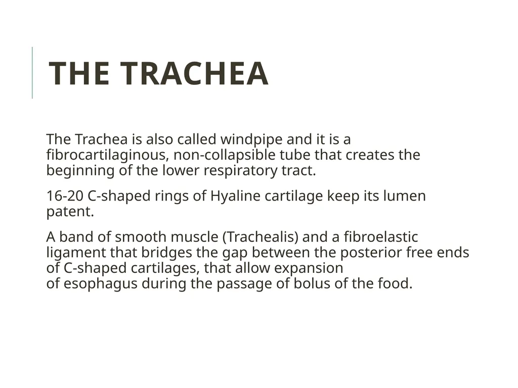
slide3. The Trachea is also called windpipe and it is a fibrocartilaginous, non-collapsible tube that creates the beginning of the lower respiratory tract.
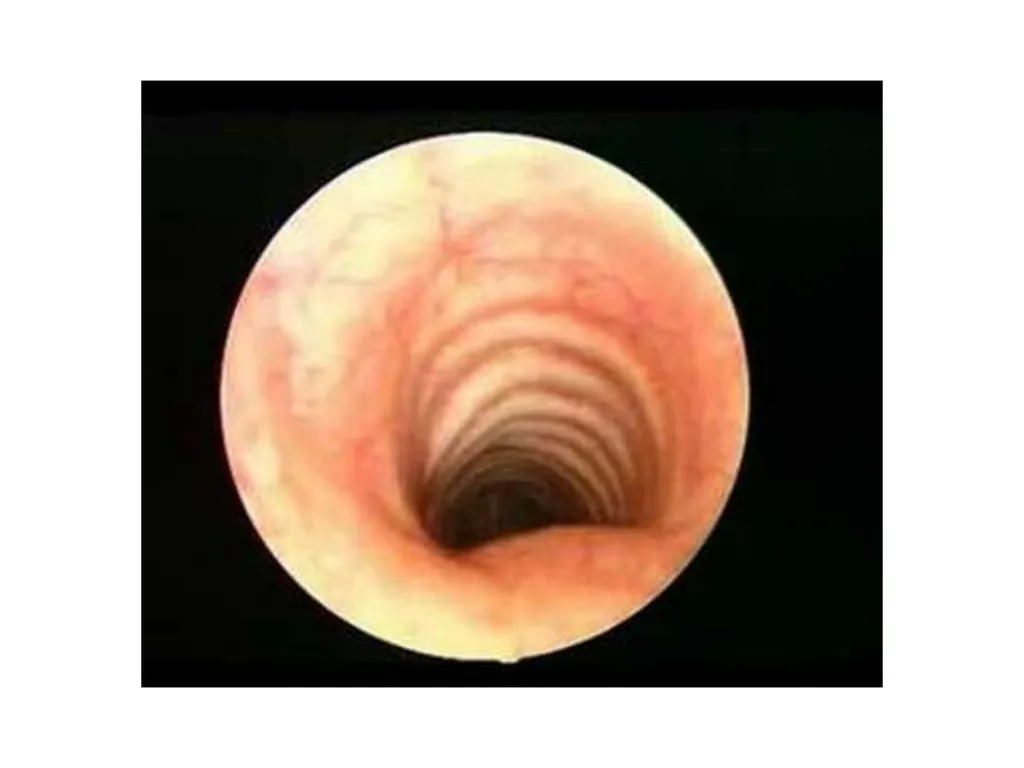
16-20 C-shaped rings of Hyaline cartilage keep its lumen patent.
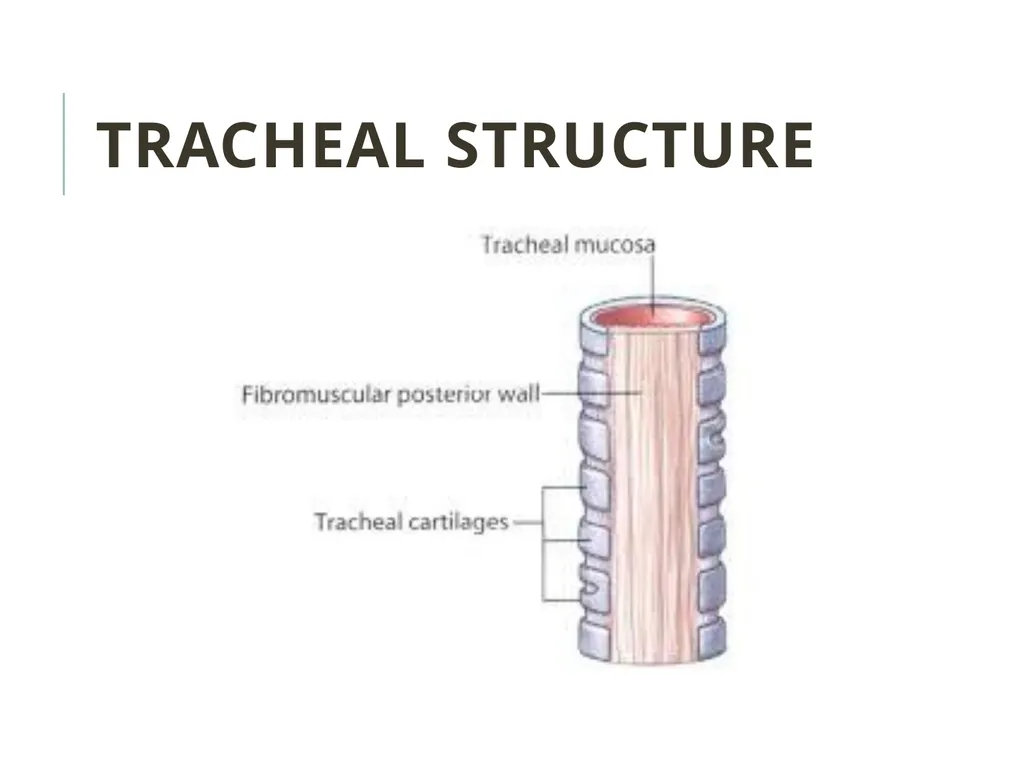
A band of smooth muscle (Trachealis) and a fibroelastic ligament that bridges the gap between the posterior free ends of C-shaped cartilages, that allow expansion of esophagus during the passage of bolus of the food. The Trachea<br>
slide4. Tracheal structure<br>
slide6. The trachea, bronchi and bronchioles form the tracheobronchial tree – a system of airways that allow passage of air into the lungs, where gas exchange occurs.
These airways are located in the neck and thorax.
The arrangement of cartilages and elastic tissue in the tracheal wall prevents its kinking and obstruction during the movements of the head and neck. The Trachea<br>
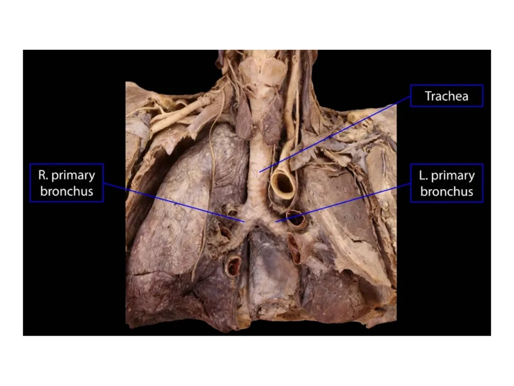
slide8. The trachea marks the beginning of the tracheobronchial tree.
It arises at the lower border of cricoid cartilage in the neck, as a continuation of the larynx.
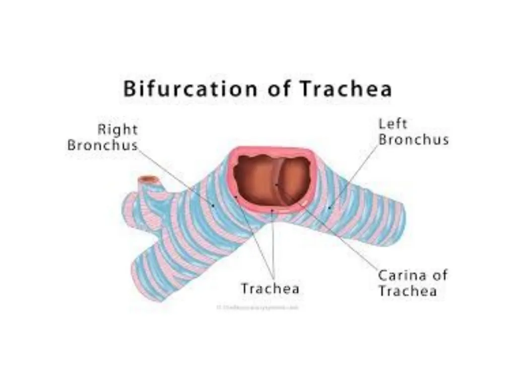
It travels inferiorly into the superior mediastinum, bifurcating at the level of the sternal angle (forming the right and left main bronchi).
As it descends, the trachea is located anteriorly to the esophagus, and inclines slightly to the right. Anatomical Position<br>
slide9. Trachea stretches from the lower border of cricoid cartilage at the lower border of the C6 vertebra to the lower border of T4 vertebra in supine position, where it ends by dividing into left and right main bronchi. Anatomical Position<br>
slide11. The upper half of trachea can be found in the neck (cervical part) on the other hand the lower half is located in the superior mediastinum of the thoracic cavity (thoracic part).
The trachea is a 4-4½ inches (10-11 cm) long tube.
The diameter of trachea is 2 cm in men and 1.5 cm in females.
The lumen is smaller in living human than that in cadavers. MEASUREMENTS<br>
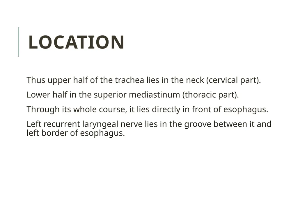
slide12. Thus upper half of the trachea lies in the neck (cervical part).
Lower half in the superior mediastinum (thoracic part).
Through its whole course, it lies directly in front of esophagus.
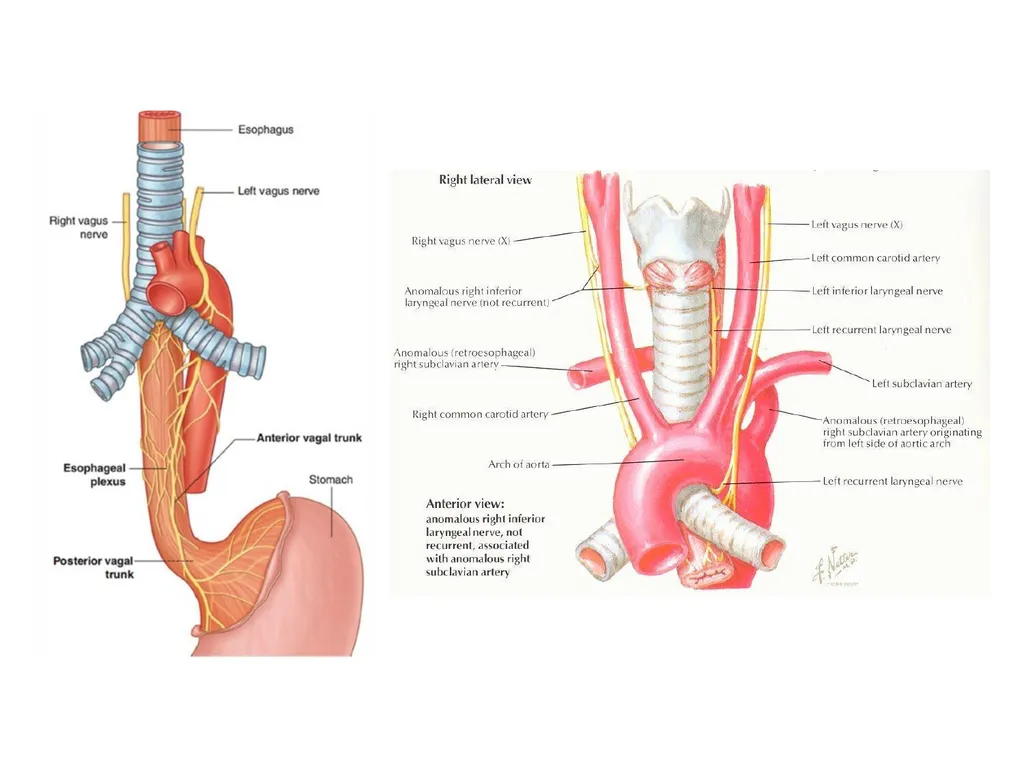
Left recurrent laryngeal nerve lies in the groove between it and left border of esophagus. LOCATION<br>
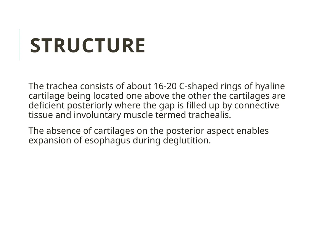
slide13. The trachea consists of about 16-20 C-shaped rings of hyaline cartilage being located one above the other the cartilages are deficient posteriorly where the gap is filled up by connective tissue and involuntary muscle termed trachealis.
The absence of cartilages on the posterior aspect enables expansion of esophagus during deglutition. STRUCTURE<br>
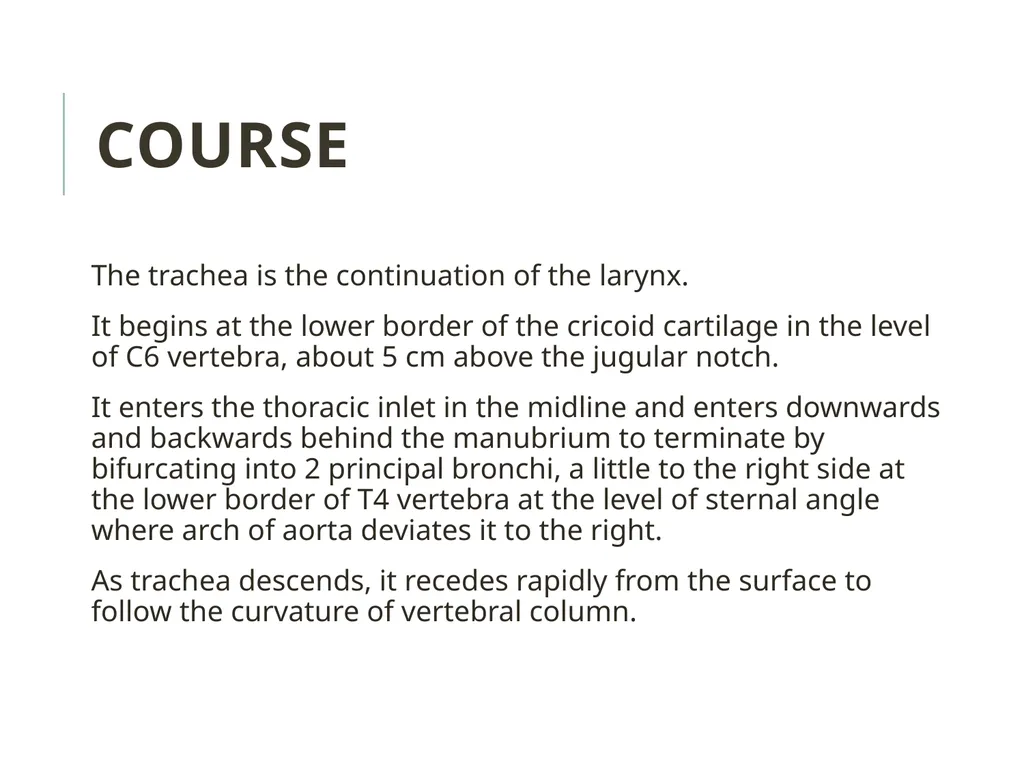
slide14. The trachea is the continuation of the larynx.
It begins at the lower border of the cricoid cartilage in the level of C6 vertebra, about 5 cm above the jugular notch.
It enters the thoracic inlet in the midline and enters downwards and backwards behind the manubrium to terminate by bifurcating into 2 principal bronchi, a little to the right side at the lower border of T4 vertebra at the level of sternal angle where arch of aorta deviates it to the right.
As trachea descends, it recedes rapidly from the surface to follow the curvature of vertebral column. COURSE<br>
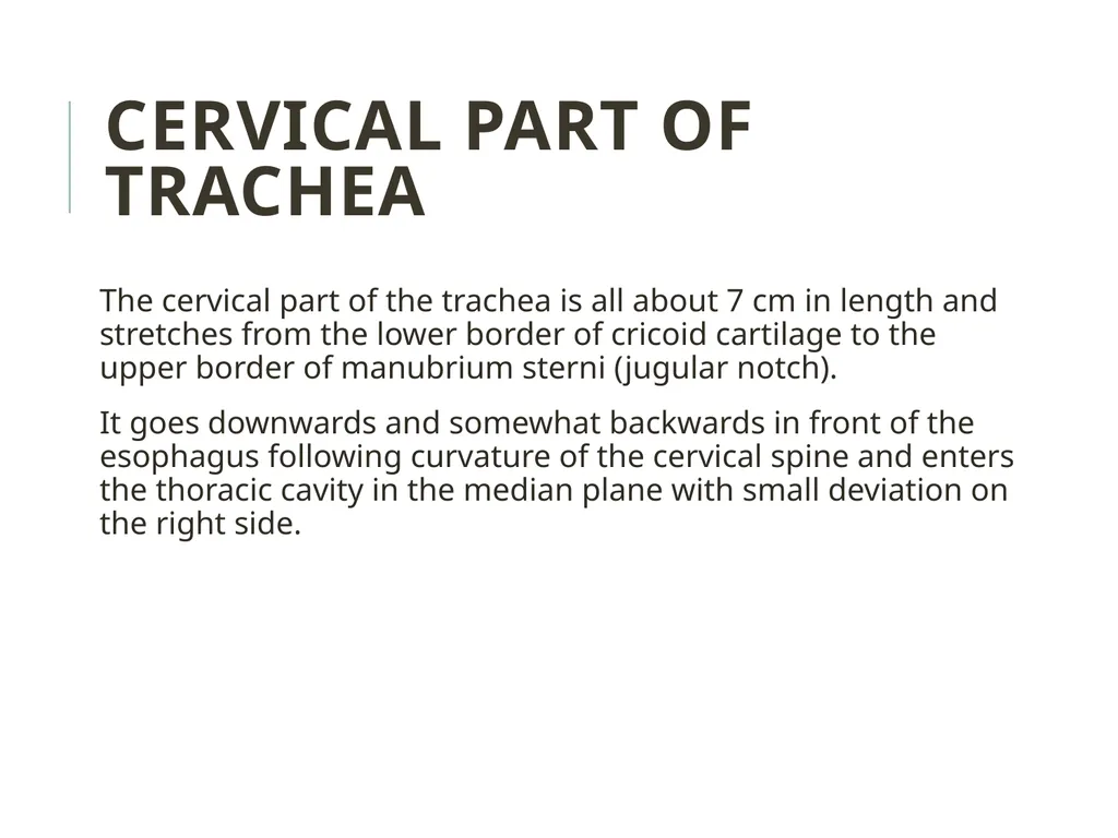
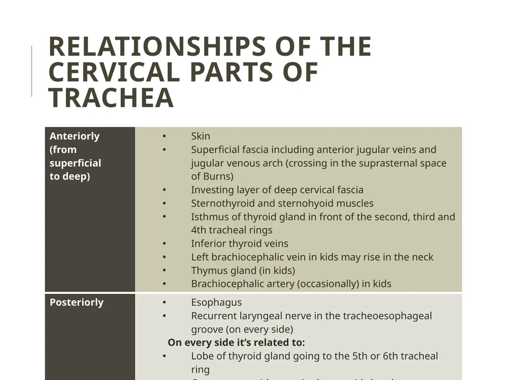
slide15. The cervical part of the trachea is all about 7 cm in length and stretches from the lower border of cricoid cartilage to the upper border of manubrium sterni (jugular notch).
It goes downwards and somewhat backwards in front of the esophagus following curvature of the cervical spine and enters the thoracic cavity in the median plane with small deviation on the right side. CERVICAL PART OF TRACHEA<br>
slide16. RELATIONSHIPS OF THE CERVICAL PARTS OF TRACHEA<br>
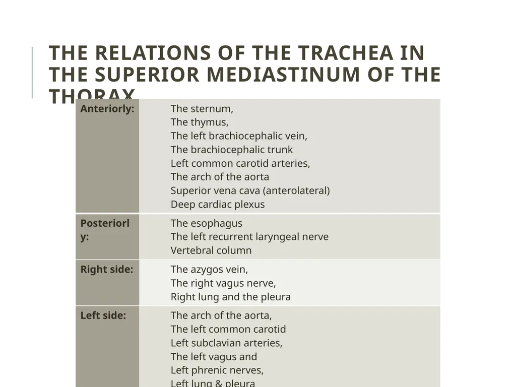
slide17. The relations of the trachea in the superior mediastinum of the thorax<br>
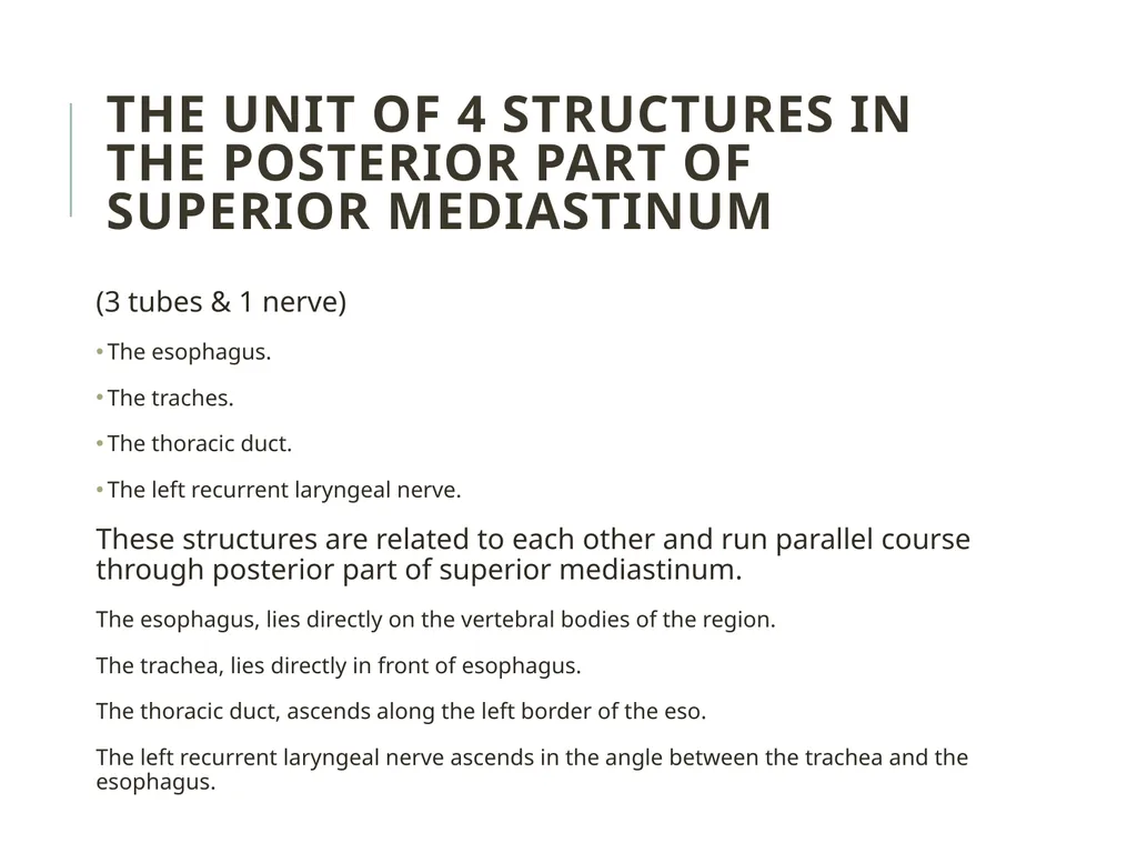
slide18. (3 tubes & 1 nerve)
The esophagus.
The traches.
The thoracic duct.
The left recurrent laryngeal nerve.
These structures are related to each other and run parallel course through posterior part of superior mediastinum.
The esophagus, lies directly on the vertebral bodies of the region.
The trachea, lies directly in front of esophagus.
The thoracic duct, ascends along the left border of the eso.
The left recurrent laryngeal nerve ascends in the angle between the trachea and the esophagus. The unit of 4 structures in the posterior part of superior mediastinum<br>
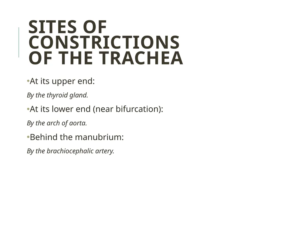
slide20. At its upper end:
By the thyroid gland.
At its lower end (near bifurcation):
By the arch of aorta.
Behind the manubrium:
By the brachiocephalic artery. Sites of constrictions
of the trachea<br>
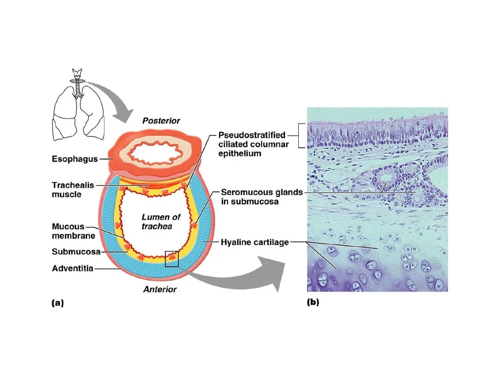
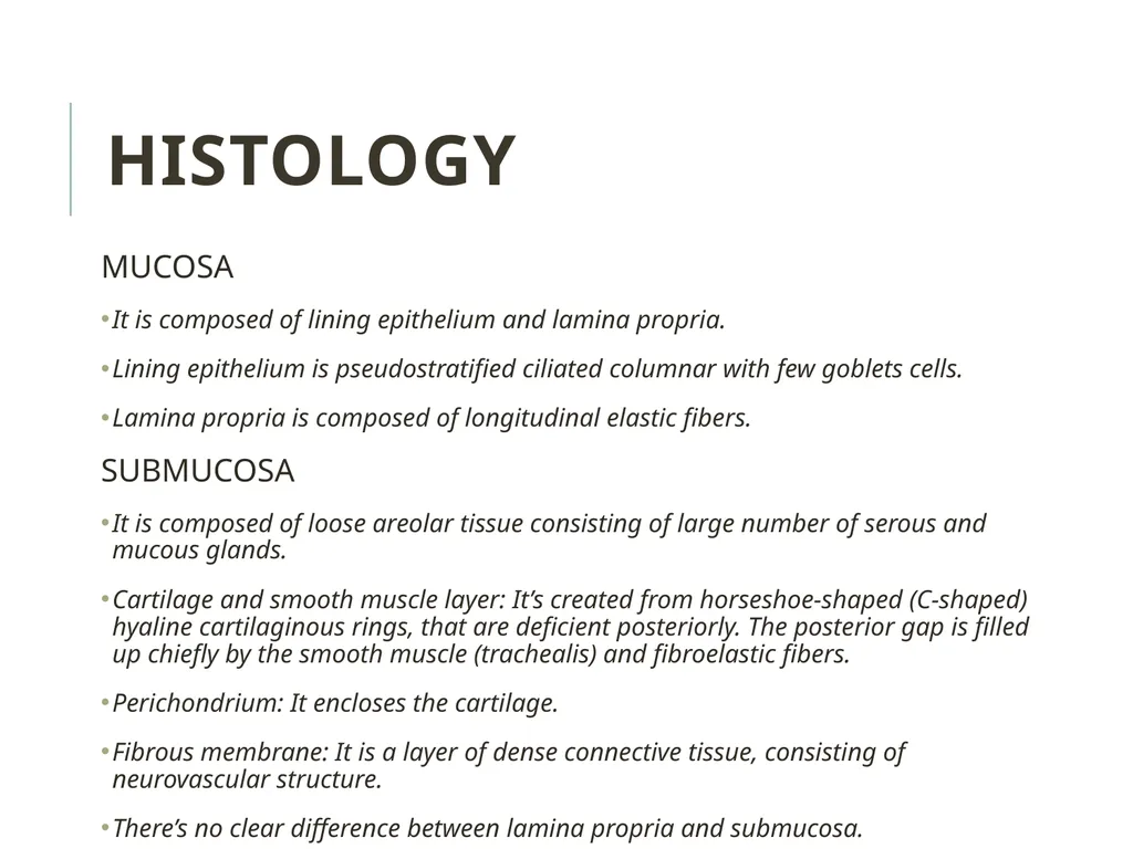
slide21. MUCOSA
It is composed of lining epithelium and lamina propria.
Lining epithelium is pseudostratified ciliated columnar with few goblets cells.
Lamina propria is composed of longitudinal elastic fibers.
SUBMUCOSA
It is composed of loose areolar tissue consisting of large number of serous and mucous glands.
Cartilage and smooth muscle layer: It’s created from horseshoe-shaped (C-shaped) hyaline cartilaginous rings, that are deficient posteriorly. The posterior gap is filled up chiefly by the smooth muscle (trachealis) and fibroelastic fibers.
Perichondrium: It encloses the cartilage.
Fibrous membrane: It is a layer of dense connective tissue, consisting of neurovascular structure.
There’s no clear difference between lamina propria and submucosa. Histology<br>
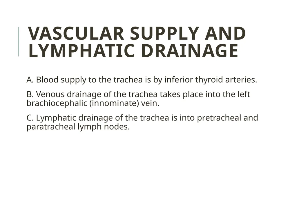
slide22. A. Blood supply to the trachea is by inferior thyroid arteries.
B. Venous drainage of the trachea takes place into the left brachiocephalic (innominate) vein.
C. Lymphatic drainage of the trachea is into pretracheal and paratracheal lymph nodes. VASCULAR SUPPLY AND LYMPHATIC DRAINAGE<br>
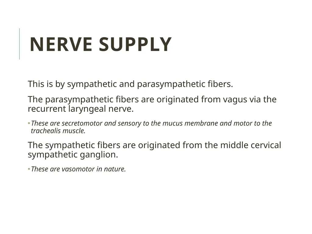
slide23. NERVE SUPPLY This is by sympathetic and parasympathetic fibers.
The parasympathetic fibers are originated from vagus via the recurrent laryngeal nerve.
These are secretomotor and sensory to the mucus membrane and motor to the trachealis muscle.
The sympathetic fibers are originated from the middle cervical sympathetic ganglion.
These are vasomotor in nature.<br>
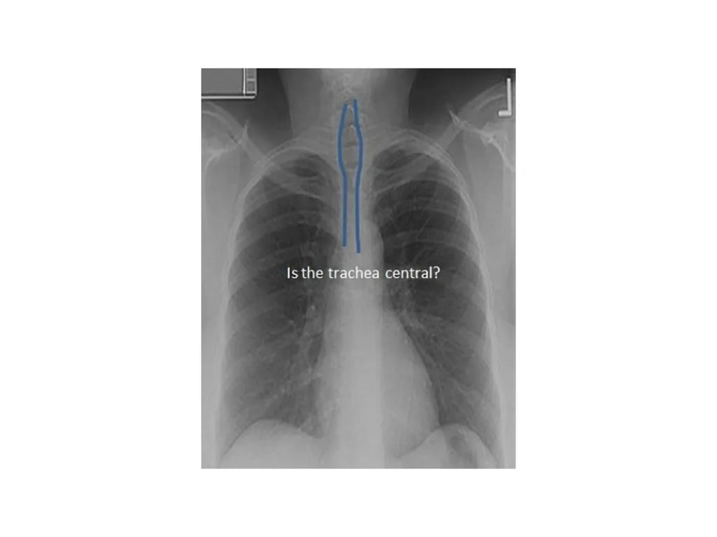
slide24. TRACHEAL SHADOW IN RADIOGRAPH It is viewed as a vertical translucent shadow in front of cervico-thoracic spine.
The translucency is because of the presence of air in the trachea.<br>
slide26. PALPATION OF TRACHEA Medically, trachea is palpated in the suprasternal notch.
Normally, it is median in position but appreciable shift of trachea to left or right side indicates the mediastinal shift.<br>
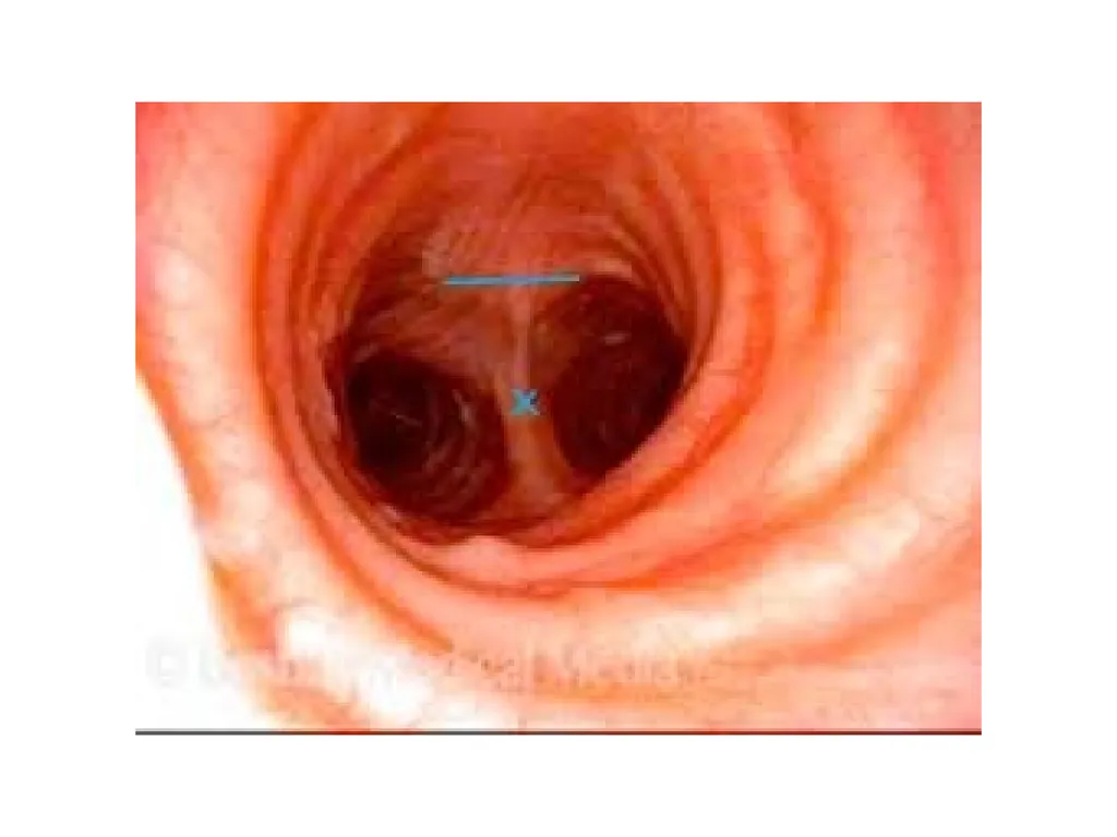
slide28. IMPORTANCE OF CARINA It is a keel-like (hook-shaped) median ridge in the lumen in the bifurcation of trachea. It is both functional and pathological importance.
Functional importance: The mucosa of trachea over the carina is most sensitive. The cough reflex is generally started here, which helps to clear the sputum.
Pathological importance: It is visible as a sharp sagittal ridge in the tracheal bifurcation during bronchoscopy, for this reason acts as a useful landmark.
It is located about 25 cm from the incisor teeth and 30 cm from the nostrils. If the tracheobronchial lymph nodes in the angle between the main (principal) bronchi are enlarged because of spread of bronchiogenic carcinoma, the carina becomes distorted and flattened.<br>
slide30. IMPORTANCE OF CARINA It is a keel-like (hook-shaped) median ridge in the lumen in the bifurcation of trachea. It is both functional and pathological importance.
Functional importance: The mucosa of trachea over the carina is most sensitive. The cough reflex is generally started here, which helps to clear the sputum.
Pathological importance: It is visible as a sharp sagittal ridge in the tracheal bifurcation during bronchoscopy, for this reason acts as a useful landmark.
It is located about 25 cm from the incisor teeth and 30 cm from the nostrils. If the tracheobronchial lymph nodes in the angle between the main (principal) bronchi are enlarged because of spread of bronchiogenic carcinoma, the carina becomes distorted and flattened.<br>
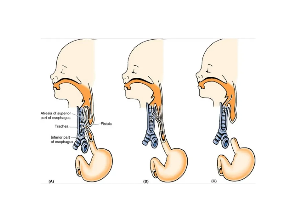
slide31. Tracheoesophageal Fistula (TEF) The most common congenital. Usually, it is combined with some form of esophageal atresia.
In the most common type of TEF (approximately 90% of cases), the superior part of the esophagus ends in a blind pouch and the inferior part communicates with the trachea (A).
In some cases, the superior esophagus communicates with the trachea and the inferior esophagus joins the stomach (B).
Sometimes, TEF exits with esophageal atresia (C).
TEFs result from abnormalities in partitioning of the esophagus and trachea by the tracheoesophageal septum.<br>
slide32. Thank You<br>
slide3. The Trachea is also called windpipe and it is a fibrocartilaginous, non-collapsible tube that creates the beginning of the lower respiratory tract.
16-20 C-shaped rings of Hyaline cartilage keep its lumen patent.
A band of smooth muscle (Trachealis) and a fibroelastic ligament that bridges the gap between the posterior free ends of C-shaped cartilages, that allow expansion of esophagus during the passage of bolus of the food. The Trachea<br>
slide4. Tracheal structure<br>
slide6. The trachea, bronchi and bronchioles form the tracheobronchial tree – a system of airways that allow passage of air into the lungs, where gas exchange occurs.
These airways are located in the neck and thorax.
The arrangement of cartilages and elastic tissue in the tracheal wall prevents its kinking and obstruction during the movements of the head and neck. The Trachea<br>
slide8. The trachea marks the beginning of the tracheobronchial tree.
It arises at the lower border of cricoid cartilage in the neck, as a continuation of the larynx.
It travels inferiorly into the superior mediastinum, bifurcating at the level of the sternal angle (forming the right and left main bronchi).
As it descends, the trachea is located anteriorly to the esophagus, and inclines slightly to the right. Anatomical Position<br>
slide9. Trachea stretches from the lower border of cricoid cartilage at the lower border of the C6 vertebra to the lower border of T4 vertebra in supine position, where it ends by dividing into left and right main bronchi. Anatomical Position<br>
slide11. The upper half of trachea can be found in the neck (cervical part) on the other hand the lower half is located in the superior mediastinum of the thoracic cavity (thoracic part).
The trachea is a 4-4½ inches (10-11 cm) long tube.
The diameter of trachea is 2 cm in men and 1.5 cm in females.
The lumen is smaller in living human than that in cadavers. MEASUREMENTS<br>
slide12. Thus upper half of the trachea lies in the neck (cervical part).
Lower half in the superior mediastinum (thoracic part).
Through its whole course, it lies directly in front of esophagus.
Left recurrent laryngeal nerve lies in the groove between it and left border of esophagus. LOCATION<br>
slide13. The trachea consists of about 16-20 C-shaped rings of hyaline cartilage being located one above the other the cartilages are deficient posteriorly where the gap is filled up by connective tissue and involuntary muscle termed trachealis.
The absence of cartilages on the posterior aspect enables expansion of esophagus during deglutition. STRUCTURE<br>
slide14. The trachea is the continuation of the larynx.
It begins at the lower border of the cricoid cartilage in the level of C6 vertebra, about 5 cm above the jugular notch.
It enters the thoracic inlet in the midline and enters downwards and backwards behind the manubrium to terminate by bifurcating into 2 principal bronchi, a little to the right side at the lower border of T4 vertebra at the level of sternal angle where arch of aorta deviates it to the right.
As trachea descends, it recedes rapidly from the surface to follow the curvature of vertebral column. COURSE<br>
slide15. The cervical part of the trachea is all about 7 cm in length and stretches from the lower border of cricoid cartilage to the upper border of manubrium sterni (jugular notch).
It goes downwards and somewhat backwards in front of the esophagus following curvature of the cervical spine and enters the thoracic cavity in the median plane with small deviation on the right side. CERVICAL PART OF TRACHEA<br>
slide16. RELATIONSHIPS OF THE CERVICAL PARTS OF TRACHEA<br>
slide17. The relations of the trachea in the superior mediastinum of the thorax<br>
slide18. (3 tubes & 1 nerve)
The esophagus.
The traches.
The thoracic duct.
The left recurrent laryngeal nerve.
These structures are related to each other and run parallel course through posterior part of superior mediastinum.
The esophagus, lies directly on the vertebral bodies of the region.
The trachea, lies directly in front of esophagus.
The thoracic duct, ascends along the left border of the eso.
The left recurrent laryngeal nerve ascends in the angle between the trachea and the esophagus. The unit of 4 structures in the posterior part of superior mediastinum<br>
slide20. At its upper end:
By the thyroid gland.
At its lower end (near bifurcation):
By the arch of aorta.
Behind the manubrium:
By the brachiocephalic artery. Sites of constrictions
of the trachea<br>
slide21. MUCOSA
It is composed of lining epithelium and lamina propria.
Lining epithelium is pseudostratified ciliated columnar with few goblets cells.
Lamina propria is composed of longitudinal elastic fibers.
SUBMUCOSA
It is composed of loose areolar tissue consisting of large number of serous and mucous glands.
Cartilage and smooth muscle layer: It’s created from horseshoe-shaped (C-shaped) hyaline cartilaginous rings, that are deficient posteriorly. The posterior gap is filled up chiefly by the smooth muscle (trachealis) and fibroelastic fibers.
Perichondrium: It encloses the cartilage.
Fibrous membrane: It is a layer of dense connective tissue, consisting of neurovascular structure.
There’s no clear difference between lamina propria and submucosa. Histology<br>
slide22. A. Blood supply to the trachea is by inferior thyroid arteries.
B. Venous drainage of the trachea takes place into the left brachiocephalic (innominate) vein.
C. Lymphatic drainage of the trachea is into pretracheal and paratracheal lymph nodes. VASCULAR SUPPLY AND LYMPHATIC DRAINAGE<br>
slide23. NERVE SUPPLY This is by sympathetic and parasympathetic fibers.
The parasympathetic fibers are originated from vagus via the recurrent laryngeal nerve.
These are secretomotor and sensory to the mucus membrane and motor to the trachealis muscle.
The sympathetic fibers are originated from the middle cervical sympathetic ganglion.
These are vasomotor in nature.<br>
slide24. TRACHEAL SHADOW IN RADIOGRAPH It is viewed as a vertical translucent shadow in front of cervico-thoracic spine.
The translucency is because of the presence of air in the trachea.<br>
slide26. PALPATION OF TRACHEA Medically, trachea is palpated in the suprasternal notch.
Normally, it is median in position but appreciable shift of trachea to left or right side indicates the mediastinal shift.<br>
slide28. IMPORTANCE OF CARINA It is a keel-like (hook-shaped) median ridge in the lumen in the bifurcation of trachea. It is both functional and pathological importance.
Functional importance: The mucosa of trachea over the carina is most sensitive. The cough reflex is generally started here, which helps to clear the sputum.
Pathological importance: It is visible as a sharp sagittal ridge in the tracheal bifurcation during bronchoscopy, for this reason acts as a useful landmark.
It is located about 25 cm from the incisor teeth and 30 cm from the nostrils. If the tracheobronchial lymph nodes in the angle between the main (principal) bronchi are enlarged because of spread of bronchiogenic carcinoma, the carina becomes distorted and flattened.<br>
slide30. IMPORTANCE OF CARINA It is a keel-like (hook-shaped) median ridge in the lumen in the bifurcation of trachea. It is both functional and pathological importance.
Functional importance: The mucosa of trachea over the carina is most sensitive. The cough reflex is generally started here, which helps to clear the sputum.
Pathological importance: It is visible as a sharp sagittal ridge in the tracheal bifurcation during bronchoscopy, for this reason acts as a useful landmark.
It is located about 25 cm from the incisor teeth and 30 cm from the nostrils. If the tracheobronchial lymph nodes in the angle between the main (principal) bronchi are enlarged because of spread of bronchiogenic carcinoma, the carina becomes distorted and flattened.<br>
slide31. Tracheoesophageal Fistula (TEF) The most common congenital. Usually, it is combined with some form of esophageal atresia.
In the most common type of TEF (approximately 90% of cases), the superior part of the esophagus ends in a blind pouch and the inferior part communicates with the trachea (A).
In some cases, the superior esophagus communicates with the trachea and the inferior esophagus joins the stomach (B).
Sometimes, TEF exits with esophageal atresia (C).
TEFs result from abnormalities in partitioning of the esophagus and trachea by the tracheoesophageal septum.<br>
slide32. Thank You<br>