Unit-5 Dr. Anil Kumar 4th Professional Asst. Professor Dept. of VCC BVC, Patna 1.Mycoplasma gallisepticum infection 2. Mycoplasma synoviae Infection 3. Mycoplasma meleagridis Infection The Mollicutes (mycoplasmas) are distinguished
"Unit-5 Dr. Anil Kumar 4th Professional Asst." is the property of its rightful owner. Permission is granted to
download and print the materials on this website for personal, non-commercial use only, and to display it
on your personal computer provided you do not modify the materials and that you retain all copyright
notices contained in the materials. By downloading content from our website, you accept the terms of this
agreement.
Presentation Transcript
01
Unit-5 Dr. Anil Kumar
4th Professional Asst. Professor
Dept. of VCC
BVC, Patna 1.Mycoplasma gallisepticum infection
2. Mycoplasma synoviae Infection
3. Mycoplasma meleagridis Infection<br>
02
The Mollicutes (mycoplasmas) are distinguished phenotypically from other bacteria by their minute size and total lack of a cell wall
Colony morphology (Broth and Agar) typically has a "fried egg" appearance under low magnification
More than 20 Mycoplasma species have been isolated from avian hosts
only 4 species are considered pathogenic in poultry– M. gallisepticum and M. synoviae are pathogenic for chicken, turkey, and other species,
M. meleagridis and M. iowae are pathogenic primarily in turkeys.
Airsacculitis due to avian mycoplasmosis is caused by MG, MS and MM Avian Mycoplasmosis<br>
03
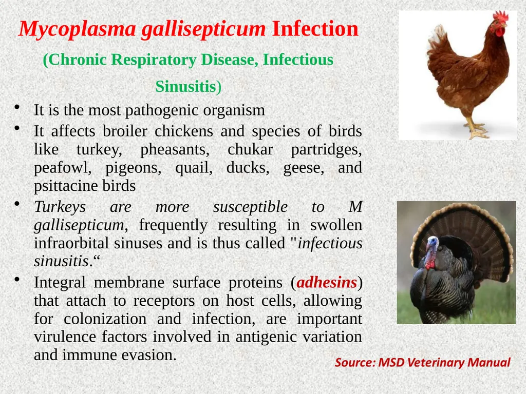
Mycoplasma gallisepticum Infection
(Chronic Respiratory Disease, Infectious Sinusitis)
It is the most pathogenic organism
It affects broiler chickens and species of birds like turkey, pheasants, chukar partridges, peafowl, pigeons, quail, ducks, geese, and psittacine birds
Turkeys are more susceptible to M gallisepticum, frequently resulting in swollen infraorbital sinuses and is thus called "infectious sinusitis.“
Integral membrane surface proteins (adhesins) that attach to receptors on host cells, allowing for colonization and infection, are important virulence factors involved in antigenic variation and immune evasion.<br>
04
Transmission :
Vertically---trans ovarian
Horizontally via infectious aerosols and through contamination of feed, water, and the environment, and by human activity on fomites (shoes, equipment, etc.)
They remain infected for life and act as carriers or reservoir for infection
Flock-to-flock transmission occurs readily by direct or indirect contact:
Movement of bird
People or
Fomites
Stressor (Cold weather, poor air quality or crowding, concurrent infections, and some live virus vaccinations) may facilitate infection, disease, and transmission.<br>
05
Initial colonization occur in epithelium of the conjunctiva, nasal passages, sinuses, and trachea
In severe, acute disease, infection may also involve the bronchi, air sacs, and occasionally lungs
Clinical Findings :
Respiratory distress, with rales, difficulty breathing, coughing, and/or sneezing
Nasal discharge and conjunctivitis with frothiness about the eyes
In turkeys the disease is more severe than in chickens, and swelling of the infra-orbital sinuses is common
Reduced feed efficiency and weight gains
May suffer high condemnations at processing due to airsacculitis
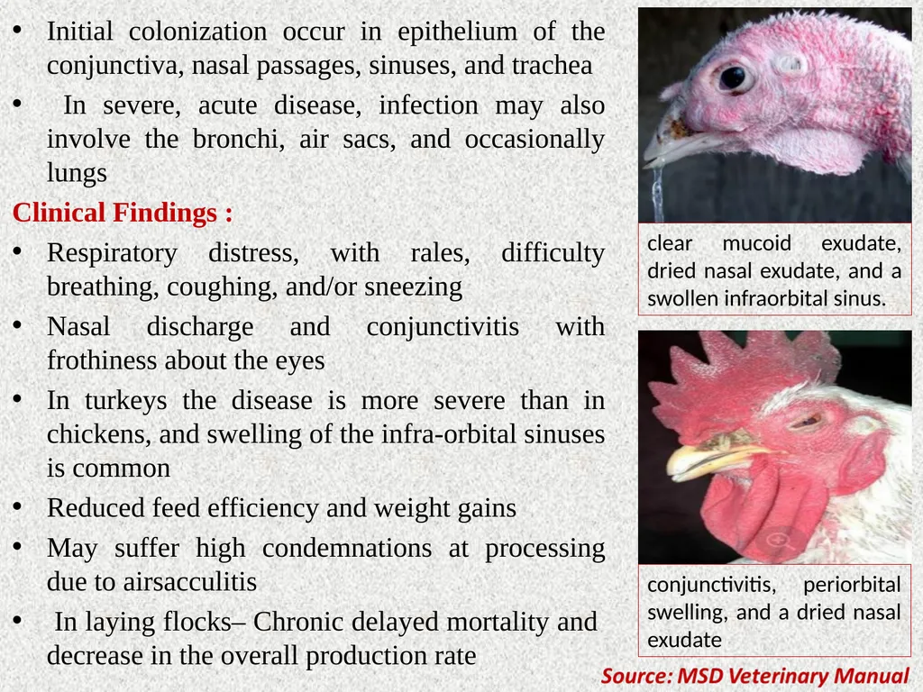
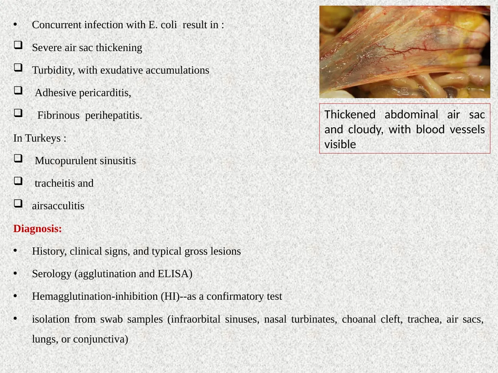
In laying flocks– Chronic delayed mortality and decrease in the overall production rate clear mucoid exudate, dried nasal exudate, and a swollen infraorbital sinus. conjunctivitis, periorbital swelling, and a dried nasal exudate<br>
06
Concurrent infection with E. coli result in :
Severe air sac thickening
Turbidity, with exudative accumulations
Adhesive pericarditis,
Fibrinous perihepatitis.
In Turkeys :
Mucopurulent sinusitis
tracheitis and
airsacculitis
Diagnosis:
History, clinical signs, and typical gross lesions
Serology (agglutination and ELISA)
Hemagglutination-inhibition (HI)--as a confirmatory test
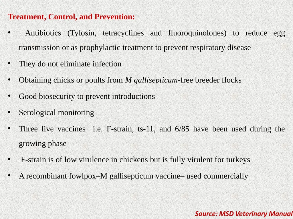
isolation from swab samples (infraorbital sinuses, nasal turbinates, choanal cleft, trachea, air sacs, lungs, or conjunctiva) Thickened abdominal air sac and cloudy, with blood vessels visible<br>
07
Treatment, Control, and Prevention:
Antibiotics (Tylosin, tetracyclines and fluoroquinolones) to reduce egg transmission or as prophylactic treatment to prevent respiratory disease
They do not eliminate infection
Obtaining chicks or poults from M gallisepticum-free breeder flocks
Good biosecurity to prevent introductions
Serological monitoring
Three live vaccines i.e. F-strain, ts-11, and 6/85 have been used during the growing phase
F-strain is of low virulence in chickens but is fully virulent for turkeys
A recombinant fowlpox–M gallisepticum vaccine– used commercially<br>
08
Mycoplasma synoviae Infection (Infectious Synovitis) Mycoplasma synoviae most commonly causes subclinical upper respiratory infections in chickens, turkeys, and other avian species world wide
It is also a complication of airsacculitis in association with Newcastle disease (ND) or infectious bronchitis(IB)
Infections of multiple-age layer flocks are common
Transmission:
Egg transmitted (transovarian)
Horizontal transmission-via the respiratory tract
Signs and Symptoms:
Slight rales
Pale-bluish head parts and lameness in many birds with a tendency to sit<br>
09
Severely affected birds are depressed and found resting around feeders and waterers
Swollen hocks and footpads
Sternal bursitis (breast blisters)
Egg shell abnormalities in egg layers
A creamy to viscous yellow-gray exudate is present in most synovial structures but most commonly seen in swollen hock and wing joints
Livers are enlarged and sometimes green
Spleens and Kidney are enlarged Swollen foot pad and swollen hock joint Viscous yellow exudate in the joint and around the tendons<br>
10
Diagnosis:
Clinical signs and gross lesions
Real-time PCR
Serum plate agglutination or ELISA are used to detect antibodies
Hemagglutination-inhibition or by culture, isolation, and identification of the organism.
Treatment, Control, and Prevention:
Stock should be obtained from disease free sources and raised with good biosecurity
Antibiotics in feed may help reduce synovitis but not very effective in established cases
A live, temperature-sensitive vaccine (MS-H) is used, but not commercially avialable<br>
11
Mycoplasma meleagridis Infection It causes an egg-transmitted (transovarian) disease of breeder turkeys (host-specific) that leads to airsacculitis in the offspring
It does not affect chickens
Transmission:
Vertical (egg) transmission
Horizontally via aerosols from the respiratory tract or to the vent
Infected semen
Manually via contaminated hands, clothing, and equipment or during vaccination
Clinical Findings:
Reduces hatchability (due to late embryo mortality), poult quality, and growth rate
Mild respiratory signs despite of high rates of airsacculitis in poults<br>
12
Egg-borne infections in turkey have early rapid growth of hock joints, periarticular tissues, cervical vertebrae, and adjacent bone, producing skeletal abnormalities such as crooked (wry) necks or leg deformities
Hatched poults may have airsacculitis with thickening, turbidity, foamy yellow exudate, and caseous flecks
Diagnosis:
Serology, culture, or real-time PCR
Airsacculitis in day-old poults suggests M meleagridis infection Large amount of caseous material indicates inflammation of airsacs Source: MSD Veterinary Manual<br>
13
Disease-free breeding stock and antibiotic treatment
Dipping eggs in tylosin or other antibiotic reduces the transmission in infected flocks
Injection of a suitable antibiotic at 1 day of age or water medication for the first 5–10 days appeared to reduce infection
Note:
Mycoplasmas are resistant to antibiotics that act on cell wall, such as penicillin, but are sensitive to tetracyclines (oxytetracycline, chlortetracycline and doxycycline), macrolides (erythromycin, tylosin, spiramycin, lincomycin, and kitasamycin), quinolones (imequil, norfloxacin, enrofloxacin and danofloxacin) or tiamulin
For better concentrations in the mucosal membranes of the respiratory and genitourinary tracts, such as tiamulin and enrofloxacin is preffered Avian Mycoplasmosis Update: Brazilian Journal of Poultry Science. 2005 / v.7 / n.1 / 01 - 09<br>