
‘Veterinary Epidemiology & Zoonoses’ VPH-321
AM
Published · 19 slides · 0 views
1 / 1

Description
Veterinary Epidemiology Zoonoses VPH-321 (Credit Hours-21) Glanders is a contagious bacterial disease affecting primarily equidae (horses, mules and donkeys) and is caused by the bacterium Burkholderia mallei. Cats, dogs, goats, camels
Related Topics
Download this presentation From Below
"‘Veterinary Epidemiology & Zoonoses’ VPH-321" is the property of its rightful owner. Permission is granted to download and print the materials on this website for personal, non-commercial use only, and to display it on your personal computer provided you do not modify the materials and that you retain all copyright notices contained in the materials. By downloading content from our website, you accept the terms of this agreement.
Share
Embed code
Presentation Transcript
01
‘Veterinary Epidemiology & Zoonoses’
VPH-321
(Credit Hours-2+1)<br>
VPH-321
(Credit Hours-2+1)<br>
02
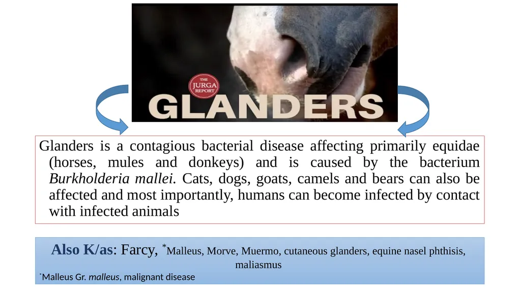
Glanders is a contagious bacterial disease affecting primarily equidae (horses, mules and donkeys) and is caused by the bacterium Burkholderia mallei. Cats, dogs, goats, camels and bears can also be affected and most importantly, humans can become infected by contact with infected animals Also K/as: Farcy, *Malleus, Morve, Muermo, cutaneous glanders, equine nasel phthisis, maliasmus
*Malleus Gr. malleus, malignant disease<br>
*Malleus Gr. malleus, malignant disease<br>
03
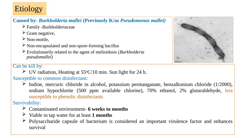
Etiology Caused by- Burkholderia mallei (Previously K/as Pseudomonas mallei)
Family -Burkholderiaceae
Gram negative,
Non-motile,
Non-encapsulated and non-spore-forming bacillus
Evolutionarily related to the agent of melioidosis (Burkholderia pseudomallei) Can be kill by:
UV radiation, Heating at 55oC/10 min. Sun light for 24 h.
Susceptible to common disinfectant:
Iodine, mercuric chloride in alcohol, potassium permanganate, benzalkonium chloride (1/2000), sodium hypochlorite (500 ppm available chlorine), 70% ethanol, 2% glutaraldehyde, less susceptible to phenolic disinfectants
Survivability:
Contaminated environment- 6 weeks to months
Viable in tap water for at least 1 months
Polysaccharide capsule of bacterium is considered an important virulence factor and enhances survival<br>
Family -Burkholderiaceae
Gram negative,
Non-motile,
Non-encapsulated and non-spore-forming bacillus
Evolutionarily related to the agent of melioidosis (Burkholderia pseudomallei) Can be kill by:
UV radiation, Heating at 55oC/10 min. Sun light for 24 h.
Susceptible to common disinfectant:
Iodine, mercuric chloride in alcohol, potassium permanganate, benzalkonium chloride (1/2000), sodium hypochlorite (500 ppm available chlorine), 70% ethanol, 2% glutaraldehyde, less susceptible to phenolic disinfectants
Survivability:
Contaminated environment- 6 weeks to months
Viable in tap water for at least 1 months
Polysaccharide capsule of bacterium is considered an important virulence factor and enhances survival<br>
04
Occurrence In post World War I era, glanders remained a serious problem in the USSR.
During world war II, it emerged as a military problem on the eastern front and in the Balkans, and was reintroduced into central Europe including Germany.
It was endemic in Chinese horses and affected 30% ponies.
At present, glanders is prevalent in Mongolian horses.
In Asia, it is said to cause high morbidity but low mortality in endemic areas.
The disease appears to be eradicated from Americas, Australia, and most of the European countries including USSR.
In India, the earliest traceable record of glanders dates back to 1881<br>
During world war II, it emerged as a military problem on the eastern front and in the Balkans, and was reintroduced into central Europe including Germany.
It was endemic in Chinese horses and affected 30% ponies.
At present, glanders is prevalent in Mongolian horses.
In Asia, it is said to cause high morbidity but low mortality in endemic areas.
The disease appears to be eradicated from Americas, Australia, and most of the European countries including USSR.
In India, the earliest traceable record of glanders dates back to 1881<br>
05
In man
Dr. Sainbel - the first principal of London veterinary College in 1723 was one of the first reported human cases of glanders.
In India, information on human glanders is scanty despite many reported cases of disease in equines.
Dr. S.H. Gaiger (1913-1916), a veterinary pathologist at Punjab Veterinary College, Lahore who contracted the disease while autopsying an infected horse Occurrence<br>
Dr. Sainbel - the first principal of London veterinary College in 1723 was one of the first reported human cases of glanders.
In India, information on human glanders is scanty despite many reported cases of disease in equines.
Dr. S.H. Gaiger (1913-1916), a veterinary pathologist at Punjab Veterinary College, Lahore who contracted the disease while autopsying an infected horse Occurrence<br>
06
Host range Animals
Equidae (horses, mules and donkey), occasionally Felidae, and other species are susceptible;
Donkeys are most susceptible, mules somewhat less and horses demonstrate some resistance manifested in chronic forms of the disease, especially in endemic areas
Susceptibility also seen in in camels, bears, wolves and dogs
Carnivores may become infected by eating infected meat
Cattle and pigs are resistant
Small ruminants may also be infected if kept in close contact to glanderous horses
Human: Veterinarians, Butchers, Groom, lab workers, Horse handlers<br>
Equidae (horses, mules and donkey), occasionally Felidae, and other species are susceptible;
Donkeys are most susceptible, mules somewhat less and horses demonstrate some resistance manifested in chronic forms of the disease, especially in endemic areas
Susceptibility also seen in in camels, bears, wolves and dogs
Carnivores may become infected by eating infected meat
Cattle and pigs are resistant
Small ruminants may also be infected if kept in close contact to glanderous horses
Human: Veterinarians, Butchers, Groom, lab workers, Horse handlers<br>
07
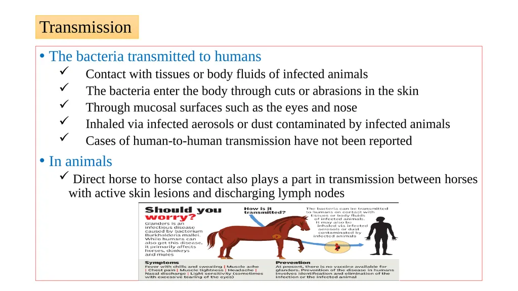
Transmission The bacteria transmitted to humans
Contact with tissues or body fluids of infected animals
The bacteria enter the body through cuts or abrasions in the skin
Through mucosal surfaces such as the eyes and nose
Inhaled via infected aerosols or dust contaminated by infected animals
Cases of human-to-human transmission have not been reported
In animals
Direct horse to horse contact also plays a part in transmission between horses with active skin lesions and discharging lymph nodes<br>
Contact with tissues or body fluids of infected animals
The bacteria enter the body through cuts or abrasions in the skin
Through mucosal surfaces such as the eyes and nose
Inhaled via infected aerosols or dust contaminated by infected animals
Cases of human-to-human transmission have not been reported
In animals
Direct horse to horse contact also plays a part in transmission between horses with active skin lesions and discharging lymph nodes<br>
08
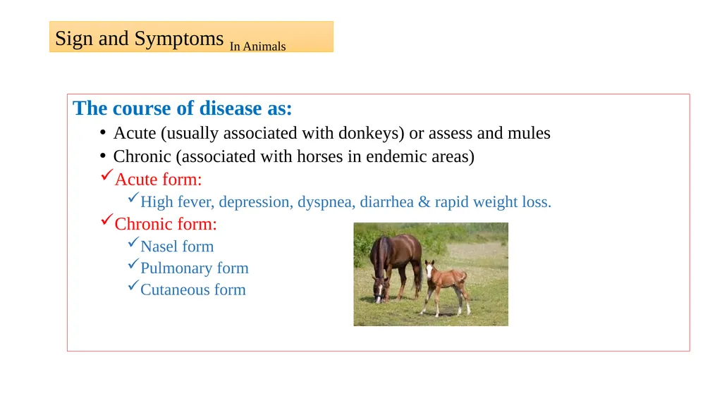
The course of disease as:
Acute (usually associated with donkeys) or assess and mules
Chronic (associated with horses in endemic areas)
Acute form:
High fever, depression, dyspnea, diarrhea & rapid weight loss.
Chronic form:
Nasel form
Pulmonary form
Cutaneous form Sign and Symptoms In Animals<br>
Acute (usually associated with donkeys) or assess and mules
Chronic (associated with horses in endemic areas)
Acute form:
High fever, depression, dyspnea, diarrhea & rapid weight loss.
Chronic form:
Nasel form
Pulmonary form
Cutaneous form Sign and Symptoms In Animals<br>
09
Sign and Symptoms In Animals Nasal form (Nasal glanders)
Ulceration of mucosa: one or both the nostril, larynx & trachea
Ulcers have: Grayish center with thick, jagged borders (star shaped)
A highly infectious, viscous, yellowish-green, mucopurulent discharge is present and this may crust around the nares
A purulent ocular discharge
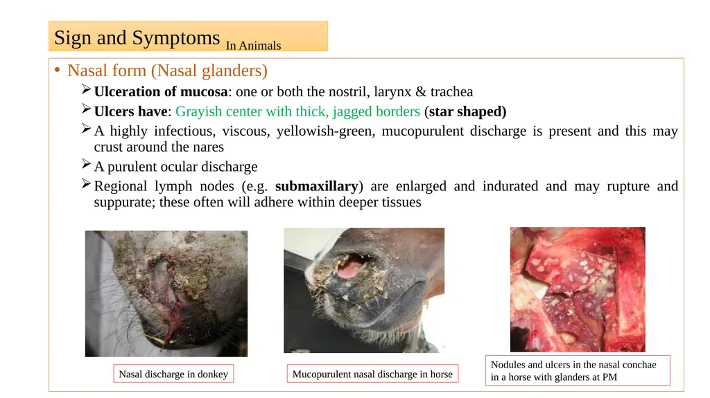
Regional lymph nodes (e.g. submaxillary) are enlarged and indurated and may rupture and suppurate; these often will adhere within deeper tissues Nasal discharge in donkey Mucopurulent nasal discharge in horse Nodules and ulcers in the nasal conchae in a horse with glanders at PM<br>
Ulceration of mucosa: one or both the nostril, larynx & trachea
Ulcers have: Grayish center with thick, jagged borders (star shaped)
A highly infectious, viscous, yellowish-green, mucopurulent discharge is present and this may crust around the nares
A purulent ocular discharge
Regional lymph nodes (e.g. submaxillary) are enlarged and indurated and may rupture and suppurate; these often will adhere within deeper tissues Nasal discharge in donkey Mucopurulent nasal discharge in horse Nodules and ulcers in the nasal conchae in a horse with glanders at PM<br>
10
Pulmonary form (Pulmonary glanders)
Usually requires several months to develop;
first manifestation: Intermittent fever, dyspnoea, paroxysmal coughing or a persistent dry cough accompanied by laboured breathing
Diarrhoea and polyuria: Progressive loss of condition
Nodules: Grayish white with red border,
Later become calcified & discharge their contents spreading the disease to the upper respiratory tract
Surrounded by: grayish, granulated or white fibers
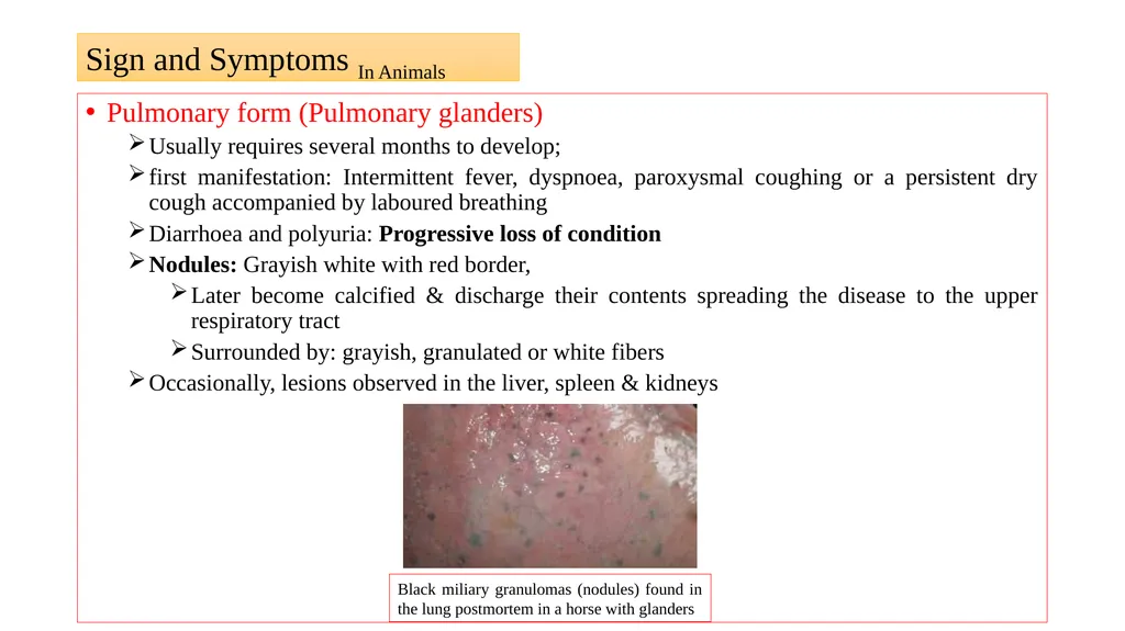
Occasionally, lesions observed in the liver, spleen & kidneys Sign and Symptoms In Animals Black miliary granulomas (nodules) found in the lung postmortem in a horse with glanders<br>
Usually requires several months to develop;
first manifestation: Intermittent fever, dyspnoea, paroxysmal coughing or a persistent dry cough accompanied by laboured breathing
Diarrhoea and polyuria: Progressive loss of condition
Nodules: Grayish white with red border,
Later become calcified & discharge their contents spreading the disease to the upper respiratory tract
Surrounded by: grayish, granulated or white fibers
Occasionally, lesions observed in the liver, spleen & kidneys Sign and Symptoms In Animals Black miliary granulomas (nodules) found in the lung postmortem in a horse with glanders<br>
11
Cutaneous form (Cutaneous glanders)
This form is also referred to as ‘farcy’.
Develops insidiously over an extended period; begins with fever, coughing, enlargement of the lymph nodes, dyspnoea usually associated with periods of exacerbation leading to progressive debilitation
Nodules: Gray center & excrete a thick, oily liquid that encrust the hair
Nodules appear under the skin along the course of lymphatics of the legs, ribs & belly
Leads to persistent ulcers connected along tortuous, thickened lymphatic vessels
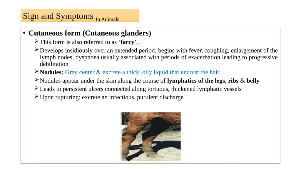
Upon rupturing: excrete an infectious, purulent discharge Sign and Symptoms In Animals<br>
This form is also referred to as ‘farcy’.
Develops insidiously over an extended period; begins with fever, coughing, enlargement of the lymph nodes, dyspnoea usually associated with periods of exacerbation leading to progressive debilitation
Nodules: Gray center & excrete a thick, oily liquid that encrust the hair
Nodules appear under the skin along the course of lymphatics of the legs, ribs & belly
Leads to persistent ulcers connected along tortuous, thickened lymphatic vessels
Upon rupturing: excrete an infectious, purulent discharge Sign and Symptoms In Animals<br>
12
Incubation period-1-14 days to month
Acute & Chronic form
The disease causes fever, malaise, fatigue, jaundice, nausea, rheumatic pain in legs and headach
Erysepelous swelling on face and limbs or painful nodules and phlegemonous inflammation
The nodular eruption is followed by pustular eruptions on the skin of face, legs, arms and other body parts
The nasal mucosa becomes congested and swollen, and conditions like severe pyaemia, metastatic pneumonia, muscular abscessation and diarrhoea set in leading to emaciation and collapse.
In chronic cases, these symptoms last for several days to months Sign and Symptoms In Human<br>
Acute & Chronic form
The disease causes fever, malaise, fatigue, jaundice, nausea, rheumatic pain in legs and headach
Erysepelous swelling on face and limbs or painful nodules and phlegemonous inflammation
The nodular eruption is followed by pustular eruptions on the skin of face, legs, arms and other body parts
The nasal mucosa becomes congested and swollen, and conditions like severe pyaemia, metastatic pneumonia, muscular abscessation and diarrhoea set in leading to emaciation and collapse.
In chronic cases, these symptoms last for several days to months Sign and Symptoms In Human<br>
13
Diagnosis In animals-
1.Physical examination of animals: Cardinal sign and symptoms of disease
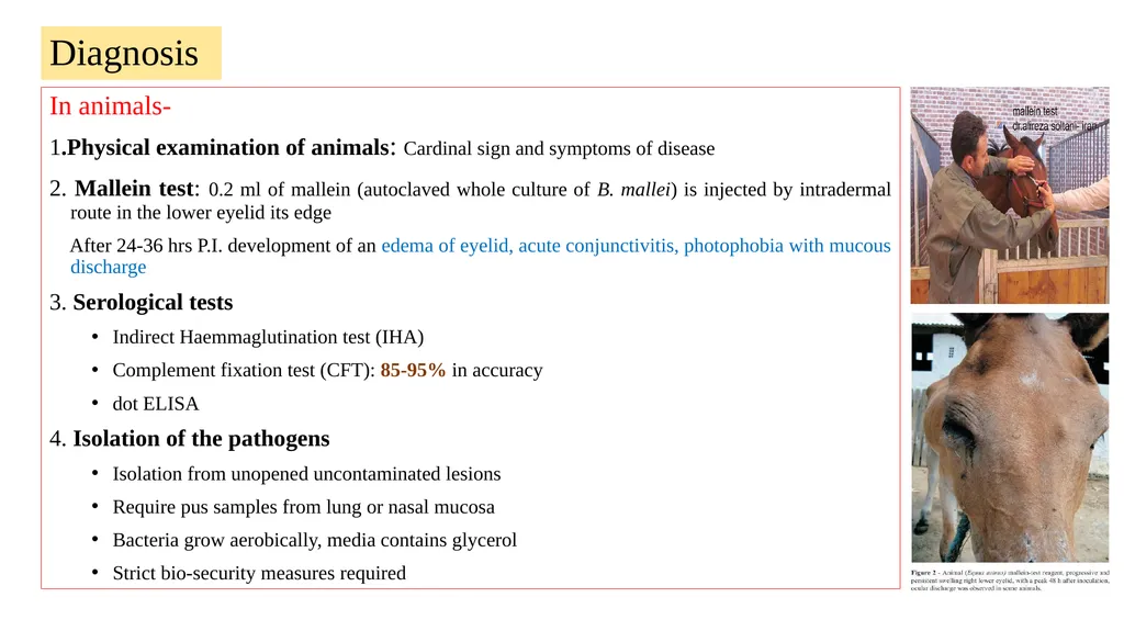
2. Mallein test: 0.2 ml of mallein (autoclaved whole culture of B. mallei) is injected by intradermal route in the lower eyelid its edge
After 24-36 hrs P.I. development of an edema of eyelid, acute conjunctivitis, photophobia with mucous discharge
3. Serological tests
Indirect Haemmaglutination test (IHA)
Complement fixation test (CFT): 85-95% in accuracy
dot ELISA
4. Isolation of the pathogens
Isolation from unopened uncontaminated lesions
Require pus samples from lung or nasal mucosa
Bacteria grow aerobically, media contains glycerol
Strict bio-security measures required<br>
1.Physical examination of animals: Cardinal sign and symptoms of disease
2. Mallein test: 0.2 ml of mallein (autoclaved whole culture of B. mallei) is injected by intradermal route in the lower eyelid its edge
After 24-36 hrs P.I. development of an edema of eyelid, acute conjunctivitis, photophobia with mucous discharge
3. Serological tests
Indirect Haemmaglutination test (IHA)
Complement fixation test (CFT): 85-95% in accuracy
dot ELISA
4. Isolation of the pathogens
Isolation from unopened uncontaminated lesions
Require pus samples from lung or nasal mucosa
Bacteria grow aerobically, media contains glycerol
Strict bio-security measures required<br>
14
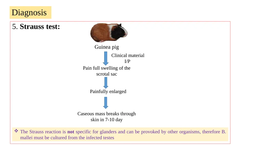
5. Strauss test: Diagnosis Clinical material
I/P Pain full swelling of the scrotal sac Painfully enlarged Caseous mass breaks through skin in 7-10 day Guinea pig The Strauss reaction is not specific for glanders and can be provoked by other organisms, therefore B. mallei must be cultured from the infected testes<br>
I/P Pain full swelling of the scrotal sac Painfully enlarged Caseous mass breaks through skin in 7-10 day Guinea pig The Strauss reaction is not specific for glanders and can be provoked by other organisms, therefore B. mallei must be cultured from the infected testes<br>
15
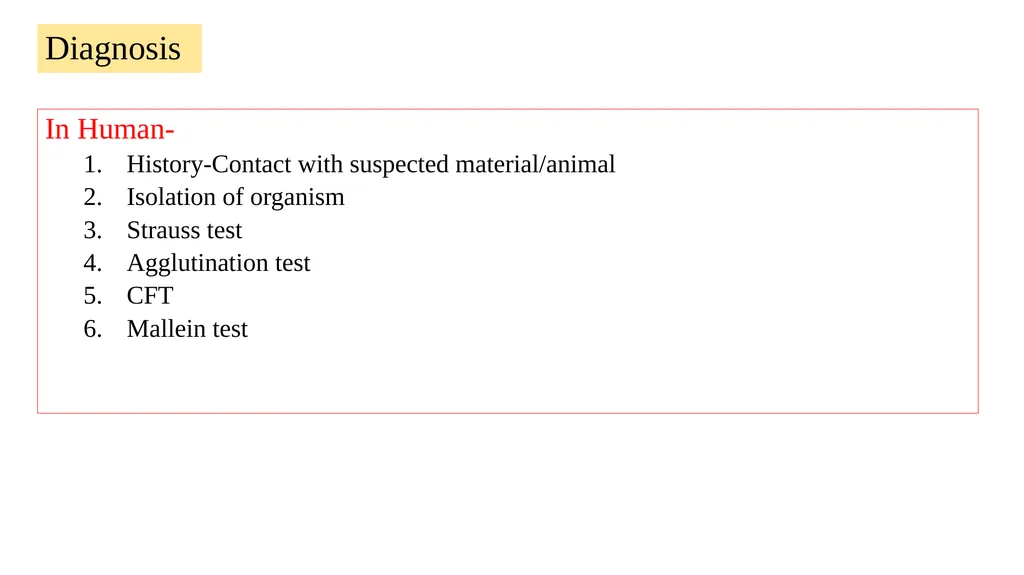
In Human-
History-Contact with suspected material/animal
Isolation of organism
Strauss test
Agglutination test
CFT
Mallein test Diagnosis<br>
History-Contact with suspected material/animal
Isolation of organism
Strauss test
Agglutination test
CFT
Mallein test Diagnosis<br>
16
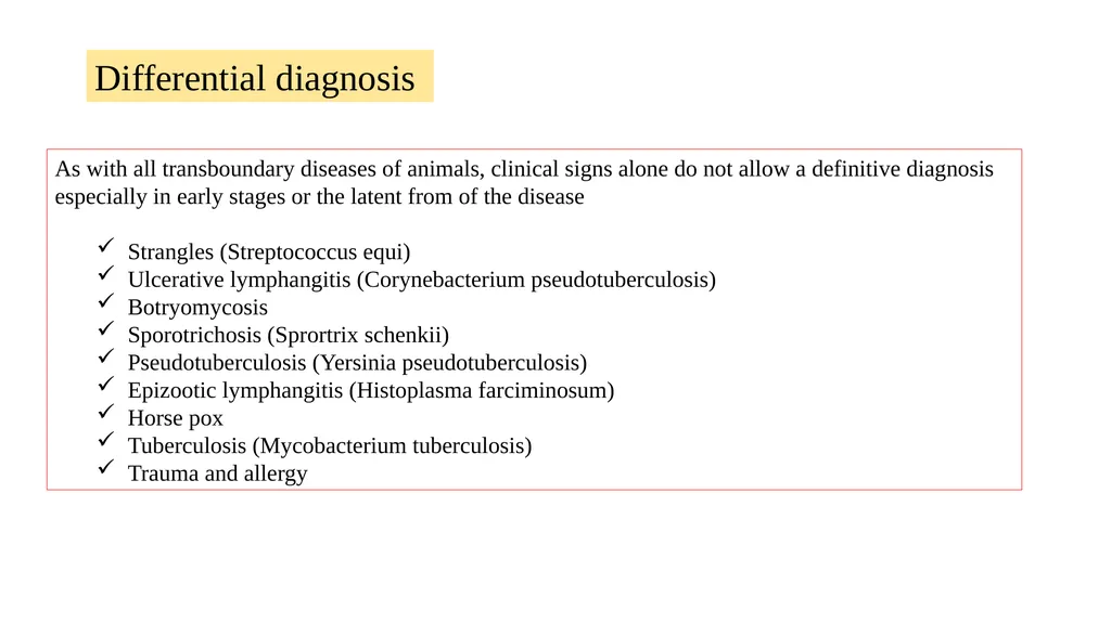
As with all transboundary diseases of animals, clinical signs alone do not allow a definitive diagnosis especially in early stages or the latent from of the disease
Strangles (Streptococcus equi)
Ulcerative lymphangitis (Corynebacterium pseudotuberculosis)
Botryomycosis
Sporotrichosis (Sprortrix schenkii)
Pseudotuberculosis (Yersinia pseudotuberculosis)
Epizootic lymphangitis (Histoplasma farciminosum)
Horse pox
Tuberculosis (Mycobacterium tuberculosis)
Trauma and allergy Differential diagnosis<br>
Strangles (Streptococcus equi)
Ulcerative lymphangitis (Corynebacterium pseudotuberculosis)
Botryomycosis
Sporotrichosis (Sprortrix schenkii)
Pseudotuberculosis (Yersinia pseudotuberculosis)
Epizootic lymphangitis (Histoplasma farciminosum)
Horse pox
Tuberculosis (Mycobacterium tuberculosis)
Trauma and allergy Differential diagnosis<br>
17
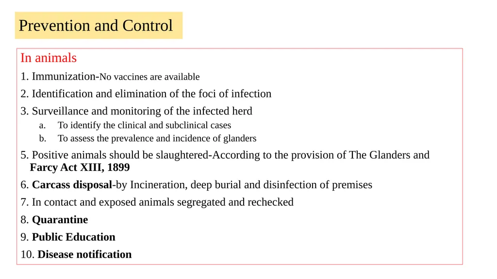
Prevention and Control In animals
1. Immunization-No vaccines are available
2. Identification and elimination of the foci of infection
3. Surveillance and monitoring of the infected herd
To identify the clinical and subclinical cases
To assess the prevalence and incidence of glanders
5. Positive animals should be slaughtered-According to the provision of The Glanders and Farcy Act XIII, 1899
6. Carcass disposal-by Incineration, deep burial and disinfection of premises
7. In contact and exposed animals segregated and rechecked
8. Quarantine
9. Public Education
10. Disease notification<br>
1. Immunization-No vaccines are available
2. Identification and elimination of the foci of infection
3. Surveillance and monitoring of the infected herd
To identify the clinical and subclinical cases
To assess the prevalence and incidence of glanders
5. Positive animals should be slaughtered-According to the provision of The Glanders and Farcy Act XIII, 1899
6. Carcass disposal-by Incineration, deep burial and disinfection of premises
7. In contact and exposed animals segregated and rechecked
8. Quarantine
9. Public Education
10. Disease notification<br>
18
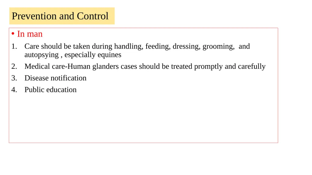
In man
Care should be taken during handling, feeding, dressing, grooming, and autopsying , especially equines
Medical care-Human glanders cases should be treated promptly and carefully
Disease notification
Public education Prevention and Control<br>
Care should be taken during handling, feeding, dressing, grooming, and autopsying , especially equines
Medical care-Human glanders cases should be treated promptly and carefully
Disease notification
Public education Prevention and Control<br>
19
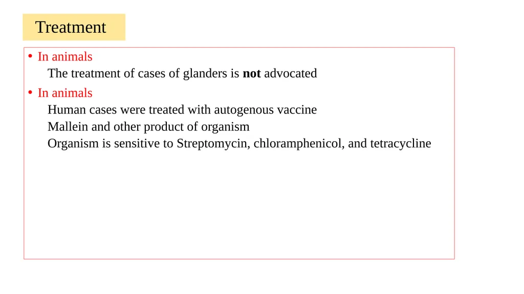
Treatment In animals
The treatment of cases of glanders is not advocated
In animals
Human cases were treated with autogenous vaccine
Mallein and other product of organism
Organism is sensitive to Streptomycin, chloramphenicol, and tetracycline<br>
The treatment of cases of glanders is not advocated
In animals
Human cases were treated with autogenous vaccine
Mallein and other product of organism
Organism is sensitive to Streptomycin, chloramphenicol, and tetracycline<br>