VETERINARY PARASITOLOGY THIRD YEAR STAGE 2018-2019 PROF. DR. GAHZI Y. AL-EMARAH PARASITOLOGEST Department of Veterinary Microbiology and Parasitology College of Veterinary Medicine University of Basrah NEMATODA FAMILY: OXYURIDEA General
"VETERINARY PARASITOLOGY THIRD YEAR STAGE 2018-2019" is the property of its rightful owner. Permission is granted to
download and print the materials on this website for personal, non-commercial use only, and to display it
on your personal computer provided you do not modify the materials and that you retain all copyright
notices contained in the materials. By downloading content from our website, you accept the terms of this
agreement.
Presentation Transcript
01
VETERINARY PARASITOLOGY
THIRD YEAR STAGE
2018-2019
PROF. DR. GAHZI Y. AL-EMARAH
PARASITOLOGEST
Department of Veterinary Microbiology and Parasitology
College of Veterinary Medicine
University of Basrah<br>
02
NEMATODA
FAMILY: OXYURIDEA
General Characteristics
- Mostly medium –sized or small nematodes.
- Mouth surrounded by three inconspicuous lips.
- No buccal capsule.
- Oesophagus has a well-developed bulb.
- Intestine may have caeca .
- Tail of female tapering, the valva is situated near the anterior end of the body.
- The female are usually much larger than the males and have long.
- Two spicules in the male and has bears a number of large papillae around the cloacal opening.<br>
03
NEMATODA
FAMILY: OXYURIDEA
General Characteristics
- The eggs are usually flattened on one side and development takes place without the need for an intermediate host.<br>
04
NEMATODA
Oxyuris equi
General Characteristics
- Occurs in the large intestine .
- The host :equines in all parts of the world.
- The males is 9-12mm long and the females up to150mm.
- The oesophagus is narrow at the middle and the bulb is nt distinctly marked off.
- The male has one pin-shape specula which is 120-150Mm long and
the tail bears two pairs of papillae.
- The young female are almost white in colour.
- The mature female have a slatey-grey or brownish colour and narrow tail which may be more than three times as long as the rest of the body.<br>
05
NEMATODA
Oxyuris equi
General Characteristics
- The eggs are elongate, slightly flattened on one side ,provided with a plug at one pole ,and measure about 90 by 42 Mm.
Life Cycle
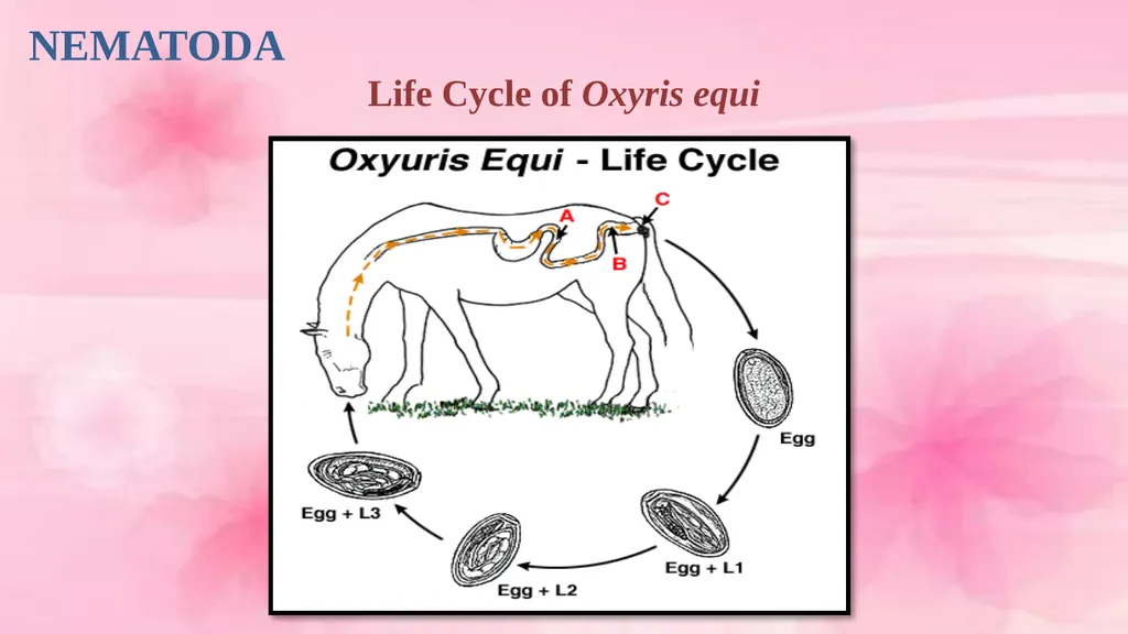
- The males and young females inhabit the caecum and large colon .
- After fertilization the mature females wander down to the rectum and crawl out through the anal opining .
- Eggs are laid in clusters on the skin in the perineal region .
- Development of the egg is rapid ,reaching the infective stage in three to five days.<br>
06
NEMATODA
Oxyuris equi
Life Cycle
- The infective stage may be reached on the perineal region (the egg falls off to the ground)
- Eggs probably survive for several weeks in moist .
- Infection by ingestion of the infective stage eggs on fodder and bedding.
- Infective larvae are laberated in the small intestine and L3 are found in the mucosal crypts of the ventral colon and caecum.
- L4 produce about 8 -10 days after infection .
- p.p is 5 months.<br>
07
NEMATODA
Life Cycle of Oxyris equi<br>
08
NEMATODA
Oxyuris equi
Pathogenicity and Pathogenesis
1- The fourth –stage larvae feeds on the intestinal mucosa of the host.
2- The chief feature of oxyuriasis in equines is the anal pruritus produced by the egg-laying females.
Clinical Signs
1-The irritation caused by the anal pruritus produces restlessness and improper feeding.
2- lose of condition.
3- Dull coat.
4- The animal rubs the base of its tail against any suitable object, the tail to acquire an un groomed ,rat tailed, appearance.<br>
09
NEMATODA
Oxyuris equi
Diagnosis
- Clinical signs
- Faecal exam.
- Rectum exam.
Treatment
Rx, mebendazole, Albendazoal and fenbendazol.
Control
- Good hygiene
- Bedding should be removed frequently.
- A clean supply of water should be available.<br>
10
NEMATODA
Strongylus sp.
General Characteristics
1- There is a globoid buccal capsule and dorsal gutter.
2- There is leaf crown or corona radiate(external and internal round the mouth opening).
3- Teeth may be present in the depth of the buccal capsule .
4- The male bursa is strongly developed and has typical rays.
5- The life cycle is direct .<br>
11
NEMATODA
Strongylus
There are three important genus:
1- S. equinus
General Characteristics
- Occurs in the caecum and colon
- The host ;equines including the zebra.
- The fresh worms are dark grey in color and sometimes the red color of the blood in the gut can be seen .
- The male is 26-35mm long and the female 37-47mm.
- The head without neck .
- The buccal capsule is oval with leaf crown .<br>
12
NEMATODA
1- S. equinus
General Characteristics
- At the base of buccal capsule there is a large dorsal tooth with a bifid tip and two smaller sub ventral teeth.
- The dorsal gutter is found.
- The male has two simple ,slender spicules .
- The vulva in the posterior extremity.
- The eggs are oval thin-shelled ,segmented when laid, and measure 70-85 by 40-47.<br>
13
NEMATODA
2- S. edentatus
General Characteristics
- Occurs in the large intestine of equines as final host.
- The male is 23-28mm long and the female 33-44mm.
- The head is somewhat wider than the following portion of the body.
- The buccal capsule is oval with leaf crown .
- At the base of buccal capsule there is no teeth<br>
14
NEMATODA
3- S. valgaris
General Characteristics
- Occurs in the large intestine of equines as final host.
- The male is 14-16mm long and the female 20-24mm.
- This worms is smaller than above species .
- The buccal capsule is oval and contains two ear-shaped dorsal teeth at the base
- with leaf crown .<br>
15
NEMATODA
Life cycle
1- S. equinus
- Exsheathed infective larvae penetrate the mucosa of the caecum and colon
- Enter the subserosa where they cause the formation of nodules .
- Eleven day after infection L4 occur in the peritoneal cavity and then to liver in 4-8 weeks
- Between 2-4 month after infection larvae leave the liver via the hepatic ligaments and pass via the pancreas to the peritoneal cavity .
- The moult L5 occurs about 118 days after infection and back to the caecum and colon.
- The prepatent period is about 260 days.<br>
16
NEMATODA
Life cycle
2- S.edentatus
- Exsheathed infective larvae penetrate the mucosa of the intestine to liver via the portal system.
- 11-12 day after infection L4 are produced.
- In nine weeks after infection larvae leave the liver and pass between the hepatic ligaments and the peritoneal layers.
- The moult L4 and L5 are found in
- The larvae migrate between the layer of the mesocolon to the wall of the caecum and colon after than to the lumen.
- P.P is 300-320 days.
.<br>
17
NEMATODA
Life cycle
3-S.valgaris
- Exsheathed infective larvae penetrate the mucosa of the intestine wall in 8 days after infection .
- 8 day after infection L4 are produced and penetrate the intima of the submucosal arterioles and migrate in these vessels towards the cranial mesentricartery .
- They are to be found in the cranial mesentricartery from 14 days after infection onwards associated with thrombi and later aneurysms.
- The L4 are found in submucosa of the caecum and colon ,after then its moult to L5in 3 months and in lumen may be maturity.
- The prepatent period is about 6-7months.
.<br>
18
NEMATODA
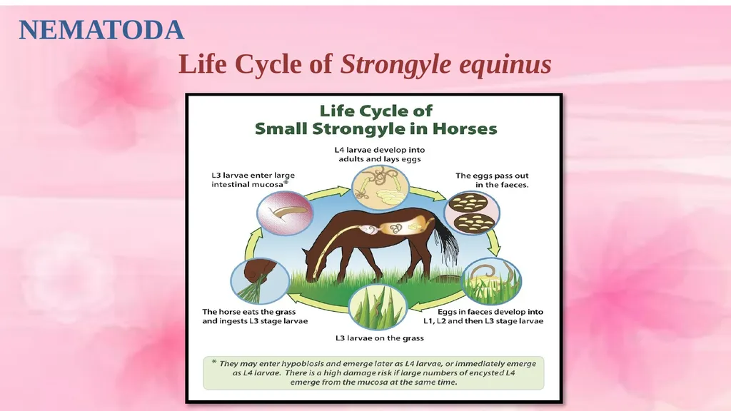
Life Cycle of Strongyle equinus<br>
19
NEMATODA
Pathogenicity and Pathogenesis
1- The adult worms from all species attach themselves to the mucosa of the large intestine and suck blood.
2- Anaemia of the normochromic normocytic type.
3- Small haemorrhagic ulcer in the large intestine.
4- Larvae migrant caused primary lesions occurred in the wall of small and large intestine and .
5- Extansive irregular inflammatory lesion occur in the media of the affected arteries producing an endarteritis and the formation of thrombi ,the larval stages may be found embedded in the thrombus, this case called aneurysms or verminous arteritis. specially of (S. valgaris).<br>
20
NEMATODA
Pathogenicity and Pathogenesis
6- haemorrhagic nodules.
Clinical Signs
1- S. equinus
1- Colic.
2- Anorexia.
3- General malaise.
2- S. edentatus
1- Peritonitis.
2- Acute toxaemia.
3- Jaundice.
4- Fever.<br>
NEMATODA
Treatment
Rx, phenothiazine, piprazine salts, Albendazoal and thiabendazol.
Control
- Routine use of Anthelmintic.
- Pasture management
- Kill the eggs and larvae by the heat of fermentation result from proper disposal of manure.<br>