
Wound Dr. Muhammad Abdullah Shwani Assistant


Description: Wound Dr. Muhammad Abdullah Shwani Assistant professorConsultant urologist Kurdistan Higher Council of Medical Specialties (KHCMS) drmalshwanimail.com WOUND DEFINITION The wound is a break or discontinuity in the integrity of skin or
Related Topics
Download Presentation
"Wound Dr. Muhammad Abdullah Shwani Assistant" is the property of its rightful owner. Permission is granted to download and print the materials on this website for personal, non-commercial use only, and to display it on your personal computer provided you do not modify the materials and that you retain all copyright notices contained in the materials. By downloading content from our website, you accept the terms of this agreement.
Presentation Transcript
slide1. Wound Dr. Muhammad Abdullah Shwani
Assistant professor/Consultant urologist
Kurdistan Higher Council of Medical Specialties (KHCMS)
drmalshwani@mail.com<br>
slide2. WOUND DEFINITION The wound is a break or discontinuity in the integrity of skin or tissues.<br>
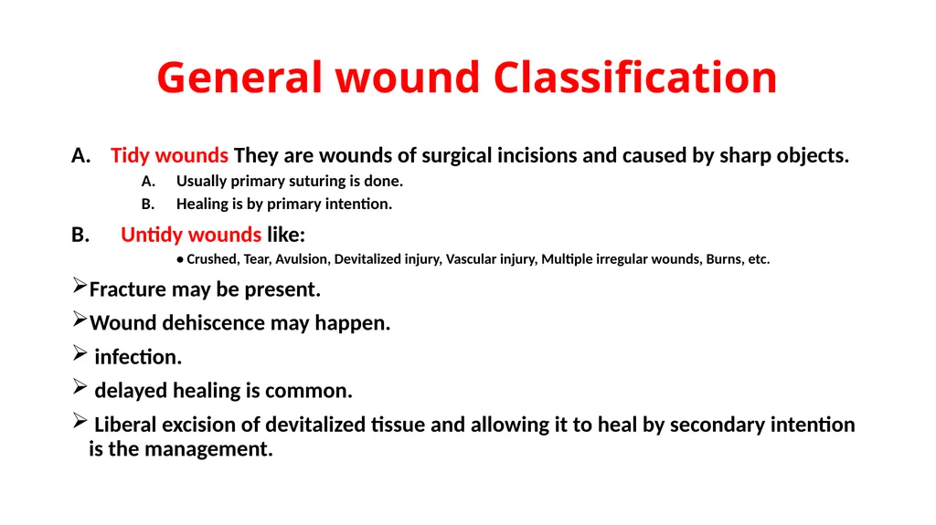
slide3. General wound Classification Tidy wounds They are wounds of surgical incisions and caused by sharp objects.
Usually primary suturing is done.
Healing is by primary intention.
Untidy wounds like:
• Crushed, Tear, Avulsion, Devitalized injury, Vascular injury, Multiple irregular wounds, Burns, etc.
Fracture may be present.
Wound dehiscence may happen.
infection.
delayed healing is common.
Liberal excision of devitalized tissue and allowing it to heal by secondary intention is the management.<br>
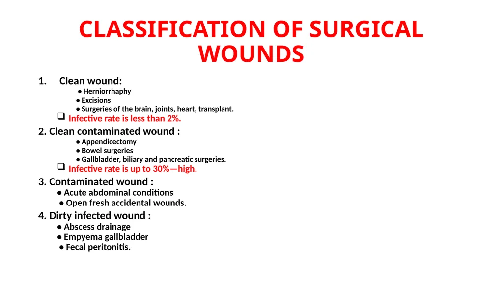
slide4. CLASSIFICATION OF SURGICAL WOUNDS Clean wound:
• Herniorrhaphy
• Excisions
• Surgeries of the brain, joints, heart, transplant.
Infective rate is less than 2%.
2. Clean contaminated wound :
• Appendicectomy
• Bowel surgeries
• Gallbladder, biliary and pancreatic surgeries.
Infective rate is up to 30%—high.
3. Contaminated wound :
• Acute abdominal conditions
• Open fresh accidental wounds.
4. Dirty infected wound :
• Abscess drainage
• Empyema gallbladder
• Fecal peritonitis.<br>
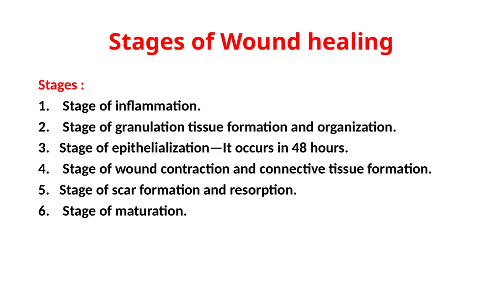
slide5. Stages of Wound healing Stages :
Stage of inflammation.
Stage of granulation tissue formation and organization.
Stage of epithelialization—It occurs in 48 hours.
Stage of wound contraction and connective tissue formation.
Stage of scar formation and resorption.
Stage of maturation.<br>
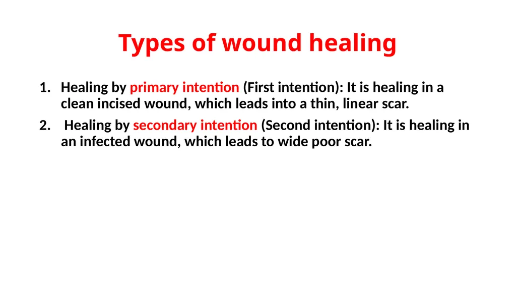
slide6. Types of wound healing Healing by primary intention (First intention): It is healing in a clean incised wound, which leads into a thin, linear scar.
Healing by secondary intention (Second intention): It is healing in an infected wound, which leads to wide poor scar.<br>
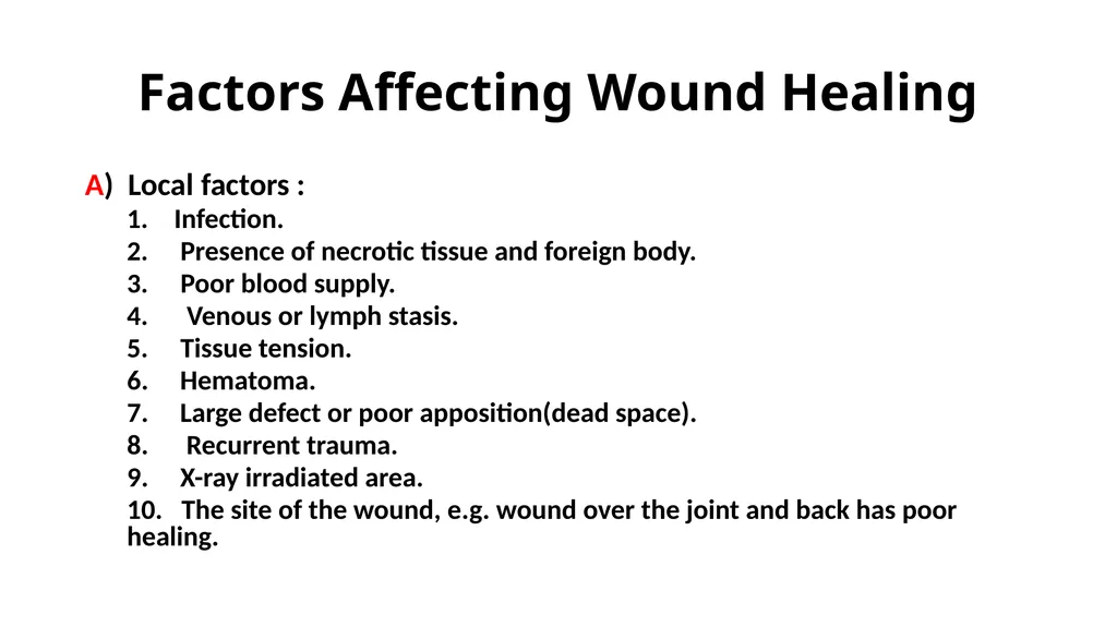
slide7. Factors Affecting Wound Healing A) Local factors :
Infection.
Presence of necrotic tissue and foreign body.
Poor blood supply.
Venous or lymph stasis.
Tissue tension.
6. Hematoma.
7. Large defect or poor apposition(dead space).
8. Recurrent trauma.
9. X-ray irradiated area.
10. The site of the wound, e.g. wound over the joint and back has poor healing.<br>
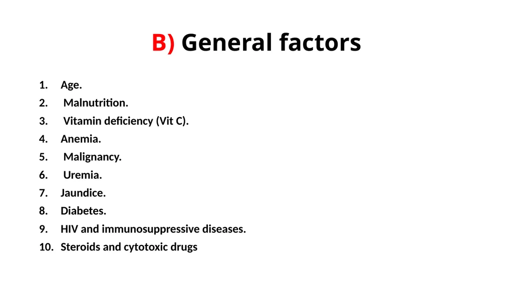
slide8. B) General factors Age.
Malnutrition.
Vitamin deficiency (Vit C).
Anemia.
Malignancy.
Uremia.
Jaundice.
Diabetes.
HIV and immunosuppressive diseases.
Steroids and cytotoxic drugs<br>
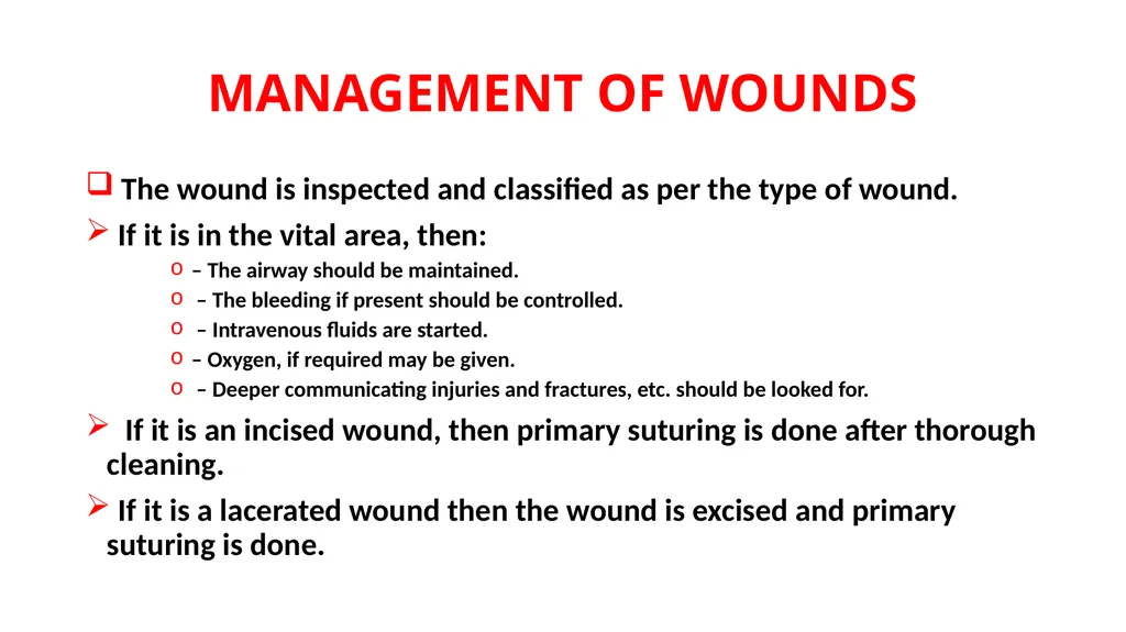
slide9. MANAGEMENT OF WOUNDS The wound is inspected and classified as per the type of wound.
If it is in the vital area, then:
– The airway should be maintained.
– The bleeding if present should be controlled.
– Intravenous fluids are started.
– Oxygen, if required may be given.
– Deeper communicating injuries and fractures, etc. should be looked for.
If it is an incised wound, then primary suturing is done after thorough cleaning.
If it is a lacerated wound then the wound is excised and primary suturing is done.<br>
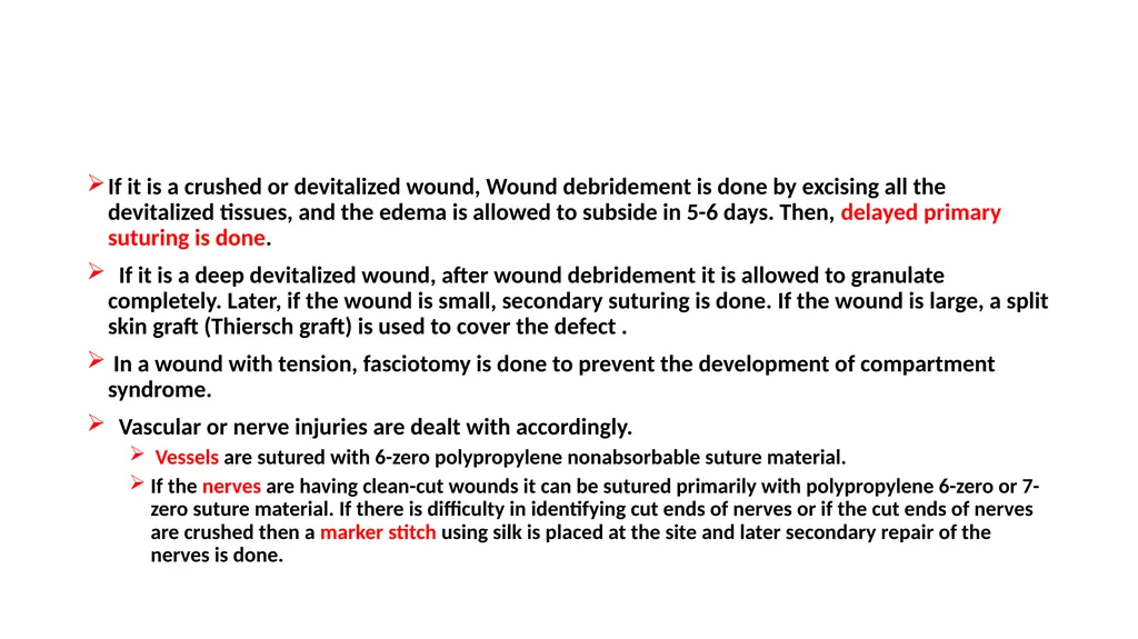
slide10. If it is a crushed or devitalized wound, Wound debridement is done by excising all the devitalized tissues, and the edema is allowed to subside in 5-6 days. Then, delayed primary suturing is done.
If it is a deep devitalized wound, after wound debridement it is allowed to granulate completely. Later, if the wound is small, secondary suturing is done. If the wound is large, a split skin graft (Thiersch graft) is used to cover the defect .
In a wound with tension, fasciotomy is done to prevent the development of compartment syndrome.
Vascular or nerve injuries are dealt with accordingly.
Vessels are sutured with 6-zero polypropylene nonabsorbable suture material.
If the nerves are having clean-cut wounds it can be sutured primarily with polypropylene 6-zero or 7-zero suture material. If there is difficulty in identifying cut ends of nerves or if the cut ends of nerves are crushed then a marker stitch using silk is placed at the site and later secondary repair of the nerves is done.<br>
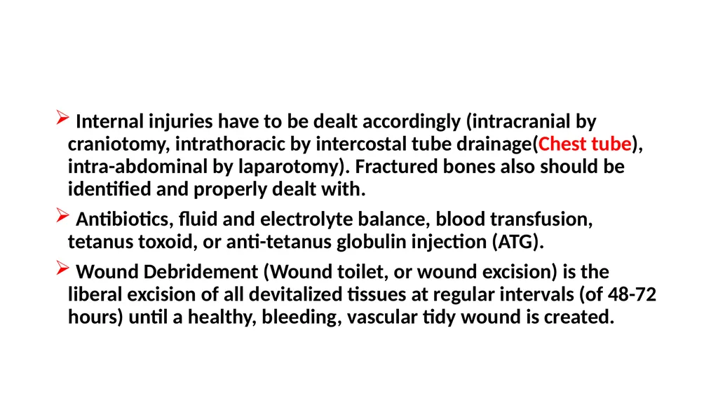
slide11. Internal injuries have to be dealt accordingly (intracranial by craniotomy, intrathoracic by intercostal tube drainage(Chest tube), intra-abdominal by laparotomy). Fractured bones also should be identified and properly dealt with.
Antibiotics, fluid and electrolyte balance, blood transfusion, tetanus toxoid, or anti-tetanus globulin injection (ATG).
Wound Debridement (Wound toilet, or wound excision) is the liberal excision of all devitalized tissues at regular intervals (of 48-72 hours) until a healthy, bleeding, vascular tidy wound is created.<br>
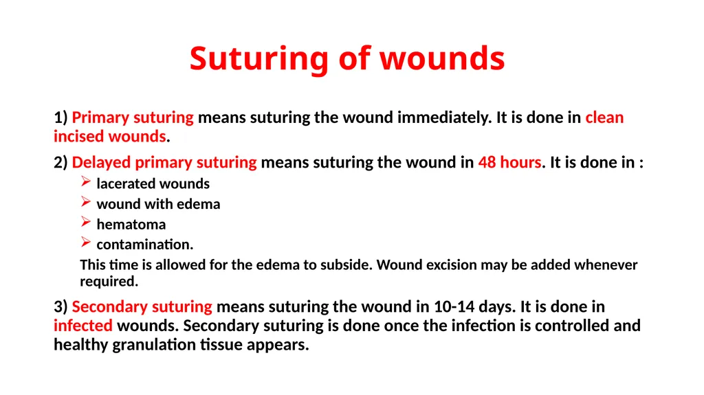
slide12. Suturing of wounds 1) Primary suturing means suturing the wound immediately. It is done in clean incised wounds.
2) Delayed primary suturing means suturing the wound in 48 hours. It is done in :
lacerated wounds
wound with edema
hematoma
contamination.
This time is allowed for the edema to subside. Wound excision may be added whenever required.
3) Secondary suturing means suturing the wound in 10-14 days. It is done in infected wounds. Secondary suturing is done once the infection is controlled and healthy granulation tissue appears.<br>
slide13. KELOID (LIKE A CLAW) Keloid is common in black patients .
Common in females.
Genetically predisposed, often familial.
There is a defect in the maturation and stabilization of collagen fibrils.
Keloid continues to grow even after 6 months, maybe for many years.
It extends into adjacent normal skin.
It is brownish black in color, painful, tender, and sometimes hyper esthetic.<br>
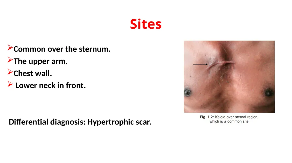
slide14. Sites Common over the sternum.
The upper arm.
Chest wall.
Lower neck in front.
Differential diagnosis: Hypertrophic scar.<br>
slide15. Treatment Modes of treatment
Excision and skin grafting.
Irradiation.
Excision and irradiation.
Steroid injection – Triamcinolone is given intra keloidally, at regular intervals, may be once in 7–10 days, of 6–8 injections.
Steroid injection → Excision → Steroid injection.
Methotrexate and vitamin A therapy into the keloid.
The recurrence rate is very high<br>
slide16. HYPERTROPHIC SCAR Occurs anywhere in the body.
Not genetically predisposed, not familial
Growth is usually limited up to 6 months.
It is limited to scar tissue only.
It will not extend to the normal skin.
It is pale brown in color, not painful, nontender.
Often, self-limiting.
It responds very well to steroid injection
Recurrence is uncommon.<br>
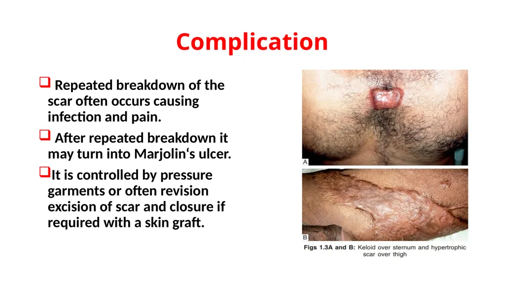
slide17. Complication Repeated breakdown of the scar often occurs causing infection and pain.
After repeated breakdown it may turn into Marjolin‘s ulcer.
It is controlled by pressure garments or often revision excision of scar and closure if required with a skin graft.<br>
slide18. THANK YOU<br>
Assistant professor/Consultant urologist
Kurdistan Higher Council of Medical Specialties (KHCMS)
drmalshwani@mail.com<br>
slide2. WOUND DEFINITION The wound is a break or discontinuity in the integrity of skin or tissues.<br>
slide3. General wound Classification Tidy wounds They are wounds of surgical incisions and caused by sharp objects.
Usually primary suturing is done.
Healing is by primary intention.
Untidy wounds like:
• Crushed, Tear, Avulsion, Devitalized injury, Vascular injury, Multiple irregular wounds, Burns, etc.
Fracture may be present.
Wound dehiscence may happen.
infection.
delayed healing is common.
Liberal excision of devitalized tissue and allowing it to heal by secondary intention is the management.<br>
slide4. CLASSIFICATION OF SURGICAL WOUNDS Clean wound:
• Herniorrhaphy
• Excisions
• Surgeries of the brain, joints, heart, transplant.
Infective rate is less than 2%.
2. Clean contaminated wound :
• Appendicectomy
• Bowel surgeries
• Gallbladder, biliary and pancreatic surgeries.
Infective rate is up to 30%—high.
3. Contaminated wound :
• Acute abdominal conditions
• Open fresh accidental wounds.
4. Dirty infected wound :
• Abscess drainage
• Empyema gallbladder
• Fecal peritonitis.<br>
slide5. Stages of Wound healing Stages :
Stage of inflammation.
Stage of granulation tissue formation and organization.
Stage of epithelialization—It occurs in 48 hours.
Stage of wound contraction and connective tissue formation.
Stage of scar formation and resorption.
Stage of maturation.<br>
slide6. Types of wound healing Healing by primary intention (First intention): It is healing in a clean incised wound, which leads into a thin, linear scar.
Healing by secondary intention (Second intention): It is healing in an infected wound, which leads to wide poor scar.<br>
slide7. Factors Affecting Wound Healing A) Local factors :
Infection.
Presence of necrotic tissue and foreign body.
Poor blood supply.
Venous or lymph stasis.
Tissue tension.
6. Hematoma.
7. Large defect or poor apposition(dead space).
8. Recurrent trauma.
9. X-ray irradiated area.
10. The site of the wound, e.g. wound over the joint and back has poor healing.<br>
slide8. B) General factors Age.
Malnutrition.
Vitamin deficiency (Vit C).
Anemia.
Malignancy.
Uremia.
Jaundice.
Diabetes.
HIV and immunosuppressive diseases.
Steroids and cytotoxic drugs<br>
slide9. MANAGEMENT OF WOUNDS The wound is inspected and classified as per the type of wound.
If it is in the vital area, then:
– The airway should be maintained.
– The bleeding if present should be controlled.
– Intravenous fluids are started.
– Oxygen, if required may be given.
– Deeper communicating injuries and fractures, etc. should be looked for.
If it is an incised wound, then primary suturing is done after thorough cleaning.
If it is a lacerated wound then the wound is excised and primary suturing is done.<br>
slide10. If it is a crushed or devitalized wound, Wound debridement is done by excising all the devitalized tissues, and the edema is allowed to subside in 5-6 days. Then, delayed primary suturing is done.
If it is a deep devitalized wound, after wound debridement it is allowed to granulate completely. Later, if the wound is small, secondary suturing is done. If the wound is large, a split skin graft (Thiersch graft) is used to cover the defect .
In a wound with tension, fasciotomy is done to prevent the development of compartment syndrome.
Vascular or nerve injuries are dealt with accordingly.
Vessels are sutured with 6-zero polypropylene nonabsorbable suture material.
If the nerves are having clean-cut wounds it can be sutured primarily with polypropylene 6-zero or 7-zero suture material. If there is difficulty in identifying cut ends of nerves or if the cut ends of nerves are crushed then a marker stitch using silk is placed at the site and later secondary repair of the nerves is done.<br>
slide11. Internal injuries have to be dealt accordingly (intracranial by craniotomy, intrathoracic by intercostal tube drainage(Chest tube), intra-abdominal by laparotomy). Fractured bones also should be identified and properly dealt with.
Antibiotics, fluid and electrolyte balance, blood transfusion, tetanus toxoid, or anti-tetanus globulin injection (ATG).
Wound Debridement (Wound toilet, or wound excision) is the liberal excision of all devitalized tissues at regular intervals (of 48-72 hours) until a healthy, bleeding, vascular tidy wound is created.<br>
slide12. Suturing of wounds 1) Primary suturing means suturing the wound immediately. It is done in clean incised wounds.
2) Delayed primary suturing means suturing the wound in 48 hours. It is done in :
lacerated wounds
wound with edema
hematoma
contamination.
This time is allowed for the edema to subside. Wound excision may be added whenever required.
3) Secondary suturing means suturing the wound in 10-14 days. It is done in infected wounds. Secondary suturing is done once the infection is controlled and healthy granulation tissue appears.<br>
slide13. KELOID (LIKE A CLAW) Keloid is common in black patients .
Common in females.
Genetically predisposed, often familial.
There is a defect in the maturation and stabilization of collagen fibrils.
Keloid continues to grow even after 6 months, maybe for many years.
It extends into adjacent normal skin.
It is brownish black in color, painful, tender, and sometimes hyper esthetic.<br>
slide14. Sites Common over the sternum.
The upper arm.
Chest wall.
Lower neck in front.
Differential diagnosis: Hypertrophic scar.<br>
slide15. Treatment Modes of treatment
Excision and skin grafting.
Irradiation.
Excision and irradiation.
Steroid injection – Triamcinolone is given intra keloidally, at regular intervals, may be once in 7–10 days, of 6–8 injections.
Steroid injection → Excision → Steroid injection.
Methotrexate and vitamin A therapy into the keloid.
The recurrence rate is very high<br>
slide16. HYPERTROPHIC SCAR Occurs anywhere in the body.
Not genetically predisposed, not familial
Growth is usually limited up to 6 months.
It is limited to scar tissue only.
It will not extend to the normal skin.
It is pale brown in color, not painful, nontender.
Often, self-limiting.
It responds very well to steroid injection
Recurrence is uncommon.<br>
slide17. Complication Repeated breakdown of the scar often occurs causing infection and pain.
After repeated breakdown it may turn into Marjolin‘s ulcer.
It is controlled by pressure garments or often revision excision of scar and closure if required with a skin graft.<br>
slide18. THANK YOU<br>