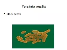
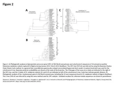
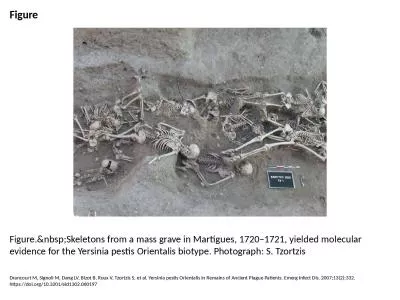
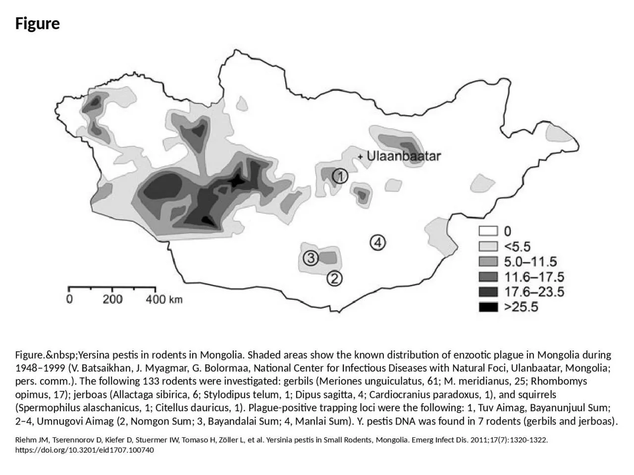
PPT-Figure Figure. Yersina pestis in rodents in Mongolia. Shaded areas show the known
Author : violet | Published Date : 2023-07-26
Riehm JM Tserennorov D Kiefer D Stuermer IW Tomaso H Zöller L et al Yersinia pestis in Small Rodents Mongolia Emerg Infect Dis 201117713201322 httpsdoiorg103201eid1707100740
Presentation Embed Code
Download Presentation
Download Presentation The PPT/PDF document "Figure Figure. Yersina pestis i..." is the property of its rightful owner. Permission is granted to download and print the materials on this website for personal, non-commercial use only, and to display it on your personal computer provided you do not modify the materials and that you retain all copyright notices contained in the materials. By downloading content from our website, you accept the terms of this agreement.
Figure Figure. Yersina pestis in rodents in Mongolia. Shaded areas show the known: Transcript
Riehm JM Tserennorov D Kiefer D Stuermer IW Tomaso H Zöller L et al Yersinia pestis in Small Rodents Mongolia Emerg Infect Dis 201117713201322 httpsdoiorg103201eid1707100740. Olayemi A, Obadare A, Oyeyiola A, Igbokwe J, Fasogbon A, Igbahenah F, et al. Arenavirus Diversity and Phylogeography of Mastomys natalensis Rodents, Nigeria. Emerg Infect Dis. 2016;22(4):694-697. https://doi.org/10.3201/eid2204.150155. Ramqvist T, Dalianis T. Oropharyngeal Cancer Epidemic and Human Papillomavirus. Emerg Infect Dis. 2010;16(11):1671-1677. https://doi.org/10.3201/eid1611.100452. Zhang Y, Zhang F, Gao N, Wang J, Zhao Z, Li M, et al. Hantaviruses in Rodents and Humans, Inner Mongolia Autonomous Region, China. Emerg Infect Dis. 2009;15(6):885-891. https://doi.org/10.3201/eid1506.081126. Adeleke A, Pruckler J, Benson R, Rowbotham T, Halablab M, Fields B. Legionella-Like Amebal Pathogens––Phylogenetic Status and Possible Role in Respiratory Disease. Emerg Infect Dis. 1996;2(3):225-230. https://doi.org/10.3201/eid0203.960311. Monroe MC, Morzunov SP, Johnson AM, Bowen MD, Artsob H, Yates T, et al. Genetic Diversity and Distribution of Peromyscus-Borne Hantaviruses in North America. Emerg Infect Dis. 1999;5(1):75-86. https://doi.org/10.3201/eid0501.990109. Pettit AC, Jahangir AA, Wright PW. Mycobacterium doricum Osteomyelitis and Soft Tissue Infection. Emerg Infect Dis. 2011;17(11):2075-2077. https://doi.org/10.3201/eid1711.110460. Delgado MJ, Gutierrez JM, Radic LB, Maretic T, Zekan S, Avšič-Županc T, et al. Imported Dengue Hemorrhagic Fever, Europe. Emerg Infect Dis. 2008;14(8):1329-1330. https://doi.org/10.3201/eid1408.080032. Drancourt M, Signoli M, Dang LV, Bizot B, Roux V, Tzortzis S, et al. Yersinia pestis Orientalis in Remains of Ancient Plague Patients. Emerg Infect Dis. 2007;13(2):332. https://doi.org/10.3201/eid1302.060197. Marcus PI, Girshick T, van der Heide L, Sekellick MJ. Super-Sentinel Chickens and Detection of Low-Pathogenicity Influenza Virus. Emerg Infect Dis. 2007;13(10):1608-1610. https://doi.org/10.3201/eid1310.061552. Béringue V, Herzog L, Reine F, Le Dur A, Casalone C, Vilotte J, et al. Transmission of Atypical Bovine Prions to Mice Transgenic for Human Prion Protein. Emerg Infect Dis. 2008;14(12):1898-1901. https://doi.org/10.3201/eid1412.080941. Salzberg SL, Kingsford C, Cattoli G, Spiro DJ, Janies DA, Aly MM, et al. Genome Analysis Linking Recent European and African Influenza (H5N1) Viruses. Emerg Infect Dis. 2007;13(5):713. https://doi.org/10.3201/eid1305.070013. Ijaz K, Yang Z, Matthews HS, Bates JH, Cave MD. Mycobacterium tuberculosis Transmission between Cluster Members with Similar Fingerprint Patterns. Emerg Infect Dis. 2002;8(11):1257-1259. https://doi.org/10.3201/eid0811.020284. Marrie TJ, Campbell N, McNeil SA, Webster D, Hatchette TF. Q Fever Update, Maritime Canada. Emerg Infect Dis. 2008;14(1):67-69. https://doi.org/10.3201/eid1401.071256. Scrimgeour EM, Johnston WJ, Al Dhahry SH, El-Khatim HS, John V, Musa M. First Report of Q Fever in Oman. Emerg Infect Dis. 2000;6(1):74-79. https://doi.org/10.3201/eid0601.000114.
Download Document
Here is the link to download the presentation.
"Figure Figure. Yersina pestis in rodents in Mongolia. Shaded areas show the known"The content belongs to its owner. You may download and print it for personal use, without modification, and keep all copyright notices. By downloading, you agree to these terms.
Related Documents