/
Option D3: Functions of the liver Option D3: Functions of the liver

Option D3: Functions of the liver - PowerPoint Presentation

violet
violet . @violet
Follow
344 views
Uploaded On 2022-02-15

Option D3: Functions of the liver - PPT Presentation

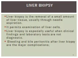
Essential Idea The chemical composition of the blood is regulated by the liver Understandings UD31 the liver removes toxins from the blood and detoxifies them UD32 components of red blood cells are recycled by the liver ID: 909256

blood liver cells hepatic liver blood hepatic cells sinusoids hepatocytes vein bile jaundice alcohol nutrients hemoglobin portal kupffer bilirubin

Share:

Link:

Embed:

Download Presentation from below link

Download Presentation The PPT/PDF document "Option D3: Functions of the liver" is the property of its rightful owner. Permission is granted to download and print the materials on this web site for personal, non-commercial use only, and to display it on your personal computer provided you do not modify the materials and that you retain all copyright notices contained in the materials. By downloading content from our website, you accept the terms of this agreement.


Presentation Transcript

Slide1

Option D3: Functions of the liver

Slide2

Essential Idea: The chemical composition of the blood is regulated by the liver.

Slide3

Understandings

U.D.3.1: the liver removes toxins from the blood and detoxifies them.

U.D.3.2: components of red blood cells are recycled by the liver.

U.D.3.3: the breakdown of erythrocytes starts with phagocytosis of red blood cells by

Kupffer

cells.

U.D.3.4: iron is carried to the bone marrow to produce hemoglobin in new red blood cells.

U.D.3.5: surplus cholesterol is converted to bile salts.

U.D.3.6: endoplasmic reticulum and Golgi apparatus in hepatocytes produce plasma proteins.

U.D.3.7: the liver intercepts blood from the gut to regulate nutrient levels.

U.D.3.8: some nutrients in excess can be stored in the liver.

Slide4

Applications

A.D.3.1: causes and consequences of jaundice.

A.D.3.2: dual blood supply to the liver and differences between sinusoids and capillaries.

Slide5

Circulation of blood to and from the liver

The liver receives blood from

two

major blood vessels, and is drained by

one

vessel.

Hepatic

artery

: branch of the aorta; carries

oxygenated

blood to the liver tissue.

Hepatic

portal vein

: brings blood to the liver from the capillaries of the villi of the small intestine.

Both the hepatic artery and hepatic portal vein drain into special capillaries of the liver, called

sinusoids

.

All sinusoids will then drain into the

hepatic vein

, which takes blood from the liver to the heart.

Slide6

Liver blood schematic

Liver (sinusoids)

Absorbed nutrients from the intestine

Oxygenated blood

Hepatic portal vein

Hepatic artery

To vena cava

Hepatic vein

Slide7

Slide8

Hepatic portal vein and hepatic vein

The

hepatic portal vein

differs in two ways from other blood vessels that come to organs:

The blood from the hepatic portal vein is

low in oxygen

and under

low pressure

, because it has come from the capillaries of the villi

The concentration of nutrients

varies

considerably, and is dependent on what was consumed and the timing of digestion

The

hepatic vein is also under low pressure and is deoxygenated, but the nutrient concentration is usually

fairly consistent, as one of the main roles of the liver is to remove and store nutrients from the blood.

Slide9

Sinusoids (A.D.3.2)

The role of the liver is to remove substances from the blood, as well as occasionally add substances. This is the role of the liver’s cells, called

hepatocytes

.

As blood comes into the liver from the hepatic artery and hepatic portal vein, it flows into the capillaries of the liver, known as

sinusoids

. Sinusoids are where the exchanges between hepatocytes and the blood occur.

Sinusoids are different from normal capillaries in several ways:

Sinusoids are

wider

Sinusoids are lined with

epithelial cells

with gaps between them, which allows large molecules to be exchanged between the bloodstream and the hepatocytes

Hepatocytes have direct contact with blood, increasing

efficiency

Kupffer cells are found in sinusoids. These cells break down hemoglobin from old red blood cells.Sinusoids receive both oxygen-rich and oxygen-poor blood

Slide10

Sinusoid structure

Slide11

Detoxification (U.D.3.1 and U.D.3.3)

One of the main roles of the liver is

detoxification

of the blood.

Two types of cells are responsible for detoxification:

Kupffer

cells

– specialized leukocytes that line the interior of the sinusoids.

Kupffer

cells remove old erythrocytes (RBCs) and bacteria through

phagocytosis

. Contain many lysosomes for intracellular digestion.

Hepatocytes

– most numerous cells in the liver; most active in removing and processing chemical toxins from the blood.

Hepatocytes are continually bathed by the liquid component of blood (plasma), and works on toxins in two steps.

The toxins are modified chemically to make them less destructiveChemical components are added to the toxins to make them

water soluble, which allows them to be removed by the kidneys.

Slide12

Kupffer cells and hepatocytes

Slide13

Alcohol vs. the liver

Alcohol can damage the liver when abused in high volumes and/or over long periods.

This is because unlike other substances, alcohol visits the liver

twice

.

First, alcohol comes to the liver from the hepatic portal vein. Then, any alcohol that was not absorbed in the first trip comes back to the liver through the

hepatic artery

.

Every time alcohol comes through the liver, the hepatocytes attempt to remove it, and thus alcohol has a magnified effect on the liver.

Slide14

Long-term alcohol abuse

Long-term alcohol abuse can damage the liver in three ways:

Cirrhosis

– a disease in which healthy liver tissue is destroyed by alcohol exposure is replaced with

scar tissue

with no function

Fat exposure

– areas that are damaged replace normal tissue with fatty deposits

Inflammation

– swelling of damaged liver tissue

Slide15

Cirrhosis

Slide16

Regulation of nutrients in the blood (U.D.3.7)

Recall that the body has a set homeostatic range for a variety of nutrients in the body (ex. glucose).

Whenever the concentration of nutrients exceeds the homeostatic set level, hormones will stimulate the liver to

store

the excess in the hepatocytes. This helps lower the nutrient concentration of the blood.

Conversely, when nutrient concentration decreases below the set homeostatic level, hormones will stimulate the release of the stored nutrients, and the hepatocytes will

release

the nutrients into the blood flowing through the sinusoids.

Slide17

Nutrients stored by the liver

Nutrient

Relevant information

Glycogen

Polysaccharide of glucose; stored

energy

Iron

Removed from hemoglobin,

later sent to the bone marrow

Vitamin

A

Provides retinal function and vision

Vitamin D

Promotes healthy bone growth

Slide18

Recycling of erythrocytes and hemoglobin (U.D.3.2 and U.D.3.4)

Erythrocytes (RBCs) are

anucleate

(they have no nucleus). This means they are unable to reproduce by mitosis, and must be created by the bone marrow.

As erythrocytes begin to break down at the end of their cell cycle, their membrane becomes weak and will eventually rupture. When ruptured, the erythrocyte will release millions of

hemoglobin

molecules into the blood.

It is the responsibility of the

Kupffer

cells

in the sinusoids to collect these hemoglobin molecules by phagocytosis (hemoglobin is large, and so must be brought in by way of active transport).

Slide19

Recycling of erythrocytes and hemoglobin cont.

Hemoglobin is made up of four polypeptides, four

heme

groups (a non-polypeptide molecule) and four iron atoms.

As hemoglobin comes into the

Kupffer

cells, it is

disassembled

.

First the polypeptides are hydrolyzed into

amino acids

. These are released back into the blood stream.

Next, the

iron is removed from the

heme group; some is stored in the liver, while the rest goes to the bone marrow for erythrocyte production.Lastly, what’s left of the heme group becomes

bilirubin, which is a key component of bile.

Slide20

Production of bile and plasma proteins (U.D.3.5 and U.D.3.6)

Recall that bile is added to the duodenum and is used to

emulsify

fats.

The way that bile works is by breaking apart fat globules, exposing more surface area for

lipase

to break down the fat molecules.

Bile is produced by the hepatocytes of the liver.

First, excess cholesterol is converted into

bile salts

by the hepatocytes.

Next, the bile salts are combined with bilirubin to create bile.

It is the bile salts that are responsible for the emulsification of lipids.

Hepatocytes are also responsible for the production of plasma proteins. Plasma proteins are found in the plasma of blood. Two plasma proteins are albumin and fibrinogen.

Albumin – regulates osmotic potential and carries bile salts and other fat soluble substances

Fibrinogen – converted to fibrin to create blood clotsReview the process of exocytosis of proteins from section 1.4.

Slide21

Causes and effects of jaundice (A.D.3.1)

Jaundice is a condition in which too much

bilirubin

circulates in the blood and body tissues. Since bilirubin has a yellowish color, people with jaundice have a yellow tint to their skin and eyes.

There are two types of jaundice:

Infant jaundice

– most commonly found in premature babies. The liver is not yet capable of processing bilirubin into bile. Excessive bilirubin levels are toxic to the

brain

. Treatment typically includes exposure to blue-green light, which stimulates a shape and structural change in the bilirubin, allowing it to be eliminated from the baby until the liver begins functioning.

Adult jaundice

– there is not one cause of adult jaundice, aside from improper liver function. The jaundice is a

symptom

of some other liver issue. It has the effects on the body as infant jaundice.

Slide22

Jaundice

Slide23

More jaundice