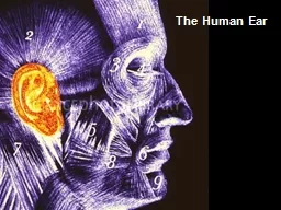
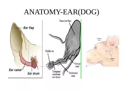
DR SANJIV KUMAR ASSISTANT PROFESSOR DEPTT OF PATHOLOGY BVC PATNA Structure of eye Anatomical features The eye ball is located the orbit It is protected by eyelids which have stratified ID: 911267
Download Presentation The PPT/PDF document "SYSTEMIC PATHOLOGY AFFECTIONS OF EYE AND..." is the property of its rightful owner. Permission is granted to download and print the materials on this web site for personal, non-commercial use only, and to display it on your personal computer provided you do not modify the materials and that you retain all copyright notices contained in the materials. By downloading content from our website, you accept the terms of this agreement.
Slide1
SYSTEMIC PATHOLOGYAFFECTIONS OF EYE AND EAR
DR. SANJIV KUMAR
ASSISTANT PROFESSOR,
DEPTT. OF PATHOLOGY, BVC, PATNA
Slide2Slide3Structure of eye
Slide4Anatomical featuresThe
eye ball is located
the
orbit
. It is protected by eyelids, which have
stratified
squamous epithelium
(the conjunctiva
) lining the surface that comes into contact with the eyeball.
Just
behind the eyelashes are a row of tiny sebaceous glands, the
meibomian
glands, the secretion of which serves to lubricate the eyelashes, preventing their adhesion.
The lens is a peculiar structure composed entirely of epithelium. It has neither
stroma
nor vascular tissue. In front it is bathed by the aqueous
humor
and is nourished by it.
Actually the anterior surface of the lens forms the posterior boundary of the anterior chamber. Its anterior surface is in contact partly with the iris.
Its
posterior surface fits into the depression of the
vitrous
- the
hyaloid
fossa.
Usually, the
conjunctival
mucosa is free of bacteria either due to the flushing action of the tears or to the bacteriostatic property of the lysozyme.
Slide5CONGENITAL ANAMOLIES
Anophthalmia
congenitus
Complete absence of one or both eyes.
Microphthalmia
One or both eyes are small. CyclopsThere is only one eye due to fusion of the orbits. AnkyloblepharonBoth the eyelids are fused together Starbismus (Squint)In animals this condition is bilateral with the two eye globules tuning inwards. EntropionTurning in of the eyelids EctropionTurning out of eyelids. Usually the lower eyelid is affected. Coloboma Failure of the closure of embryonic ocular cleft.
Slide6Dermoids of cornea
Due to the
sublethal
factor. The cornea of one or both eyes is partly covered by skin.
Congenital anterior
synechia
There is adhesion
between iris and the posterior surface of the cornea.Microphakia The lens is small and is spherical. Luxation of the lens The dislocated lens is opaque. CataractA condition in which the lens becomes opaque. Congenital aplasia of retina and hypoplasia of the optic nerveMay be met with in calves and they are born blind.
Slide7Cyclops
Entropion
Ectropion
Dermoids
of cornea
Slide8PATHOLOGY OF THE EYELIDSTrichiasis: Turning in of the
eyelashes.
Blepharitis
:
Inflammation
of eyelids.
Hordeolum
or stye: Inflammation or even abscess formation of the follicles of an eyelid.Chalazion: Abscess formation of the meibomian glands.
Slide9PATHOLOGY OF ORBITExophthalmos:Exophthalmos means
protrusion of the eyeball
.
Enophthalmos
:
Enophthalmos
means
sinking of the eyeball into the orbit.Orbital cellulitis: Inflammation of the orbit is called orbital cellulitis.
Slide10PATHOLOGY OF LACHRYMAL GLANDDacryoadenitis: Dacryoadenitis is the inflammation of the lachrymal glands.
Occlusion
of lachrymal canal
PATHOLOGY OF THE CONJUNCTIVAConjunctivitis: Inflammation of the
conjunctiva is
called conjunctivitis
.
Clinical signs
There
is congestion of the conjunctiva and increased production of tears which flow over the face as the lachrymal canal may be closed due to the swelling of the
membrane. Croupous or diphtheritic conjunctivitis is mostly encountered in fowls. In cattle infection by S. necrophorous causes croupous conjunctivitis.
Slide12PATHOLOGY OF THE CORNEAPannus:
Pannus
is a condition in
which
vascular granulation tissue is found
between
the corneal epithelium and the Bowman’s membrane.Keratitis: Inflammation of the cornea is called keratitis.Corneal ulceration occurs during acute or chronic conjunctivitis. There may be prolapse of the iris through the rupture (Staphyloma), followed by dislocation of the lens.Infectious keratoconjunctivitis in cattle (Pink eye)The causative organism is Moraxella bovis which is gram negative and is found in the tears. An endotoxin that causes necrosis of the skin is produced by this organism.
Slide13PATHOLOGY OF THE LENSLuxation of the lens
The
lens is anchored by the suspensory ligaments to the
ciliary
body. If
these ligaments are ruptured, the lens may be displaced into the anterior chamber or into the
hyaloid
fossa or into the vitreous.Cataract : Opacity of the lens is known as cataract.Congenital: Failure of the hyaloid artery to regress and disappear completely or Impairment of translucence of the lens due to abnormal arrangements of the lens fibres .Acquired: Degeneration of the lens due to Trauma, Luxation, Senility, Diabetes mellitus, deficiency of vitamin D; deficiency of vitamin C in the lens; deficiency of cystein
Slide14Types of CataractCataract may be partial or complete depending on its situation.Depending on the nature of the lesion, cataract is classified as follows:
Subcapsular
cataract
: In
this condition there is abnormal proliferation of the lens epithelium.
Cortical cataract
: This
is the most common form and involves the lens
fibres.Lamellar cataract : It may be congenital or acquired and results due to some injury during development.Nuclear cataract: Are probably the results of senile changes in which the fibres at the centre become denser thereby making the nucleus dull or hazy.
Slide15PATHOLOGY OF THE UVEAL TRACTAnterior synechia: Anterior synechia is the condition in which there is adhesion of the iris to the posterior surface of the cornea
.
Posterior
synechia
:
Posterior
synechia
is the adhesion of the posterior surface of the iris to the anterior surface of the lens capsule
.Iridocyclitis: This is the inflammation of iris and ciliary body and is also known as anterior uveitis. This condition in horses is known as periodic opthalmia.
Slide16PATHOLOGY OF THE IRISMydriasis: Dilatation of the pupil is known as mydriasis. This can be brought about by various drugs like atropine,
hyocyamine
and
stramonium
, cocaine, adrenaline and amphetamine.
Myasis
: Constriction
of the pupil is known as
myasis. This can be brought about by pilocarpine, physocarpine and ergotamine.
Slide17PATHOLOGY OF THE RETINAProgressive retinal atrophy in dogsDetachment of retina
Slide18PATHOLOGY OF THE OPTIC NERVEAtrophyEtiologyo Congenital
o
Acquired: Retinitis
,
glaucoma
;
choroidoretinitis
, trauma on occiput, hemorrhages; poisons; morphine,
deficiency of vitamin A.Sequelaeo The optic papillae become thinned with disappearance of the interstitial capillaries.o Retinal degeneration follows atrophy of the optic nerve fibre.o Total blindness results ultimately.
Slide19GLAUCOMAGlaucoma is a condition in which there is increased intraocular pressure leading to secondary changes in the eyeball
like
enlargement of eye ball, opaque cornea and increase aqueous
humor
.
It may be unilateral or bilateral
Increased
intraocular pressure may result from a) too excessive a secretion of the aqueous humor or b) hindrance in its drainage.Primary glaucoma: If the causes that give rise to obstruction of the flow, leading to glaucoma cannot be determined with certainty, the condition is known as primary glaucoma.Secondary glaucoma: If the causes for such obstruction can be determined, the condition is known as secondary glaucoma .
Slide20PATHOLOGY OF EYE BALLOphthalmitis is Inflammation of the eye ball.Xerophthalmia is abnormal dryness of the
eye ball,
with inflammation and ridge formation, typically associated with vitamin A deficiency.
Slide21NEOPLASMS OF THE EYEPrimary neoplasms Squamous cell carcinoma, especially in the bovines, is the most common neoplasm. Adenomas and adenocarcinomas of the lachrymal gland and Harderian glands may be met with.
Adenoma and Adenocarcinoma of the
ciliary
epithelium and iris may occur
.
Secondary
matastases
Secondary
matastases of carcinoma, sarcomas, melanoma, lymphosarcoma, meningioma and the venereal tumor may be met with.
Slide22POSTMORTEM CHANGESPostmortem changes in the eye includes dryness of the eyeball, pupillary dilatation and detachment of the retina.
Slide23Slide24Structure of ear
Slide25External earConsists of the concha, the external auditorymeatus and the ceruminous glands
Otitis
externa
:
Inflammation
of the external
ear.
Macroscopic
featuresSwelling and congestion leading to obstruction of ear canal.Excessive production of thick, tenacious and brownish wax.Granulomatous lesions filling the external auditory meatus Microscopic featuresGranulomatous lesions of actinomycosis in subcutaneous region around the cartilage
Slide26AURICULAR HAEMATOMA
Slide27Slide28MIDDLE EARConsists of the tympanic cavity, the ossicles and the eustachian tubes. In horse, guttural pouches are diverticula of the
eustachian
tubes
The
epithelium lining the tympanic cavity is continuous with the nasal mucosa through the
eustachian
tubes and so infection from the nose and pharynx can extend into the middle ear
.Otitis media: Inflammation of the middle ear. FeaturesOcclusion of Eustachian tubePurulent inflammation
Slide29INTERNAL EAR (LABYRINTH OF THE EAR)The bony labyrinth, a cavity in the temporal bone, is divided into three sections: the vestibule, the semicircular canals, and the cochlea. Within the bony labyrinth is a membranous labyrinth, which is also divided into three parts: the semicircular ducts; two saclike structures, the
saccule
and utricle, located in the vestibule; and the cochlear duct, which is the only part of the inner ear involved in hearing.
Otitis
interna
:
Inflammation of internal ear.
Macroscopic features
Disturbance in equilibriumDeafnessMicroscopic features Suppurative inflammation