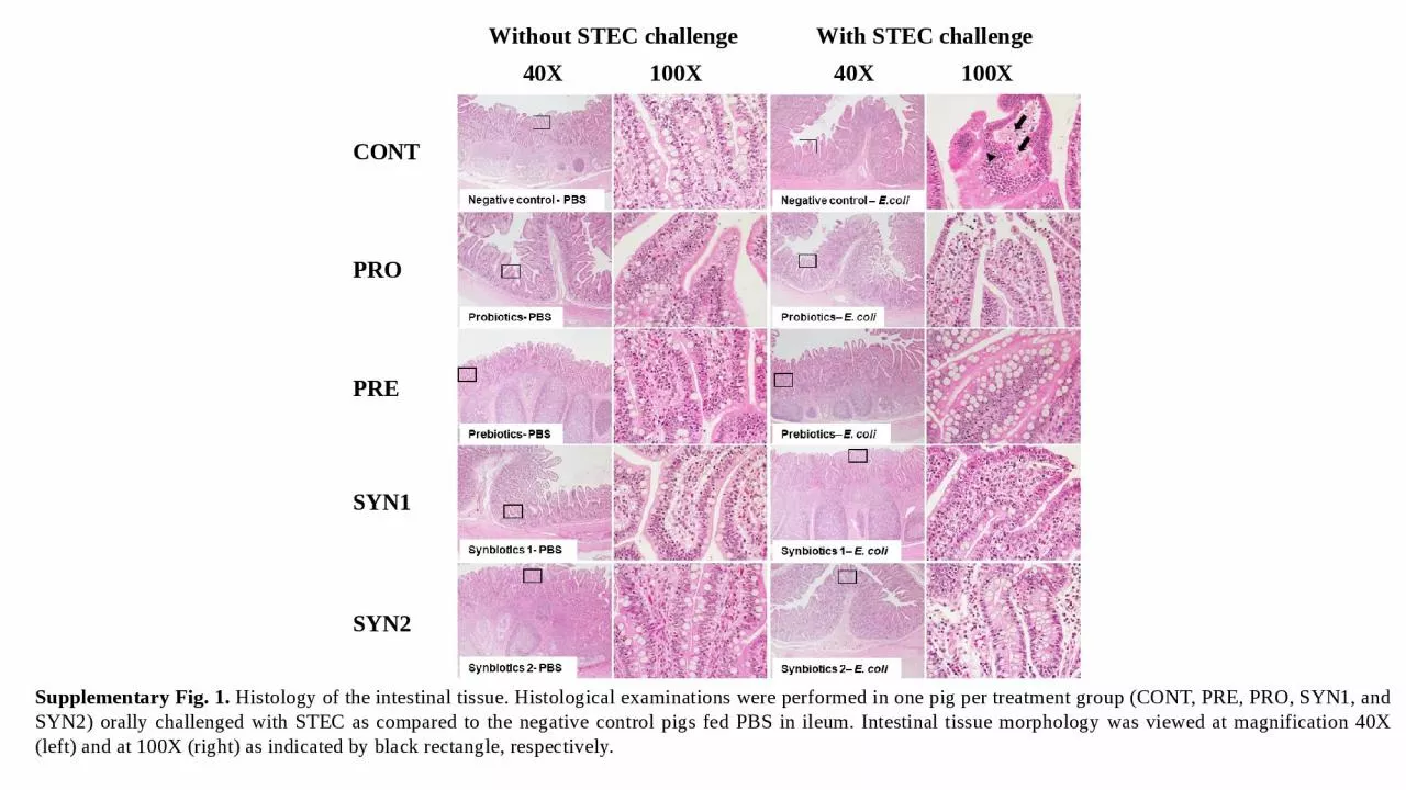
PPT-Supplementary Fig. 1. Histology of the intestinal tissue. Histological examinations were
Author : williams | Published Date : 2023-11-15
Without STEC challenge With STEC challenge 40X 100X CONT PRO PRE SYN1 SYN2 40X 100X Supplementary Fig 2 Histology of the intestinal tissue Histological examinations
Presentation Embed Code
Download Presentation
Download Presentation The PPT/PDF document "Supplementary Fig. 1. Histology of the ..." is the property of its rightful owner. Permission is granted to download and print the materials on this website for personal, non-commercial use only, and to display it on your personal computer provided you do not modify the materials and that you retain all copyright notices contained in the materials. By downloading content from our website, you accept the terms of this agreement.
Supplementary Fig. 1. Histology of the intestinal tissue. Histological examinations were: Transcript
Without STEC challenge With STEC challenge 40X 100X CONT PRO PRE SYN1 SYN2 40X 100X Supplementary Fig 2 Histology of the intestinal tissue Histological examinations were performed in one pig per treatment group CONT PRE PRO SYN1 and SYN2 orally challenged with STEC as compared to the negative control pigs fed PBS in cecum Intestinal tissue morphology was viewed at magnification 40X left and at 100X right as indicated by black rectangle respectively. By. Professor Abdel-Majeed Safer. 1. Professor Abdel-Majeed Safer Histology & Histochemistry . Professor Abdel-Majeed Safer Histology & Histochemistry . 2. Chapter 1. What is HISTOLOGY ?. By. Professor Abdel-Majeed Safer. 1. Professor Abdel-Majeed Safer Histology & Histochemistry . Professor Abdel-Majeed Safer Histology & Histochemistry . 2. Chapter 1. What is HISTOLOGY ?. The . diaphysis. is the shaft or . body of a long bone.. The . epiphyses. form the distal . and proximal ends of a . long bone.. The . metaphyses. are the areas . where the epiphyses and . diaphysis join.. VIBS 289 lab. . . Larry Johnson Texas A&M University. Epithelium - function. Epithelium. forms the . outer protective surface . of the body and . all the glands. Colon Cancer. KCR Abstractor’s Training . April 21-23, 2015. Nicole Catlett, CTR. Multiple Primary & Histology Coding Rules. It is very important to use the MPH rules for each case you are abstracting. . VIBS 289 lab. . . Larry Johnson Texas A&M University. Epithelium - function. Epithelium. forms the . outer protective surface . of the body and . all the glands. , human. , plant. Sources - medical, veterinary, research . Grossed. , fixed, processed, embedded, sectioned, and . stained . I. mportant to . the scientific investigation process . T. o . establish and confirm patient . organs. Literally, histology means tissue or web. To study tissues, certain techniques an instruments are used to study tissues. Tissue Preparation. For conventional bright field light microscopy, we follow the following steps:. SYSTEMIC HISTOLOGY . II. ATIBA, P.M . OVARY. MESOTHELIAL EPITHELIUM. Simple cuboidal or Columnar. TUNICA ALBUGENIA. Dense connective tissue . Histology of Female Reproductive System. 2. OVARY. CORTEX . HISTOLOGICAL STRUCTURE. HISTOLOGICAL STRUCTURE OF TESTIS. Each testis is enclosed in a think connective tissue capsule . called the tunica albuginea,. inner to which is a vascular layer of loose connective tissue called the . . VAN-607. TOPIC-. . HISTOLOGY OF MALE GENITAL-TUBULAR PART URETHRA PENIS & PREPUCE. Dr. SANJAY KUMAR BHARTI. HEAD,VETERINARY ANATOMY. Male Genital Organs Contains following organs 1.-One Pair Gonads 2.- Tubular Organs . Immunofluorescene. assay was used to analyze cell proliferation marker Ki67 and its expression in UVA and BR treated cells for 48 hours.. . Supplementary Fig. 2 Co-treatment has no effect on keratinocyte apoptosis.. Consort flow chart summarizing the number of patients screened, enrolled, treated and followed on the study. *one patient was re-assessed for eligibility at recurrence and re-enrolled on the study. Created with BioRender.com.. A Female . B Male . Supplementary fig. S2.
Download Document
Here is the link to download the presentation.
"Supplementary Fig. 1. Histology of the intestinal tissue. Histological examinations were"The content belongs to its owner. You may download and print it for personal use, without modification, and keep all copyright notices. By downloading, you agree to these terms.
Related Documents