/
Analysis of nonsquamous vulvar cancer cases A 21year Nonskuamz v Analysis of nonsquamous vulvar cancer cases A 21year Nonskuamz v

Analysis of nonsquamous vulvar cancer cases A 21year Nonskuamz v - PDF document

willow
willow . @willow
Follow
343 views
Uploaded On 2022-08-20

Analysis of nonsquamous vulvar cancer cases A 21year Nonskuamz v - PPT Presentation

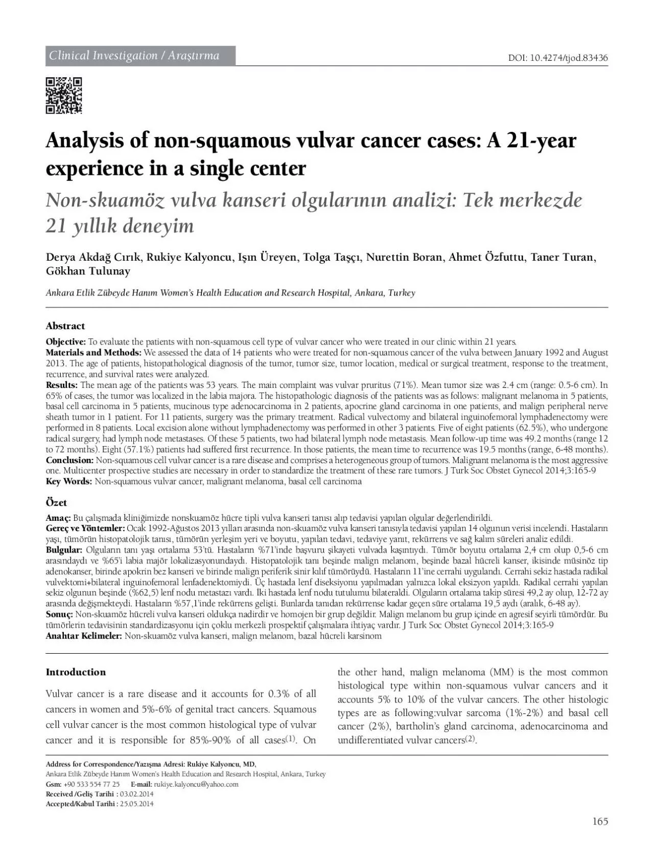
165DOI 104274tjod83436 Address for CorrespondenceYaz3130ma Adresi Clinical Investigation Ara31t30rma hypertensive diabetic obese heavy smoker and chronic immunosuppressive wome ID: 938826

vulvar patients tumor cell patients vulvar cell tumor node cancer lymph recurrence cases case treatment basal surgery study squamous

Share:

Link:

Embed:

Download Presentation from below link

Download Pdf The PPT/PDF document "Analysis of nonsquamous vulvar cancer ca..." is the property of its rightful owner. Permission is granted to download and print the materials on this web site for personal, non-commercial use only, and to display it on your personal computer provided you do not modify the materials and that you retain all copyright notices contained in the materials. By downloading content from our website, you accept the terms of this agreement.


Presentation Transcript

165DOI: 10.4274/tjod.83436 Analysis of non-squamous vulvar cancer cases: A 21-year Non-skuamöz vulva kanseri olgularnn analizi: Tek merkezde Ankara Etlik Zübeyde Hanm Women’s Health Education and Research Hospital, Ankara, Turkey Address for Correspondence/Yazma Adresi: Clinical Investigation / Aratrma hypertensive, diabetic, obese, heavy smoker and chronic immunosuppressive women, the etiology is not fully . Vulvar cancer is usually a post-menapousal disesaseandhas a slow clinical course. If it is diagnosed at an earlier stages, the treatmentis more succesfull. However, an In this study, we aimed to analyze thepatients who were treated for non-squamous cell type vulvar cancer between January The clinico-pathological datas of the women, referred to our hospital with a diagnosis of non-squamous cell type vulvar cancer, between January 1992 and August 2013 was retrospectively for the study. Non-squamous cell vulvar cancer were diagnosed in 14 patients and they eligible for the study.colposcopy if necessary. Chest radiograph, upper abdominal and pelvic ultrasonography, abdominal computerized operative radiologic examinations. For patients diagnosed tomography and if necessary cranial magnetic resonance imaging was also performed. Clinico-pathologic data of the patients were reviewed from patient’s les, and computerized database.the tumor 2.5 cm far from the midline. Tumor size was calculatedby measuringthe largest diameter of the tumor. nodes, lymp node positivity, were reviewed from pathology records of the patients.When lymph node or distant metastasis were detected after surgery, patients were given systemic chemotherapy (vincristine, bleomycin) or concomitant chemoradiotherapy, as an adjuvant therapy. Primary radiotherapy or concomitant chemoradiotherapy undergone surgery due to additional diseases.as; ‘complete clinical response’; havingmore than 50% shrinkage between 25%-50% of tumor was dened as; ‘stable years. Disease free survival was dened as the time period from diagnosis to recurrence. The overall survival was dened

as; the 68 years. The most frequent complaint was vulvar pruritis 2 withmucinous type adenocarcinoma, one with apocrine gland carcinoma and one with malignant peripheral nerve sheat tumor.were diagnosed as MM. Of those patients, tumor site was labia majora in two patients, clitoris in two and labia minora in one case. In these patients, the mean tumor diameter was 1.9 cm, patients, the treatment and follow-up protocols were presented in (Table 1). Three of malignant melanoma cases were lymph node dissection as primary therapy. In two of these three cases, lymph node metastases were detected and adjuvant involvement (patient no #1). Other lymph node positive up (patient no #12). Other two MM patients, had either had lymph node,measuring 10 cm in the right groin (patient no #11 case thatunderwentsurgery, disease progressionwas detected during therapy. Complete clinical response was obtained in the follow-up. In 3 patients who were treated with radical surgery, recurrence was developed at 12, 12 and 24 months after surgery. Recurrence localization was as following; the breast, cervix, pelvic bones, urethra and vagina. For breast recurrence surgery was the choice of treatment, for cervical recurrence chemoterapy + bleomycin + cisplatin + methotrexate. BOMP) was given. In three recurrent cases who had undergone radical surgery, tumor diagnosis. The other MM case who was given radiotherapy, died 9 months after diagnosis.Five patients were diagnosed with basal cell cancer of the vulva. to 4 cm. In two of the patients diagnosed with basal cell cancer, 167J Turk Soc Obstet Gynecol 2014;3:165-9Derya Akda Crk Analysis of non-squamous vulvar cancer Table 1. Tumor characteristics, treatment and follow-up schemes of patients with non-squamous vulvar cancerFollow-bladderPelvic boneRT: Radiotherapy, KT: Chemotherapy, BCC: Basal cell carcinoma, MM: Malign melanoma, Operation: Radical vulvectomy and bilateral inguinofemoral lymp node dissection performed. In one of the patients who underwent radical surgery, that case, recurrence occured in vagina 30 months after diagnosis and chemotherapy (BOMP) was given for

treatment. There was complete response to treatmet for recurrence.carcinoma (patient no #3). In this case, tumor was 4 cm in diameter and localized in labia majora. Radical surgery was doneand adjuvant chemoradiotherapy was given for this case. tumor (patient no #5) was 22 years old, the tumor was 3 cm in for this case. No lymph node metastasis was detected after was no recurrence at 28 months follow-up in this case.In two cases of mucinous adenocarcinoma, tumor localization was labia majora and labia minora, 5 cm and 6 cm in diameter, respectively. Radical vulvectomy and bilateral inguinofemoral lymph node dissection was performed for that cases. First case had unilateral lymph node involvement (one lymph node) and resection of recurrent tumor was done in that patients. This patient was disease-free during 66 months period of follow-up. was not appropriate treatment due to presence of tumoral recurrence was occured in the bladder after 12 months following the rst recurrence. This patient did not accept the treatment of second recurrence and died 25 months after diagnosis.In our case series, inguinal lymph node metastasis was detected in 5 of 8 patients who were undergone radical surgery. and bilaterally in two patients. In one case with bilateral lymph node involvement, the tumor was located in midline and in lateral side in the other one. Lesion was located laterally in all three cases with unilateral lymph node involvement.months. Recurrence developed in eight patients. The average 12 days). Surgical site infection and inguinal wound disruption was observed in two patients as early complications.including MM, basal cell cancer, bartholin gland cancer, sarcoma and lymphoma. These tumors extend from basal cell cancer, presence of different clinico-pathologic characteristics for each, there is no standardized treatment regimen for these tumors. Depending on the tumor histology, surgical management and if Contrary to squamous cell cancer, non-squamous vulvar cancer is not only diagnosed in postmenopausal women but also young women. In presented series, patients’ mean age was 53 rangingfrom 22 to 68

years, 78% of these patients were postmenopausal and 28% of them were over the age of sixty. Tumor is often localized in labia majora for tumors with non-squamous cell types, but clitoris is a special localization . In our study, tumor were localized in clitoris in 40% of MM cases. Similar to squamous cell vulvar cancer, the most common complaint islong-lasting vulvar itchingIn accordance with the literature, 71% of cases in this series admitted to hospital with vulvar itching.literature. Therefore there is no consensus about treatment modalities and follow-up schedules of these cases. For vulvar melanoma which has very poor prognosis, surgical treatment is cancers and cutaneous melanom (8). Localized disease, negative lymph node involvement, young age are good prognostic factors for malign melanoma in the Breslow microstaging system. For and inguinofemoral lymph node disection should be preferred treatment approach in those cases. For tumors more than 4 mm depth of invasion, regional lymphadenectomy is a correct treatment choice due to risk of distant metastasis. Overall 5 same within last 40 years. Among patients with malign melanoma, 3 of them hadtumoral invasion depth between 1-2 mm underwent radical surgery. Two of these three patients had lymph node metastasis. In these three patients with MM, recurrence developed during follow-up. And recurrence was Basal cell cancer, which is seen in 2% of all vulvar cancers has generally good prognosis. However, if left untreated it can be locally destructive. Generally, local excisionis the preferred . However, lymphnode positivity or hematogenous metastasis has been reported in basal cell cancers in the our study, two ofve patients diagnosed with basal cell cancer underwent radical surgery, local excision was performed for other three cases. In one case with basal cell carcinoma, vaginal and there was complete clinical response. After 60-72 months of follow-up period, all ve patients were alive with no disease. In our study, mean duration of hospitalization was 15 days, it is similar to study of Brinton et al.. The most common infection and wound disru

ption, more rarely leg edema and . In this study two patients had early post-op wound disruption in the groin).of tumor and seen in extremely rare. Malign melanomais the stages. Forthe treatment of vulvar malignant melanomas, there the literature. Contribution of current treatment on survivalis necessary.Cavanagh D, Fiorica JV, Hoffman MS, Roberts WS, Bryson SC, LaPolla JP, et al. Invasive carcinoma of the vulva: changing trends in surgical Oonk MH, de Hullu JA, Hollema H, Mourits MJ, Pras E, Wymenga AN, et al. The value of routine follow-up in patients treated for Madsen BS, Jensen HI, van der Brule AJ, Wohlfahrt J, Frisch M. Risk factors for invasive squamous cell carcinoma of the vulva and vagina- population-based case control study in Denmark. Int J Canser Finan MA, Barre G. Bartholin’s gland carcinoma, malignant melanoma and other rare tumours of the vulva. Best Pract Res Clin Obstet Gynaecol 2003;17:609-33. Sugiyama VE, Chan JK, Shin JY, Berek JS, Osann K, et al. Vulvar melanoma: a multi-variable analysis of 644 patients. Obstet Gynecol Dunton JD, Berd D. Vulvar melanom, biologically different from other cutaneous melanomas. Lancet 1999;354:2013-4. Aydn Ç, Göl M, Balolu A, Gezgin Z, Vulva Kanseri Nedeniyle Radikal Vulvektomi Uygulanan Hastalarin Deerlendirilmesi. Ege Trone JC, Guy JB, Mery B, Langrand Escure J, Lahmar R, Moncharmont C, et al. Melanomas of the female genital tract: state of the art. Bull Balch CM, Urist MM, Karakousis CP, Smith TJ, Temple WJ, et al. Efcacy of 2 -cm surgical margins for intermediate - thickness melanoma (1-4 mm):results of a multi-institutional randomized Tcheung WJ, Selim MA, Herndon JE 2nd, Abernethy AP, Nelson KC. Clinicopathologic study of 85 cases of melanoma of the female De Giorgi V, Salvini C, Massi D, Raspollini MR, Carli P. Vulvar basal cell carcinoma: retrospective study and review of literature. Gynecol Mulayim N, Foster Silver D, Tolgay Ocal I, Babalola E. Vulvar basal cell carcinoma: two unusual presentations and review of the literatüre. Gynecol Oncol 2002;85:532-7.Jimenez HT, Fenoglio CM, Richart RM. Vulvar basal cell carcinoma