/
Energy Dispersive  spectroscopy Energy Dispersive  spectroscopy

Energy Dispersive spectroscopy - PowerPoint Presentation

winnie
winnie . @winnie
Follow
0 views
Uploaded On 2024-03-13

Energy Dispersive spectroscopy - PPT Presentation

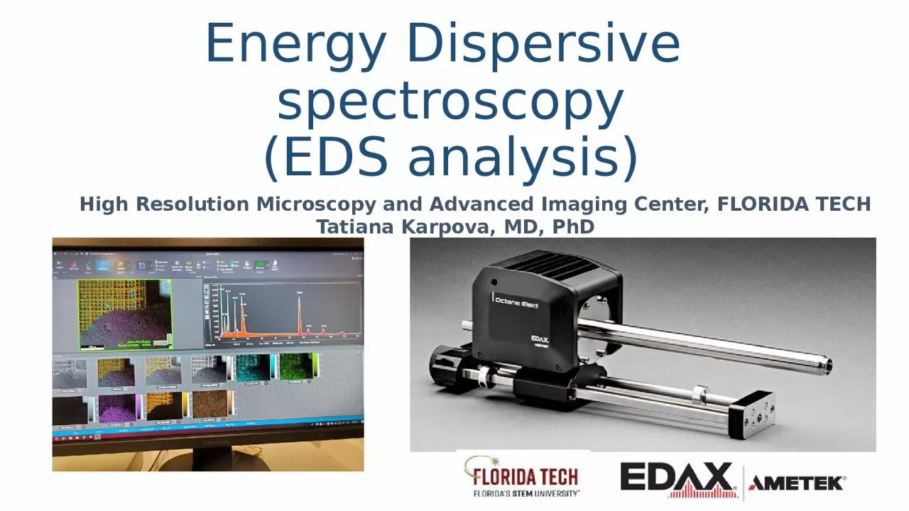
EDS analysis High Resolution Microscopy and Advanced Imaging Center FLORIDA TECH Tatiana Karpova MD PhD What is energy dispersive spectroscopy Energy Dispersive Spectroscopy EDS also known as energy dispersive Xray spectroscopy EDX and sometimes ca ID: 1046897

ray energy sample rays energy ray rays sample electron shell electrons elements eds beam analysis element characteristic kev quantitative

Share:

Link:

Embed:

Download Presentation from below link

Download Presentation The PPT/PDF document "Energy Dispersive spectroscopy" is the property of its rightful owner. Permission is granted to download and print the materials on this web site for personal, non-commercial use only, and to display it on your personal computer provided you do not modify the materials and that you retain all copyright notices contained in the materials. By downloading content from our website, you accept the terms of this agreement.


Presentation Transcript

1. Energy Dispersive spectroscopy(EDS analysis)High Resolution Microscopy and Advanced Imaging Center, FLORIDA TECH Tatiana Karpova, MD, PhD

2. What is energy dispersive spectroscopy?Energy Dispersive Spectroscopy (EDS, also known as energy dispersive X-ray spectroscopy, EDX, and sometimes called X-ray energy dispersive spectroscopy XEDS) is a qualitative and quantitative X-ray microanalytical technique that can provide information on the chemical composition of a sample for elements with atomic number (Z) >3, although for specific sample and equipment configurations (Z) >2 is now achievable.An electron beam is focused on the sample in either a scanning electron microscope (SEM) or a transmission electron microscope (TEM). The electrons from the primary beam penetrate the sample and interact with the atoms from which it is made. Two types of X-rays result from these interactions: Bremsstrahlung X-rays, which means ‘braking radiation’ and are also referred to as Continuum or background X-rays, and Characteristic X-rays.The X-rays are detected by an Energy Dispersive detector which displays the signal as a spectrum, or histogram, of intensity (number of X-rays or X-ray count rate) versus Energy. The energies of the Characteristic X-rays allow the elements making up the sample to be identified, while the intensities of the Characteristic X-ray peaks allow the concentrations of the elements to be quantified.https://myscope.training/#/EDSlevel

3. Generation of X-rays in the electron microscopeThe specimens examined in an electron microscope are made up of atoms. When electrons from the primary beam of the electron microscope are focused on a specimen, they penetrate it and interact with the atoms that make up the sample material. In the scanning electron microscope (SEM) the electrons from the primary beam spread out in the sample to form the interaction volume. The size of the interaction volume depends on the accelerating voltage of the primary electron beam and the mean atomic number (or density) of the sample. The interaction volume will be larger for a larger accelerating voltage, but smaller for samples with a higher mean atomic number.In the SEM secondary electrons are produced from the surface of the sample or topmost part of the interaction volume; backscattered electrons come from the top half of the interaction volume; and X-rays are generated within the whole of the interaction volume. Therefore, the typical spatial resolution for X-ray microanalysis in the SEM is of the order of a few microns.In the TEM, the sample is very thin (~10-200 nm) and there is minimal spreading of the electron beam as it passes through the sample. Therefore, the spatial resolution for X-ray microanalysis in the TEM is approximately of the same order as the sample thickness.https://myscope.training/#/EDSlevel

4. Bremsstrahlung X-ray generationTwo types of X-rays are produced by interaction of the electron beam with the sample in both the SEM and TEM: Bremsstrahlung (which means ‘braking radiation’) and Characteristic X-rays. Bremsstrahlung X-rays are produced by slowing down of theprimary beam electrons by the electric field surrounding the nuclei of the atoms in the sample. Note: Bremsstrahlung X-rays are also referred to as continuum or background X-rays. The primary-beam electrons lose energy and change direction due to inelastic scattering in the sample. Some of the lost energy is converted to X-rays which have a range of energies, from ~0 up to E –the energy of the electrons in the primary beam. Bremsstrahlung X-rays cannot have energies greater than the energy of the electrons in the primary beam so this energy forms the upper energy limit of the Xray spectrum and is known as the Duane-Hunt limit.The primary beam electrons are slowed down or deflected by the electric field around the atoms in the specimen. Part of the energy that they lose is converted to Bremsstrahlung X-rays with energies between ~0 and the Duane-Hunt limit.https://myscope.training/#/EDSlevel

5. Origin of Characteristic X-rays Core electronic energy levels of an atom.Adapted from Introduction to Energy Dispersive X-ray Spectroscopy (EDS), micron.ucr.edu/public/manuals/EDS-intro.pdfThe atoms of each element in the sample consist of a nucleus made up of neutrons and positively charged protons, and a cloud of negatively charged electrons that surrounds the nucleus. The number of protons in the nucleus of the atom defines its atomic number, Z, while in a neutrally charged atom the number of protons is matched by the number of electrons.The electrons in the electron cloud have a stable set of energy levels, also known as electron shells. The shell closest to the nucleus is known as the K shell, followed outwards by the L, M, N, O, P and Q shells. EDS microanalysis is mostly concerned with electrons in the inner shells, i.e., the K, L and M shells. The maximum number of electrons in each shell is governed by quantum mechanics, with a maximum of two electrons in the K shell; eight electrons in the L shell; 18 electrons in the M shell, and so on. Each shell, apart from the K shell, is split into subshells, with the electrons in related subshells having slightly different energies. The L shell has three subshells; the M shell has five subshells, and so on. The K shell has the highest ionisation energy or critical ionisation energy in the atom. That is, more energy is needed to remove an electron from this shell than from subshells further from the nucleus. The further from the nucleus the electron is, the lower its ionisation energy. Characteristic X-rays are produced by electron transitions between the inner electron shells. The electrons in each shell and subshell have specific ionisation energies, and these are different for every element, that is, the ionisation energy for the K shell in Si (1.84 keV) is different from the ionisation energy of the K shell in Pt (78.4 keV).https://myscope.training/#/EDSlevel

6. Characteristic X-raysThe production of Characteristic X-rays is a two-stage process: ionisation followed by relaxation. Firstly, an electron is removed from one of the inner shells of the atom by an electron from the primary beam so that the atom is ionized and in an unstable state. Secondly, the atom regains stability when an electron from an outer shell fills the inner shell vacancy and an X-ray photon is emitted. The energy of the emitted X-ray is equal to the difference between the ionisation energies of the electrons involved in the transition.https://myscope.training/#/EDSlevel

7. Characteristic X-rays ProductionAs each element has specific ionisation energies for each subshell, so the difference between the energies is characteristic of the element involved in producing the X-ray photon. For Si, the ionisation energy of the K shell is 1.84 keV, the ionisation energy of the L shell is ~0.10 keV and the ionisation energy of the M shell is ~0.01 keV.The Characteristic X-ray spectrum for Si shows three spectral lines. The line at low energy (~0.09 keV) results from ionisation of the L shell with an electron from the M shell filling the vacancy: E = 0.10 – 0.01 keV. (This line would be at or below the limit of detection for most EDS detectors.) The line at ~1.74 keV results from ionisation of the K shell with an electron from the L shell filling the vacancy (E = 1.84 – 0.10 keV), whereas the smaller peak at higher energy (~1.83 keV) results from ionisation of the K shell and an electron from the M shell filling the vacancy (E = 1.84 – 0.01 keV).

8. NomenclatureIn spectroscopy, the most commonly used naming convention for Characteristic X-ray lines is the Siegbahn notation. The first component of the name is the element involved, e.g. Si. The second component is the electron shell that was ionized to produce the X-ray, e.g. K, L or M. The third component reflects the relative intensity of the line within each shell, e.g. α is the most intense line, followed by β and γ. The lines within each shell make up a family, or series, of lines for that shell, e.g., the K family comprises the Kα and Kβ X-ray lines.In the Si spectrum, the lowest energy X-ray line is the Si Lα line; the line at 1.74 keV is the Si Kα line and the line at ~1.83 keV is the Si Kβ line.For each element, the electrons in the K shell have the highest ionisation energies while the ionisation energies of electrons in outer shells are lower. More energy is required to ionize the K shell, and it follows that the energies of the K family X-ray lines for each element are greater than those of the L family, which are greater than those of the M family. That is, for every element: EK > EL > EM

9. The X-ray spectrumThe spectrum seen in the output from the Energy Dispersive Spectrometer (EDS) shows the Characteristic X-ray lines superimposed on the Bremsstrahlung X-rays. Although the Characteristic X-rays have discrete energies, in measuring them the signal is averaged and the lines become peaks which are superimposed on the continuum.https://myscope.training/#/EDSlevel

10. Accelerating voltage or overvoltage ratioThe accelerating voltage used in the electron microscope controls the energy of theelectrons in the primary beam. In the SEM, accelerating voltages are typically 5-30 keVbut in TEMs much higher accelerating voltages, 100-400 keV or more, are used. In orderto generate Characteristic X-rays the electrons in the primary beam must have enoughenergy to overcome the ionization energy, also called the critical ionization energy, of theinner-shell electrons in the atoms of the sample. With the high accelerating voltages usedin TEMs this is not a problem, but in SEMs care must be taken to use a sufficiently highaccelerating voltage to stimulate X-rays from all elements in the sample.https://myscope.training/#/EDSlevel

11. Accelerating voltage or overvoltage ratioThe overvoltage ratio is the ratio of the energy of the electrons in the primary beam, E , tothe critical ionization energy, E , needed to ionize an inner shell of an atom in the sample.For example, if the accelerating voltage is 15 kV, the energy of the electrons in the primarybeam is 15 keV. The critical excitation energy of the Fe Kα X-ray is 7.11 keV, andtherefore the overvoltage ratio, U = E /E , is 2.11. For efficient generation of X-rays, theovervoltage ratio should be at least 2 (the optimum value is ~2.7).https://myscope.training/#/EDSlevel

12. Spatial resolution High spatial resolution energy dispersive X-ray spectrometry in the SEM and the detection of light elements including lithium Simon Burgess, Xiaobing Li, and James Holland Oxford Instruments NanoAnalysis, High Wycombe, UK

13. Qualitative microanalysis means that the elements present in the sample are identified from their Characteristic X-ray peaks, but their abundances are not determined. While commercial peak-identification software is improving all the time, it is not yet 100% accurate. Elements that are present in the sample may be missed, and elements that are not present may be falsely identified.For qualitative microanalysis of an unknown sample, consideration must be given to the operating parameters of the microscope and also the properties of the sample to be analysed. What elements are present in the sample?

14. Microscope operating parametersIn the SEM, if the sample is stable under high-vacuum in the electron microscope and is not susceptible to damage by the electron beam, then an accelerating voltage of 15-20 kV is recommended for SEM analysis. This is sufficient to efficiently generate at least one family of X-ray lines for all elements.If the sample is likely to be damaged by a high-energy primary electron beam in either the SEM or TEM, then it may be necessary to use a lower accelerating voltage. If this is the case in the SEM, the higher energy X-ray lines may not be efficiently generated and low-energy X-rays will have to be used for element identification, e.g., the L or M lines for elements with Z > 20.The electron beam current (probe current or spot size) will control the X-ray count rate or intensity of the generated X-rays. The beam current should be adjusted to minimize damage to the sample but generate sufficient X-rays to allow reliable identification of peaks. At the same time, the beam current needs to be adjusted to minimize spectral artefacts and achieve system dead times of 20-50%.

15. Characteristic X-rays

16. X-ray peak identificationThe energies of the Characteristic X-rays emitted by the elements in a sample allow them to be identified. However, the presence of overlapping peaks from different elements means that correct identification depends on being able to recognize the peaks in the different X-ray families. The relative weights of the Characteristic X-ray lines in each family are consistent and this, along with their energies, allows related peaks in the X-ray spectrum to be recognized. By measuring the energies of the major X-ray peaks in each family, the corresponding element can be identified. Commercial peak-identification software can do this, but the results need to be verified.At energies above 4 keV, the peaks of the K and L families are resolved.The K family of X-rays has two peaks, Kα and Kβ, with intensity ratios of approximately 10:1. Note: There are actually two X-ray lines in the Kα X-ray peak: Kα1 and Kα2, but they cannot be resolved by ED spectrometers so the combined peak is referred to as Kα.https://myscope.training/#/EDSlevel

17. X-ray peak identificationThe L family of X-rays is more complicated with six lines commonly visible in the spectrum. The Ll (ell-ell) line results from electron transitions between the M I and L III subshells, and forms a small peak on the low-energy side of the family. The Lα, Lβ1 and Lβ2 lines form a series of three peaks with descending magnitudes and relative intensities of ~10:7:2. The Lγ1 and Lγ3 lines form small peaks on the high-energy side of the family. M family peaks are not present at energies above 4 keVhttps://myscope.training/#/EDSlevel

18. X-ray peak identificationThe Mα and Mβ peaks may not be fully resolved, but the Mζ (M zeta) peak on the low-energy side of the family and the Mγ peak on the high-energy side are usually present and allow M family peaks to be identified.https://myscope.training/#/EDSlevel

19. Quantitative EDS - overviewHow much of each element is present in the sample?In quantitative EDS microanalysis in SEM, the mass fractions or weight percents of the elements present in the sample are calculated. The spectra are processed to remove Bremsstrahlung X-rays and spectral artefacts, and then the Characteristic X-rays are compared with data measured from standard reference materials. In so called Standardless Quantitative analysis, or semi-quantitative analysis, the spectra are compared with data collected from standards in the factory of the manufacturer of the EDS system and stored with the system software. In fully Standardized Quantitative analysis the spectra from the standards are collected on the same instrument as the spectra from the sample being analysed, which allows for more accurate analyses.Qualitative analysis to identify the elements present in the sample is a good prelude to quantitative analysis as it will allow the best operating parameters for the microscope to be selected.

20. Limitations of quantitative analysisSome of the limitations of quantitative EDS analysis are listed below:Light elements (Z < 11) cannot be routinely analysed by EDS.Hydrogen (Z = 1) and He (Z = 2) do not have Characteristic X-rays, and the Li (Z = 3) K X-rays are of too low energy to be detected by EDS.Beryllium (Z = 4) to Ne (Z = 10) X-rays can be detected by EDS, but there are two problems. Firstly, they are low energy X-rays subject to strong absorption by the specimen. Secondly, the electrons involved in generating the Characteristic X-rays are also the valence electrons involved in the chemical bonding of the element, therefore the shapes and positions of the peaks may change in different compounds. The samples and the standards must be closely matched for best results.https://myscope.training/#/EDSlevel

21. Standardized quantitative analysisFor quantitative analysis in the SEM the sample must be bulk, flat and polished.Different combinations of electron microscope and ED X-ray detector will involve different protocols for quantitative microanalysis, but there are six basic steps required for Standardized Quantitative analysis that should be common to all systems:Define the list of elements that you want to analyse for. Use qualitative analysis to identify all of the elements present in the samples that you want to analyse. Older software packages may only derive quantitative data for a specified element list but newer systems will derive quantitative data for all of the elements detected in the spectrum to be processed.Perform any calibration of the EDS system that is necessary for quantitative analysis. At a minimum, it will be necessary to measure the beam current or the X-ray count rate on a pure element standard.Measure spectra from standards for the elements that you want to analyse for. Ideally, the spectra will be collected under the same conditions, and on the same instrument, that will be used for collecting spectra from the samples to be analysed. If no standards are available, it may be necessary to use default spectra, and this will affect the accuracy of the analysis.Collect spectra from the samples to be analysed. Be sure to monitor the beam current for drift between analyses.Process the spectra to calculate the mass fraction or weight percent of the elements present or the percentages of the elements defined in an element list.Assess the quality of the analysis. Is it a ‘good’ analysis? If not, why not?

22. Standardless or semi-quantitative analysisMost commercial EDS X-ray analysis systems will be installed with default spectra for all elements and the most commonly used X-ray analysis lines. This data allows an estimation of composition to be made for spectra collected on different instruments, and this information can be helpful in identifying different phases in a sample, for example. However, differences between the instrument and conditions for collection of the default spectra and those being used to undertake analysis elsewhere will limit the accuracy and precision of the results, so that they cannot be published as quantitative analyses.

23. Detection limitsThe minimum detection limit is the concentration corresponding to a peak that can just be distinguished statistically from background fluctuations. This is generally taken to be a peak height equal to three times the standard deviation of the background count. This will vary between different elements and analytical lines, and for the same element in different matrixes. For routine EDS analysis, the detection limits are about 1000 ppm or 0.1 wt%.EDS can detect major and minor elements with concentrations higher than 10 wt% (major) and minor concentrations (concentrations between 1 and 10 wt%). The detection limit for bulk materials is 0.1 wt% therefore EDS cannot detect trace elements (concentrations below 0.01 wt%)the smallest detectable elemental concentration and the minimum number of detectable atoms, which correspond to the smallest X-ray signal that can be measured above the background, are usually defined as the minimum detectable limithttps://myscope.training/#/EDSlevel

24. How detectors workThe energy of the incoming X-ray, in this case Ca Kα, generates electron-hole pairs in a silicon crystal detector. A bias voltage across the detector causes movement of electrons and holes to opposite sides of the crystal, generating a charge signal. The detector consists of:A collimator to ensure that only X-rays generated from where the primary electron beam interacts with the sample will be collected.An electron trap to ensure that X-rays, but no electrons, enter the detector.A window to isolate the detector crystal, under high vacuum, from the chamber of the microscope. Older windows were composed of Be which did not allow low-energy X-rays (< ~0.9 keV) to pass through it, but more modern windows are composed of polymers that will allow low-energy X-rays (down to ~0.1 keV) to pass. There is also a windowless configuration that removes the absorption issue.A semiconductor crystal detector.Electronics to detect the charge recorded by the detector, convert it to a voltage pulse and pass it to the pulse processor.https://myscope.training/#/EDSlevel

25. The detectorEDS analysis in our Microscopy center is achieved with Octane Elect EDS System. The Octane Elect EDS System is an enhanced Energy Dispersive Spectroscopy (EDS) platform with the latest advancements in Silicon Drift Detector (SDD) technology and high speed electronics. Tailored for users who demand higher performance and functionality than the options available in entry-level systems, the Octane Elect EDS System provides excellent resolution and high throughput at an optimal value with a remarkable low energy sensitivity for light element detection and low voltage (kV) microanalysis. The Octane Elect SDDs incorporate a silicon nitride (Si3N4) window, high speed electronics and a manual slide.Best Light Element PerformanceThe Octane Elect SDD with a Si3N4 window offers major transmission improvements compared to detectors with a polymer window, leading to greatly improved light element performance and significantly more critical data for the materials analyst.Low kV PerformanceThe mechanical properties of silicon nitride allow the use of thinly fabricated windows with a low aspect ratio support grid, offering a great benefit in terms of low energy sensitivity and optimal low voltage analysis.

26. Mapping informationX-ray mapping provides images of elemental distributions in a sample. The information that X-ray maps provide can be readily assimilated and, in many cases, a problem can be solved by knowing the distribution of a particular element without requiring quantitative point analysis.Screen view during mapping data collection with Apex software at our Microscopy CenterImportant parameters of mapping: CPS 19655, dead time (DT) 15.5, Resolution 126.6

27. Mapping informationAn image is generated by converting the number of X-ray photons of specified energy detected at each point into a brightness value for a pixel on the screen or digital image. The raw data for the image is a matrix of integers corresponding to the number of X-rays counted at each point and these are mapped to a single-band image with 0-255 levels. The image can be displayed as a grayscale image or assigned false colors based on a look-up table.

28. Mapping informationThree single-band images, representing X-ray maps for three different elements, can be assigned different colors (red, green and blue) and combined to form a colored three-band image. Combining maps from different images can highlight different phases in the area being scanned.

29. Parameters for X-ray mappingCounts Per Second (CPS) – The Most Important Factor. Ultimately, EDS performance and accuracy is dictated by the number of characteristic x-ray counts that are collected. The more x-rays that are collected, the better the EDS results. Acceptable CPS is ranging between thousands and 100 thousands for high quality map generation.In more recent packages, a complete ED spectrum is collected at every pixel in the image. This allows a map to be generated for all elements in the area mapped. More detailed mining of the data cube is possible after the data have been collected, for example changing the color of the selected elements.Maps are generally collected at magnifications of 50 to 1000 times. At magnifications less than 50 times, geometrical error is possible, while at magnifications greater than 1000 times oversampling is likely.The dwell time for each pixel can range from a millisecond to a second or more. Dwell time – time the electron beam stays on a pixel while capturing an image during acquiring EDS map, or during acquiring EDS line scan.These times are much less than those used for point analysis (30-60 s), so mapping is ideal for major elements but is not going to reveal minor or trace element distributions as not enough X-rays will be collected.The total acquisition time for the map will depend on the resolution of the collected maps (the number of pixels) and the dwell time per pixel. The acquisition time may range from ~0.5 h to >12 h (an overnight run). A very stable beam is required for long acquisition times, although means of adjusting for drift of the beam are available in newer software packages.

30. Influence of sample topographyA: X-rays are blocked from reaching the detector by sample topography. B: Rotating the stage presents the region of interest in sight of the EDS detector allowing the X-rays to be detected. Image Credit: JEOL USA, Inc.The electron beam – sample – detector geometry should be optimized. Set the sample stage to the microscope's manufacturer's recommended working distance. The X-ray EDS detector have been installed with an optimal working distance also, and this will vary from microscope to microscope. The X-ray detector should be as close as possible to the sample to maximize collection of the generated X-rays, but this may be limited by the presence of other detectors and safe operation of the microscope. There should be a clear path between the sample and the X-ray detector.Black areas in the “element distribution” map (left image), correlate to the lack of X-ray detection and absence of counts in the “counts per seconds” map (right image) due to sample topography.Overlapping of multiple element distribution mapsMap of CPS (counts per seconds) parameter