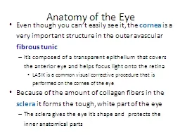
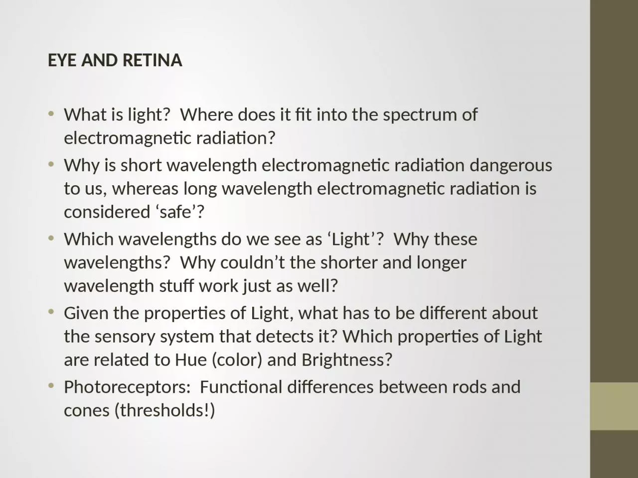
/
EYE AND RETINA   What  is light?  Where does it fit into the spectrum of electromagnetic EYE AND RETINA   What  is light?  Where does it fit into the spectrum of electromagnetic

EYE AND RETINA   What is light? Where does it fit into the spectrum of electromagnetic - PowerPoint Presentation

winnie
winnie . @winnie
Follow
0 views
Uploaded On 2024-03-15

EYE AND RETINA   What is light? Where does it fit into the spectrum of electromagnetic - PPT Presentation

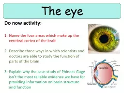
Why is short wavelength electromagnetic radiation dangerous to us whereas long wavelength electromagnetic radiation is considered safe Which wavelengths do we see as Light Why these wavelengths Why couldnt the shorter and longer wavelength stuff work just ID: 1048526

color light receptive eye light color eye receptive wavelength fields wavelengths threshold vision cones rods opponent retina pigments structure

Share:

Link:

Embed:

Download Presentation from below link

Download Presentation The PPT/PDF document "EYE AND RETINA   What is light? Where..." is the property of its rightful owner. Permission is granted to download and print the materials on this web site for personal, non-commercial use only, and to display it on your personal computer provided you do not modify the materials and that you retain all copyright notices contained in the materials. By downloading content from our website, you accept the terms of this agreement.


Presentation Transcript

1. EYE AND RETINA What is light? Where does it fit into the spectrum of electromagnetic radiation? Why is short wavelength electromagnetic radiation dangerous to us, whereas long wavelength electromagnetic radiation is considered ‘safe’? Which wavelengths do we see as ‘Light’? Why these wavelengths? Why couldn’t the shorter and longer wavelength stuff work just as well?Given the properties of Light, what has to be different about the sensory system that detects it? Which properties of Light are related to Hue (color) and Brightness? Photoreceptors: Functional differences between rods and cones (thresholds!)

2. Light and the Spectrum of Electromagnetic RadiationThe duality of EMR – ‘packets’ of energy400-700 nm wavelengths = Light. Why these wavelengths?Photons: wavelength (color) and number (brightness)Since light comes in ‘packets’, limited capacity to absorbthe eye must continuously ‘regulate and regenerate’Sun and stars emit all of theseSHORTMEDIUMLONGHIGH ENERGYLOW ENERGYWAVELENGTH (nm)WAVELENGTH (nm)ReflectedBy GasesIncreasinglyAbleToPassThroughSolidsE = mc2

3. S M LSUNEARTH

4. Pigments and reflected lightColor vision requires abundant lightSo, we have TWO eyes (‘duplex’ eye: rods, cones)Primaries for color vision (RGB)Across-fiber pattern coding for color (using just three broadly-tuned receptors we can perceive an enormous number of different colors)For example:‘white’ = R-ON, G-ON, B-ON‘yellow’ = R-ON, G-ON, B-OFFVision

5. BlueGreenRedThe Three Cone Pigments and the Rod PigmentVisual system: pigments are characterized by wavelength that is absorbedEverywhere else: pigments are characterized by wavelength that is reflected

6. Rod vs. Cone VisionRods and Cones Differ in Sensitivity to Light (note that these ‘threshold’ curves are just inverted ‘absorbance’ curves)Rods most sensitive to ‘green’ light (i.e. 510 nm)The amount of light required for Photopic (Cone) vision is generally TOO MUCH light for Scotopic (Rod) vision.Dark AdaptationLog of light intensity for threshold vision(arbitrary units)Wavelength (nm)

7. EYE AND RETINAThe basic structure and function of the human eye/retinaAnatomy of the Eye (which are the moving parts?) Function of curved optical elements of the eye (cornea, lens) How does variation in the shape of the eye lead to poor eyesight?

8. Structure of the EyeNote: only 2 moving parts (iris and lens)

9. Structure of the EyeThe ‘curved’ optical elements of the eye – cornea and lens. A microscope in reverse.

10. Structure of the Eye IEyeglasses and Contact Lenses ‘correct’ variation in the structure of the eye

11. EYE AND RETINAAnatomy of Retina (photoreceptors, bipolar cells, ganglion cells) The Blind Spot (s) Fovea vs. Periphery of the human retina How is the trade-off between detection and identification expressed in the eye (rods vs. cones)?Acuity/Cones (Identification) vs. Sensitivity to Light/Rods (Detection)

12. Optic NerveblindspotThe retina is ‘installed’ backwards!?lightlightlightphotoreceptorsphotoreceptors

13. Retinal Cell Types(typical mammal retina)LIGHTBack of EyeManyFewerFewest

14. Human Retina

15.

16. EEEEEEEEFineDetailLow DetailLow Threshold for Light, MovementLow DetailLow Threshold for Light, Movement

17. EEEEEEEEFineDetailLow DetailLow Threshold for Light, MovementLow DetailLow Threshold for Light, Movement

18. Periphery FoveaTo Detect, Or To Identify,That Is The QuestionYou see:You see:Fine detail, but only works when light is abundantLow threshold for light, but lacks fine detail

19. EYE AND RETINAHow does phototransduction occur? In other words, how is a photon turned into the closing of Na+ channels? Photoreceptor responses to light vs. Ganglion Cell responses to light (opponent process, contrast detection) Color Vision (Trichromacy vs. Opponent Process) and Color Mixing (Subtractive vs. Additive Mixing).

20. Phototransduction

21. Light CLOSES Na+ Channels in PhotoreceptorsPhotons are absorbed by the disks

22. Disks are continuously shed and addedPhotons are absorbed by the disks

23. When struck by a photon, 11-cis retinal is converted to all-trans retinal (i.e., the photon changes the ‘shape’ of retinal).This, in turn, alters the shape of rhodopsin, allowing it to couple to a G-protein and activate a ‘second messenger’.

24. 2nd Messenger Systems:G-Protein CoupledReceptorsThe end result is similar to ‘1st Messenger’ systems

25. Visual Pigments are Metabotropic Receptors!A ‘second messenger’ system closes the Na+ channel

26. Inside a photoreceptor synaptic terminal….

27. Inhibitory NeurotransmitterRodBipolarDisinhibited!!

28. Receptive Fields of ‘Parasol’ RGCsCenter/surround organization – ‘Opponent Process’Many (~200) photoreceptors (RODS) connect to one RGCImagine a sombrero (Mexican cowboy hat)Edge enhancementWhat ‘leaves’ the eye are dots of contrast (light/dark, or two-color)RGCExcitatory CenterInhibitory SurroundThe RGC only fires if there is more light on the center than on the surround (i.e., contrast)

29. Receptive Fields of ‘Parasol’ RGCsCenter/surround - on/off or off/on – ‘Opponent Process’Illuminating the entire receptive field has no effect

30. Receptive Fields of ‘Parasol’ RGCsCenter/surround - on/off or off/on – ‘Opponent Process’RGC responses to ‘spatial frequencies’

31. Excitatory CenterInhibitory Surround

32.

33. Theories of Color VisionTrichromatic TheoryLight of three wavelengths sufficient to produce entire visible spectrumColor determined at level of CONES

34. Receptive Fields of ‘Midget’ RGCsOne photoreceptor (CONE) connects to one RGCContrast EnhancementDecreased sensitivity to light, movementIncreased acuity (resolution) Fovea

35. Advantages of Color

36. Theories of Color VisionOpponent-Process Theoryblue-yellowred-greenwhite-blackReturn of the Sombrero (inhibitory process, afterimages)Color Determined at the level of CORTEX

37. Neurons with ‘Double Opponent Process’Receptive Fields are found in CORTEX. Notice that the connectivity of the fovea cannot support these types of receptive fields. FoveaThe purpose of these receptive fields is to use COLOR as an added form of CONTRAST – to highlight the borders between objects of different colors.

38. The artist Liu Bolin demonstrates how we depend on color contrasts to define the borders between objects.

39. Color MixingSubtractive Mixing (Ink on Paper)Additive Mixing (Computers, TVs)

40. Color MixingAdditive MixingTelevisions, Computers‘Adding’ together various amounts of RGB light produces thousands of colors

41. Color MixingSubtractive MixingMust have ‘white’ lightPigments‘Subtracting’ wavelengths from the white light produces thousands of colors