/
PEDIATRIC COLLAGENOUS GASTRITIS: PRESENTING FEATURES AND CLINICAL EVOLUTION PEDIATRIC COLLAGENOUS GASTRITIS: PRESENTING FEATURES AND CLINICAL EVOLUTION

PEDIATRIC COLLAGENOUS GASTRITIS: PRESENTING FEATURES AND CLINICAL EVOLUTION - PowerPoint Presentation

ximena
ximena . @ximena
Follow
64 views
Uploaded On 2023-12-30

PEDIATRIC COLLAGENOUS GASTRITIS: PRESENTING FEATURES AND CLINICAL EVOLUTION - PPT Presentation

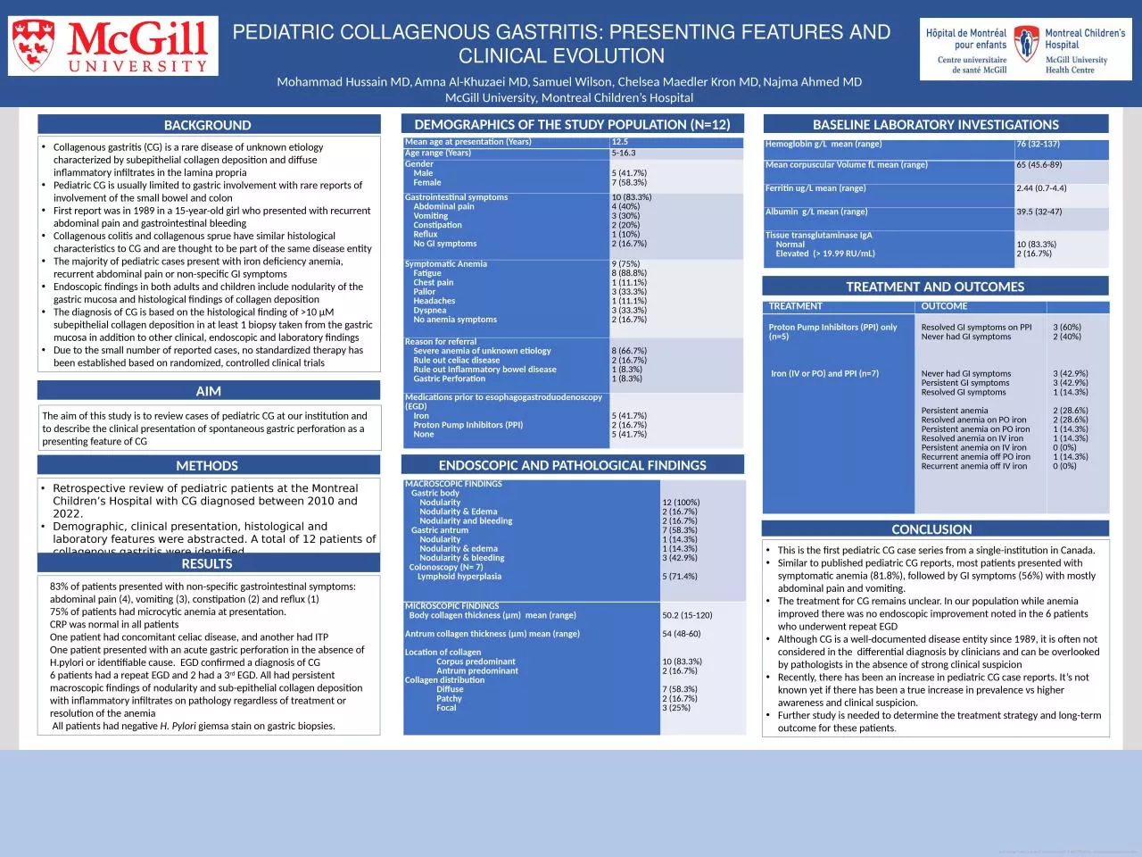
Mohammad Hussain MD Amna Al Khuzaei MD Samuel Wilson Chelsea Maedler Kron MD Najma Ahmed MD McGill University Montreal Childrens Hospital Collagenous gastritis CG is a rare disease of unknown etiology characterized by subepithelial collagen deposition and diffuse ID: 1036728

patients anemia pediatric gastric anemia patients gastric pediatric collagen clinical range nodularity collagenous symptoms iron abdominal presentation egd treatment

Share:

Link:

Embed:

Download Presentation from below link

Download Presentation The PPT/PDF document "PEDIATRIC COLLAGENOUS GASTRITIS: PRESENT..." is the property of its rightful owner. Permission is granted to download and print the materials on this web site for personal, non-commercial use only, and to display it on your personal computer provided you do not modify the materials and that you retain all copyright notices contained in the materials. By downloading content from our website, you accept the terms of this agreement.


Presentation Transcript

1. PEDIATRIC COLLAGENOUS GASTRITIS: PRESENTING FEATURES AND CLINICAL EVOLUTIONMohammad Hussain MD, Amna Al-Khuzaei MD, Samuel Wilson, Chelsea Maedler Kron MD, Najma Ahmed MD McGill University, Montreal Children’s Hospital Collagenous gastritis (CG) is a rare disease of unknown etiology characterized by subepithelial collagen deposition and diffuse inflammatory infiltrates in the lamina propriaPediatric CG is usually limited to gastric involvement with rare reports of involvement of the small bowel and colon First report was in 1989 in a 15-year-old girl who presented with recurrent abdominal pain and gastrointestinal bleeding Collagenous colitis and collagenous sprue have similar histological characteristics to CG and are thought to be part of the same disease entity The majority of pediatric cases present with iron deficiency anemia, recurrent abdominal pain or non-specific GI symptomsEndoscopic findings in both adults and children include nodularity of the gastric mucosa and histological findings of collagen deposition The diagnosis of CG is based on the histological finding of >10 µM subepithelial collagen deposition in at least 1 biopsy taken from the gastric mucosa in addition to other clinical, endoscopic and laboratory findings Due to the small number of reported cases, no standardized therapy has been established based on randomized, controlled clinical trials BACKGROUND AIMRetrospective review of pediatric patients at the Montreal Children’s Hospital with CG diagnosed between 2010 and 2022. Demographic, clinical presentation, histological and laboratory features were abstracted. A total of 12 patients of collagenous gastritis were identified.METHODS This is the first pediatric CG case series from a single-institution in Canada. Similar to published pediatric CG reports, most patients presented with symptomatic anemia (81.8%), followed by GI symptoms (56%) with mostly abdominal pain and vomiting. The treatment for CG remains unclear. In our population while anemia improved there was no endoscopic improvement noted in the 6 patients who underwent repeat EGDAlthough CG is a well-documented disease entity since 1989, it is often not considered in the  differential diagnosis by clinicians and can be overlooked by pathologists in the absence of strong clinical suspicionRecently, there has been an increase in pediatric CG case reports. It’s not known yet if there has been a true increase in prevalence vs higher awareness and clinical suspicion.Further study is needed to determine the treatment strategy and long-term outcome for these patients.CONCLUSION The aim of this study is to review cases of pediatric CG at our institution and to describe the clinical presentation of spontaneous gastric perforation as a presenting feature of CGDEMOGRAPHICS OF THE STUDY POPULATION (N=12)BASELINE LABORATORY INVESTIGATIONSMean age at presentation (Years)        12.5 Age range (Years) 5-16.3 Gender    Male     Female  5 (41.7%)7 (58.3%)Gastrointestinal symptoms     Abdominal pain    Vomiting     Constipation    Reflux     No GI symptoms10 (83.3%)4 (40%)3 (30%)2 (20%)1 (10%)2 (16.7%)Symptomatic Anemia     Fatigue    Chest pain    Pallor     Headaches    Dyspnea    No anemia symptoms 9 (75%) 8 (88.8%)1 (11.1%)3 (33.3%)1 (11.1%)3 (33.3%)2 (16.7%)Reason for referral     Severe anemia of unknown etiology    Rule out celiac disease    Rule out Inflammatory bowel disease         Gastric Perforation8 (66.7%)2 (16.7%)1 (8.3%)1 (8.3%)Medications prior to esophagogastroduodenoscopy (EGD)    Iron    Proton Pump Inhibitors (PPI)     None5 (41.7%)2 (16.7%)5 (41.7%)RESULTS 83% of patients presented with non-specific gastrointestinal symptoms: abdominal pain (4), vomiting (3), constipation (2) and reflux (1)75% of patients had microcytic anemia at presentation.CRP was normal in all patientsOne patient had concomitant celiac disease, and another had ITPOne patient presented with an acute gastric perforation in the absence of H.pylori or identifiable cause. EGD confirmed a diagnosis of CG6 patients had a repeat EGD and 2 had a 3rd EGD. All had persistent macroscopic findings of nodularity and sub-epithelial collagen deposition with inflammatory infiltrates on pathology regardless of treatment or resolution of the anemia All patients had negative H. Pylori giemsa stain on gastric biopsies. Hemoglobin g/L  mean (range)76 (32-137)Mean corpuscular Volume fL mean (range)    65 (45.6-89)Ferritin ug/L mean (range)   2.44 (0.7-4.4)Albumin  g/L mean (range)39.5 (32-47)Tissue transglutaminase IgA      Normal      Elevated  (> 19.99 RU/mL)10 (83.3%)2 (16.7%)ENDOSCOPIC AND PATHOLOGICAL FINDINGSMACROSCOPIC FINDINGS    Gastric body       Nodularity        Nodularity & Edema       Nodularity and bleeding   Gastric antrum        Nodularity        Nodularity & edema       Nodularity & bleeding   Colonoscopy (N= 7)      Lymphoid hyperplasia 12 (100%)2 (16.7%)2 (16.7%)7 (58.3%)1 (14.3%)1 (14.3%)3 (42.9%)5 (71.4%)MICROSCOPIC FINDINGS   Body collagen thickness (μm)  mean (range)            Antrum collagen thickness (μm) mean (range)               Location of collagen                Corpus predominant                Antrum predominant Collagen distribution                Diffuse               Patchy               Focal 50.2 (15-120)54 (48-60)10 (83.3%)2 (16.7%)7 (58.3%)2 (16.7%)3 (25%)TREATMENT AND OUTCOMESTREATMENT OUTCOME  Proton Pump Inhibitors (PPI) only (n=5)    Iron (IV or PO) and PPI (n=7)  Resolved GI symptoms on PPINever had GI symptoms   Never had GI symptomsPersistent GI symptoms Resolved GI symptoms Persistent anemia Resolved anemia on PO ironPersistent anemia on PO iron Resolved anemia on IV ironPersistent anemia on IV iron Recurrent anemia off PO iron Recurrent anemia off IV iron 3 (60%)2 (40%)   3 (42.9%)3 (42.9%)1 (14.3%) 2 (28.6%)2 (28.6%)1 (14.3%)1 (14.3%)0 (0%)1 (14.3%)0 (0%)