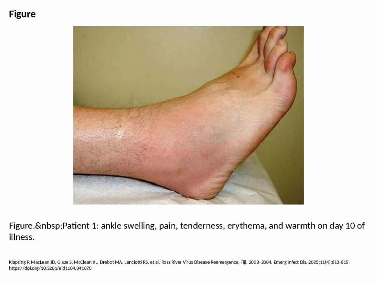
PPT-Figure Figure. Patient 1: ankle swelling, pain, tenderness, erythema, and warmth

Author : yahya | Published Date : 2024-10-04

Klapsing P MacLean JD Glaze S McClean KL Drebot MA Lanciotti RS et al Ross River Virus Disease Reemergence Fiji 20032004 Emerg Infect Dis 2005114613615 httpsdoiorg103201eid1104041070

Presentation Embed Code

Download Presentation

Download Presentation The PPT/PDF document "Figure Figure. Patient 1: ankle..." is the property of its rightful owner. Permission is granted to download and print the materials on this website for personal, non-commercial use only, and to display it on your personal computer provided you do not modify the materials and that you retain all copyright notices contained in the materials. By downloading content from our website, you accept the terms of this agreement.

Figure Figure. Patient 1: ankle swelling, pain, tenderness, erythema, and warmth: Transcript


Klapsing P MacLean JD Glaze S McClean KL Drebot MA Lanciotti RS et al Ross River Virus Disease Reemergence Fiji 20032004 Emerg Infect Dis 2005114613615 httpsdoiorg103201eid1104041070. Sarah Rayner. Extended Scope Practitioner Physiotherapist. Anatomy. The ankle and foot is a complex structure comprised of 28 bones (including 2 sesamoid bones) and 55 articulations (including 30 synovial joints), interconnected by ligaments and muscles. injuries. SECTA. Sports Medicine. Common Injuries of the . Foot & Ankle. Ankle sprains:. The most common injury. Mostly due to excessive inversion and plantar flexion. Most commonly injured ligament is the anterior talofibular ligament. Tyler Beauchamp. Definitions. Cry/o/therapy – “cry” meaning cold. ROM – range of motion. MMT – manual muscle testing. RTP – return to play. Edema – swelling. Inversion – foot rolls in. Notes Quiz. 1. What are the long bones of the foot called?. Notes Quiz. 2. What are the three arches of the foot?. Notes Quiz. 3. What is the ligament on the medial side of the ankle?. Notes Quiz. Bony anatomy. Tibia. Medial . mallelous. Fibula. Lateral . mallelous. Talus. Calcaneous. Soft Tissue Anatomy. Ligaments. Lateral aspect. Anterior . tibiofibular. Posterior . tibiofibular. Anterior . talofibular. Presented by:. Jonathan Reynolds, PT, PhD. 1/30/19. 1. 22. nd. Annual Orthopaedic and Sports Medicine Conference . Diagnostic Excellence. Detailed history. Thorough examination. Radiology dependence. There are 28 bones in the lower leg/foot including:. Tibia & Fibula: Malleolus – (medial & lateral) ends of the tibia & fibula (commonly referred to as the ankle bones). Talus: sits on top of the calcaneus (forms the ankle joint along with tibia & fibula). What to Expected after Ligament Repair Surgery149 Outpatient Day Surgery149 Splint for 1 week149 Boot for 4-6 weeks149 Brace when walking until 12 weeks149 Physical therapy begins after boot comes o29 Most common lower leg injury. Classified as to degree of . ligamentous. severity. Lateral. ankle sprain . Mechanism:. Inversion and plantar flexion. Anterior . talofibular. ligament (ATF) usually first structure to injury. . Assessing the Knee Joint. Determining the mechanism of injury is critical. History- Current Injury. Past history. Mechanism- what position was your body in?. Did the knee collapse?. Did you hear or feel anything?. What are the aims of this leaflet? This leaflet has been written to help you understand more about erythema multiforme. It tells you what it is, what causes it, what can be done about it, and where What is Ankle Osteoarthritis?. What are the causes?. What are the symptoms?. How is it diagnosed?. What is the management?. How can I manage it?. What . is Ankle . Osteoarthritis?. Arthritic or degenerative changes within the ankle joint.. HOPS. History. Observation. Palpation . Special Tests. Evaluation of the ANKLE. History. MAPPS. M = . Mechanism. of injury. A = . Acute. or chronic. P = . Previous. . history. of injury. P = . Pain. What are the causes?. What are the symptoms?. How is it diagnosed?. What is the management?. How can I manage it?. What is an ankle sprain?. Ankle sprain is characterised by an injury to at least one of the ligaments supporting your ankle. .

Download Document

Here is the link to download the presentation.
"Figure Figure. Patient 1: ankle swelling, pain, tenderness, erythema, and warmth"The content belongs to its owner. You may download and print it for personal use, without modification, and keep all copyright notices. By downloading, you agree to these terms.

Related Documents