/
Female reproductive anatomy Female reproductive anatomy

Female reproductive anatomy - PowerPoint Presentation

yoshiko-marsland
yoshiko-marsland . @yoshiko-marsland
Follow
421 views
Uploaded On 2017-03-18

Female reproductive anatomy - PPT Presentation

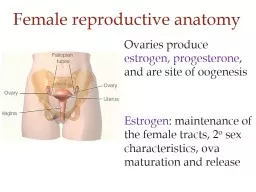
Ovaries produce estrogen progesterone and are site of oogenesis Estrogen maintenance of the female tracts 2 o sex characteristics ova maturation and release Oogonia divide mitotically ID: 525913

phase estrogen follicular progesterone estrogen phase progesterone follicular cells embryo oocyte follicle endometrium fsh uterus corpus contractions levels placenta

Share:

Link:

Embed:

Download Presentation from below link

Download Presentation The PPT/PDF document "Female reproductive anatomy" is the property of its rightful owner. Permission is granted to download and print the materials on this web site for personal, non-commercial use only, and to display it on your personal computer provided you do not modify the materials and that you retain all copyright notices contained in the materials. By downloading content from our website, you accept the terms of this agreement.


Presentation Transcript

Slide1

Female reproductive anatomy

Ovaries produce estrogen, progesterone, and are site of oogenesisEstrogen: maintenance of the female tracts, 2o sex characteristics, ova maturation and releaseSlide2

Oogonia

divide mitotically Meiosis I produces a primary oocyte (diploid), surrounded by follicle cells These oocytes develop into secondary oocytes on a cyclical basis

Oogenesis overviewSlide3

Follicular phase

- first half of cycle when follicles mature and are ovulatedLuteal phase - second half of cycle when uterus is prepared for implantationOvarian cycleSlide4

Primary follicle

oocyte

Follicle cells

Looking within the ovary…follicular phase

FSH and LH levels are increasingSlide5

Follicle cells secrete estrogen

Dividing

follicular cells

Thecal cells

Primary

oocyte

Looking within the ovary…follicular phaseSlide6

antrum

Antrum collects fluid with estrogen

Estrogen inhibits FSH and LH, so FSH

Looking within the ovary…follicular phaseSlide7
Slide8

Thecal cells

Granulosa cells

Antrum

primary oocyte

Late follicular phase

High estrogen levels promote LH secretion w/ positive feedback loopSlide9
Slide10

Late follicular phase

Mature

follicle

Antrum

Oocyte finishes meiosis I

it now is a 2

o

oocyteSlide11

Follicle

remains

secondary oocyte

Egg is flushed out

Pop! Ovulation!Slide12

corpus

luteum

Luteal phase

Corpus luteum secretes

progesterone

and estrogenSlide13

Estrogen and progesterone

Estrogen initiates preparation of endo- and myometrium, ‘prime’ uterus for progesterone (follicular phase)Progesterone endometrium vascularization, glycogen, decrease contractions (luteal phase)

Progesterone

inhibits LH and FSH

(this is how birth control pills work)Slide14

If the corpus luteum degenerates, progesterone drops and menstruation occurs

Changes in endometrium

Degenerating

corpus luteumSlide15
Slide16

Fertilization normally occurs within a day of ovulation

Contractions of the myometrium help some sperm reach the oviduct

But if there are sperm around…

acrosome

in actionSlide17

Embryo

Trophoblast

Implants the embryo

Blastocyst

Cleavage

Fertilization

Ovulation

Implantation

Embryo forms before reaching the uterusSlide18

Blastocyst secretes

chorionic gonadotropinEndometrium has glycogen, and becomes more vascularized from progesterone40% of blastocysts never implant

Settling into the uterus…Slide19

Trophoblast

enzymes digest proteins of the endometrium. This carves a hole for implantation

.

Settling into the uterus…

Endometrium

Trophoblast

Embryo

Embryo

EndometriumSlide20

Embryo

Eventually forms

chorionSlide21

Placenta =

chorion from embryo and uterine lining from mother. These tissues interlock like ‘fingers’ Projections of chorion have capillaries to form placental villi. They extend into the mother’s blood.

Placenta developmentSlide22

Gasses, wastes, nutrients diffuse

bw capillaries of mother and fetusDrugs, pollutants, chemicals also diffuse

Placenta development

mother’s blood

Placental

villus

Amniotic sac

ChorionSlide23

Human chorionic gonadotropin (hCG)

Estrogen

Progesterone

Fertilization

DeliverySlide24

Questions

Is reproduction different for a female with a single ovary?Abortion via chemical meansDoes scoliosis affect the ability to carry/deliver a child?Does the fact that female genitals are inside the body allow for wider range of lower body movement? Athletes want to knowSlide25

Relaxin

from placenta softens the cervix and pelvisUterus has mild contractions. Baby’s head is down in pelvisHigh levels of estrogen make the uterus more sensitive to oxytocin. Progesterone levels drop

Getting ready for birth....Slide26

Stretching the cervix causes more oxytocin to be released

Oxytocin induces stronger contractionsPositive feedback with oxytocinSlide27

LactationSlide28

If students are interested, I will go over the extra credit information at the end of this class.I will need to hear some feedback via email to know if students are interested in this so I’ll prepare extra slides…thanks