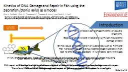
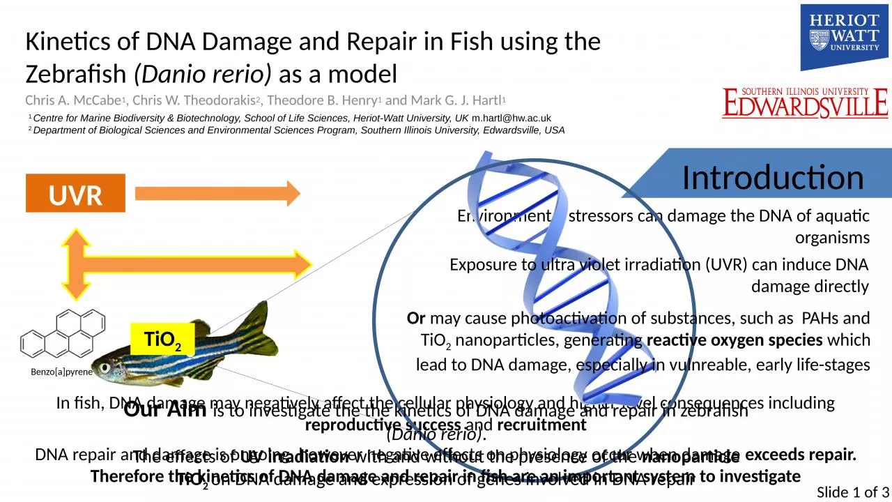
Danio rerio as a model Chris A McCabe 1 Chris W Theodorakis 2 Theodore B Henry 1 and Mark G J Hartl 1 Introduction Environmental stressors can damage the DNA of aquatic organisms ID: 932040
Download Presentation The PPT/PDF document "Kinetics of DNA Damage and Repair in Fis..." is the property of its rightful owner. Permission is granted to download and print the materials on this web site for personal, non-commercial use only, and to display it on your personal computer provided you do not modify the materials and that you retain all copyright notices contained in the materials. By downloading content from our website, you accept the terms of this agreement.
Slide1
Kinetics of DNA Damage and Repair in Fish using the Zebrafish
(Danio rerio) as a model
Chris A. McCabe1, Chris W. Theodorakis2, Theodore B. Henry1 and Mark G. J. Hartl1
Introduction
Environmental stressors can damage the DNA of aquatic organisms
In fish, DNA damage may negatively affect the cellular physiology and higher-level consequences including reproductive success and recruitment
1 Centre for Marine Biodiversity & Biotechnology, School of Life Sciences, Heriot-Watt University, UK m.hartl@hw.ac.uk2 Department of Biological Sciences and Environmental Sciences Program, Southern Illinois University, Edwardsville, USA
Exposure to ultra violet irradiation (UVR) can induce DNA damage directly
Or
may cause
photoactivation
of substances, such as PAHs and TiO
2
nanoparticles
, generating
reactive oxygen species which lead to DNA damage, especially in vulnreable, early life-stages
Benzo
[a]
pyrene
TiO
2
UVR
Our Aim
is to investigate the the kinetics of DNA damage and repair in zebrafish (Danio rerio).The effects of UV irradiation with and without the presence of the nanoparticle TiO2 on DNA damage and expression of genes involved in DNA repair
DNA repair and damage is ongoing, however negative effects on physiology occur when damage exceeds repair.Therefore the kinetics of DNA damage and repair in fish are an important system to investigate
Slide 1 of 3
Slide2Methods
and Optimisations
A Comet Assay method was developed to identify and assess DNA Damage and Repair
Slide 2 of 3
Individual Larvae (72 hr post- fertilisation) were exposed to UVR with the aim to damage the DNA and assess repair.
Comet Head Start
Comet Nucleus
Comet Tail End
Spectromicroscopy
and specialised software is used to assess the comet cells from the assay
A comet cell with tail, indicating damage and subsequent loss of DNA fragments from the cell nucleus.
DNA Damage was assessed by the intensity of Comet ‘tails’- a quantification of the loss of fragmented DNA from the cell post-damage
Repair was quantified by the disappearance of damage, and a reduction in this measure of intensity
The differences in DNA damage were compared between treatments in order to
optimise
the variables within the experiment.
Electrophoresis Duration
Optimisations
have included:
Ultraviolet Radiation Types
Damage Specific Enzyme dilutions
-
Formamidopyrimadine
glycosyase
(
FaPy
): Makes single-strand nicks wherever there is an oxidized
purine
or an
abasic
site.
-
Endonuclease
III
(
EndoIII
): Makes single-strand nicks wherever there is an oxidized
pyrimidine
- T4
pyrimadine
dimer
glycosylase
(PDG)
(Shown)
Makes single-strand nicks wherever there is a UV photoproduct
UV Optimisation
Larvae were exposed to 10 J/cm2 UVA or 0.333 J/cm2 UVC. Larvae were subjected to mixture of
Fpg
(1:400) and
Endonuclease
III (1:100) enzymes in buffer dilution to detect oxidized bases. The results show that both UVA and UVC induced oxidised base DNA damage. Although the amount of UVC damage was lower, the dose was also significantly lower. Therefore UVC may be more effective in causing base oxidative damage than UV-A. (n=2 in all treatments)
Electrophoresis Duration
Assessment of
dimer
-specific damage to different electrophoresis times. Graph depicts the difference between samples that were incubated with and without T4 PDG enzyme (1:80 dilution). Larvae were either exposed to 1 min UVC (0.333 J/cm2) (red) or unexposed (blue). The relative difference between control and exposed larvae was greatest at 35 min. (n= 2 for all treatments)
Enzyme Dilutions
In this optimisation larvae were exposed to 1 min UVC (0.333 J/cm2) and treated with various T4 PDG enzyme dilutions in buffer (5x PDG buffer). Slides were incubated with 50
uL
of enzyme dilutions. From the results, the optimal appears at 1:100. At higher dilutions fewer
dimers
were detected in exposed fish; while at 1:50 dilution, there were more apparent “
dimers
” in the control. This may be due to non-specific cutting of the DNA by the enzyme if the concentration is too high. (n=2 in all treatments)
Slide3Conclusions
A method to assess DNA Damage in larval fish was established, using
Danio
rerio as a model.
Slide 3 of 3
Initial experimental results investigating a
recovery or repair period, post-exposure, suggest the amount of DNA damage decreases in proportion to recovery time. Most damage was observed to be repaired after a 1- hour period.Future StudiesAim to
analyse the kinetics of expression of genes involved in DNA repair
using qRT
-PCR in fish exposed to photoactivated TiO2 nanoparticles to establish the potential effects of this substance in the environment.
Thank You for watching
AcknowledgementsI would like to thank Heriot-watt Univeristy and for the use of the Centre for Marine Biodiversity & Biotechnology for experimentation,
Mihalis Panagiotidis for the use of scientific equipment, and Jade Middlemiss for her tireless help in lab work and comet scoring.
I would like to take this opportunity to thank my Co-Authors:Dr. Theodore Henry, Dr. Chris Theodorakis, and Dr. Mark Hartl for their continuous support and guidance.
http
://markhartl.sls.hw.ac.uk/