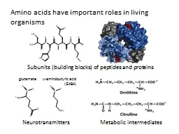
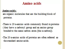
Building blocks for peptides proteins Some individually important or converted to important molecules Gly Glu Tyr neurotransmitters Tyr parentprecursor for epinephrine adrenaline ID: 933705
Download Presentation The PPT/PDF document "Amino Acids General Amino Acids:" is the property of its rightful owner. Permission is granted to download and print the materials on this web site for personal, non-commercial use only, and to display it on your personal computer provided you do not modify the materials and that you retain all copyright notices contained in the materials. By downloading content from our website, you accept the terms of this agreement.
Slide1
Amino Acids
General
Slide2Amino Acids:
Building blocks for peptides, proteins
Some individually important (or converted to important molecules)
Gly
,
Glu
, Tyr
neurotransmitters
Tyr
parent/precursor
for epinephrine (adrenaline)
His stomach secretes
HCl
, symptoms for inflammation, colds.
Essential (10)
needed for normal health
not synthesized by the body
must be supplied by diet
Complete (animal) vs. Incomplete (vegetable) protein
Slide3Amino Acids
Structure
Slide4Amino Acid Structure:
Amide, CA, R-group (variable)
D/L Isomers
Slide5Amino Acids
Side Chains
Slide6AA – Side Chains:
Side chains determine the functionality of the AA b/c the –COOH and –NH
2
groups react to form the backbone
3 letter abbreviations (given on cheat sheet)
Classification
Functional Group
Property
Nonpolar
-R (aliphatic or aromatic)
Hydrophobic
Polar
-COOH, -NH
2
, -OH
Hydrophilic
Acidic
-COOH (extra)
Lose H
2
anion Salt
Bridges
Basic
-NH
2
(extra)
Gain H
2
cation
Salt
Bridges
Slide7Zwitterion
Slide8Zwitterion:
dipolar form of AA, found at biological pH’s (internal acid/base
Rxn
)
Slide9Amphoteric
Slide10Amphoteric:
molecules with properties
of both acid and base
Slide11Formation of
Polypeptides
Slide12Formation Reaction:
Dehydration reaction
CA + Amine
Amide
Amide structure/Peptide bond/Peptide linkage
Slide13Amide/Peptide
Bonds
Slide14Slide15Polypeptides
Slide16Polypeptides:
Small chains of AA (40-50 units)
Many ways to connect together (N!)
~30 biologically relevant ones
Hormones or Nerve transmitters
Small changes structure
HUGE changes in functionality
Slide17Protein
Structure
Slide18Proteins – General:
> 50 AA
Linus Pauling – 1954 Nobel Prize
α-helix and β-pleated sheetFredrick Sanger – 1958 Primary structure of beef insulin
Classification
Description
Examples
Primary
#, kind, type and sequence of AA
Secondary
Regular
3D structure, held together by H-bonds in backbone
α
-helix
β
-pleated sheet
triple helix
Tertiary
Distinct
3D structure due to interactions between R-groups
H-bonds
Ionic bonds (Salt
Bridges)
Disulfide bonds
Hydrophobic
Hydrophilic
Quaternary
Complex
proteins
Multiple units
Non-protein parts
Metal ions
Slide19Primary Structure
Slide20Primary Structure:
#, kind, type, and sequence of AA
Fredrick Sanger (1958 Nobel Prize) Beef Insulin
Several years of work to sequence 51 AA
Hydrolyzed proteins into smaller fragments to analyze
Edman
Degradation – split AA at N-Terminal End
Gly
-
Glu
-
Arg
-
Gly
-
Phe
-
Phe
-Tyr-
Thr
-Pro-Lys
Gly
-
Phe
-
Phe
-Tyr-
Thr
-Pro-Lys
Gly-Glu-Arg-Gly-Phe-Phe
-
Fragment 1:
Combined:
Fragment 2:
Overlap
Slide21Secondary Structure
Slide22Secondary Structure:
Determined by
H-bonds
between AA-backbone
α
-helix
AA 4 residues apart, R-groups towards outside
β
-pleated sheet
AA far apart, R-groups face outwards
Slide23Tertiary Structure
Slide24Tertiary Structure:
Determined by interactions between R-groups
H-bonds: -COOH and –OH
Ionic/Salt Bridges
Disulfide Bonds
Hydrophobic (form core of protein)
Hydrophilic (face outwards to interact with water)
Slide25Quaternary Structure
Slide26Quaternary Structure:
Multiple protein units
Non-protein parts
Metal ions
Ex: Hemoglobin4 subunits
Fe atoms
Slide27Protein Structure
Summary
Slide28Slide29α
-Helix
Slide30α
-helix Structure
:
Secondary
Determined
by
H-bonds
between
AA-backbone
α
-helix
AA 4 residues apart, R-groups towards outside
Slide31β
-Pleated
S
heet
Slide32β
-pleated sheet
s
tructure
:Secondary
Determined
by
H-bonds
between
AA-backbone
β
-pleated sheet
AA far apart, R-groups face outwards
Slide33H-bonds
Slide34Secondary H-bonds:
Between the C=O and NH of backbone
Responsible for secondary structure
Tertiary H-bonds:
Between the C=O and -NH or -OH of R-groups
Responsible for tertiary structure
Slide35Salt Bridges
Slide36Ionic Bonds/Salt Bridges:
Tertiary Structure
Between –COO
-
and –NH3+ groups
Slide37Disulfide Bonds
Slide38Disulfide bonds:
Tertiary Structure
Between -SH and –SH groups
Mainly between
Cys-Cys
Slide39Hydrophobic
Interactions
Slide40Hydrophobic Interactions:
Tertiary Structure
Between –R groups (Alkane and Aromatic)
Interior of proteins to avoid water
Slide41Hydrophilic
Interactions
Slide42Hydrophilic Interactions:
Tertiary Structure
E
xterior
of proteins to
interact with water
Polar groups (OH)
Acidic groups (COOH)
Basic groups (NH
2
)
Slide43Identify 2°/3°
Structure
Slide44Slide45Protein Functions
Slide46Protein Functions:
Structural Support – skin, connective tissue
Storage – Fe in Liver
Transport – O
2 in HemoglobinDefense – antibodies, venomMotion/Movement – muscles
Regulation – blood/glucose/insulin
Catalysis – Enzymes (Ch. 30!)
Slide47Denaturation
Slide48Denaturation:
Loss of 3D conformation in a protein
Disruption of 2°/3°/4° interactions
Does NOT break 1° structure (hydrolysis)
Loss of biological activityCauses of Denaturation
Cause
Example
1.
Heat
Cooking
2.
Acids/Base (pH)
Lactic Acid
3.
Organic Molecules
Ethanol/Isopropanol
4.
Heavy Metals
Pb
, Hg
5.
Agitation
Stirring
6.
UV Light
7.
Enzymes
Digestion
8.
Salts
Water purification
Slide49Xanthoproteic
Test
Slide50Xanthoproteic
Test:
Detects Benzene rings
Yellow color
Phe, Try, Tyr
Slide51Biuret
Test
Slide52Biuret Test:
Detects tri-peptides (must have at least 2 peptide bonds)
Cu
2
SO4Violet color
Slide53Ninhydrin
Test
Slide54Ninhydrin
Test:
General test for AA
All AA
bluePro, hydroxyproline yellow
Very sensitive 1
μ
g (10
-6
)
Slide55Chromatography
Slide56Chromatography:
separation technique for AA
Difference in distribution between two phases
○ Solubility
○ ChargeTLC (thin-layer) – solid/liquid phase ○ Solvent Front (rate solvent moves) ○ Differences in
solubility
cause AA to
travel at different rates in the solvent
Column chromatography (variation of above)
Slide57Electrophoresis
Slide58Electrophoresis:
separation technique for AA
Charged particles separate in electric field (zwitterions)
Separation based on
○ Size – friction (sieve) ○ Charge – electric fieldTypes
○ SDS – masks charge/separate by mass/size
○ Isoelectric Focusing – AA separated by charge
○ 2D – separate on both.
Slide59Fredrick Sanger
Slide60Fredrick Sanger:
Solved structure of beef
insulin (1955)
Nobel prize 1958
51 AA in two chains held together by disulfide bondsDFNB + N-terminal end + hydrolysis to solve structure“Paper shredder”