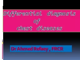
Differential diagnosis of chest diseases Micronodular miliary disease TB Histoplasmosis Chicken box Sarcoidosis LCH Pneumoconiosis Alveolar microlithiasis Metastasis ID: 935910
Download Presentation The PPT/PDF document "Dr Ahmed Refaey , FRCR" is the property of its rightful owner. Permission is granted to download and print the materials on this web site for personal, non-commercial use only, and to display it on your personal computer provided you do not modify the materials and that you retain all copyright notices contained in the materials. By downloading content from our website, you accept the terms of this agreement.
Slide1
Dr Ahmed Refaey , FRCR
Differential diagnosis
of
chest diseases
Slide2Micronodular(miliary)disease TBHistoplasmosisChicken boxSarcoidosisLCHPneumoconiosisAlveolar microlithiasisMetastasis
Slide3Septal thickeningBeaded / nodular * lymphangitic carcinomatosa * sarcoidosis * silicosisSmooth *
lymphangitisc
carcinomatosis
* pulmonary edema
* alveolar
proteinosis
Slide4Bilateral perihilar air-space diseasePulmonary edemaPulmonary hemorrhageAlveolar proteinosisPCP
Slide5Acute alveolar infiltratePulmonary edemaPneumoniaPulmonary hemorrhageAspiration
Slide6Chronic alveolar diseaseAlveolar cell carcinomaAlveolar sarcoidosisLymphomaAlveolar proteinosis
Slide7Unilateral hyperlucent lungObstructive emphysemaMcleoid syndrome ( Swyer James syndrome)PneumothoraxPulmonary embolismBullous diseaseAir trapping e.g. FB , massPoland syndrome
Slide8Multiple lung nodules (macronodules)MetastasisWegner’s granulomatosisRheumatoid n nodulesAVMsSeptic emboli
Slide9Small cavitating lung lesionsSeptic emboliRheumatoid nodulesWegner’s granulomatosisSq. or transitional cell ca metastasis
Slide10Upper lober disease( CASSET P )Cystic fibrosisAnkylosing spondylitisSarcoidosisSilicosisEsinophilic
granuloma
PCP
Slide11Basilar interstitial lung disease ( BADASS )BronchiectasisAspiration Drugs / DIP-UIPAsbestosisSickle cell diseaseScleroderma
Slide12Masses with air bronchogramLymphomaAlveolar cell carcinomaPseudolymphoma ( maltoma )
Slide13Anterior mediastinal massThymic tumorsSubsternal ThyroidLNsGerm cell tumor
Slide14Slide15Middle mediastinal massLNsAneurysmEsophageal duplicationBronchogenic cyst
Slide16Posterior mediastinal massNeurogenic tumorsParaspinal hematomaParaspinal abscessExtraskeletal hematopoiesisLNs enlargementLateral meningeocele
Slide17Opacified hemithoraxAtelectasisPneumoniaPleural effusionPost pneumonectomy
Slide18PneumomediastinumRuptured esophagusRuptured trachea / bronchusIatrogenicBroncheal asthmapneumoperitoneum
Slide19Pleural calcification Old TB empyemaAsbestos exposureHemithorax
Slide20Cardiopherenic angle massPericardial cystDiaphragmatic hernia ( Morgagni )Lung sequestration Pericardial fat bad
Slide21Unilateral pulmonary edemaAspirationDisease in other lung e.g. COPDPosturalRapid expansion pulmonary edema “ Look for ICT”
Slide22Long segment tracheal narrowingTracheopathia osteoplasticaTracheo-broncheal amyloidosisWegner’s granulomatosisIntubation injuryTracheobronchomalaciaRelapsing polychondritisPapillomatosis ( extensive )
Slide23Peripheral distributed lung diseaseEsinophilic pneumonia --- upper lobesBOOP ----- lower lobesHypersensitivity pneumonitisMultiple infarctsDIPAlveolar cell carcinoma
Slide24Bronchiectasis CT criteria of bronchiectasis1- lack of tapering of bronchi2-internal diameter of bronchus larger than the adjacent artery3- bronchi are visible within 1 cm of pleura4- mucus-filled , dilated bronchi
Slide25Bronchiectasis Chronic airway infectionPulmonary fibrosis –traction bronchiectasisABPA ------- central bronchiectasisCystic fibrosis ------- apical bronchiectasisMounier-Kuhn syndrome – tracheobronchomegallyKartagner syndrome --- situs inversus + sinusitis
Slide26Mounier-Kuhn syndrome
Slide27ABPA
Slide28Cystic fibrosis
Slide29Kartagener’s syndrome
Slide30Dilated esophagusSclerodermaAchalasiaObstructing tumorDrugs ( atropine)Surgury ( gastric pull-up)