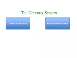
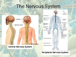
J Matthew Velkey PhD mattvelkeydukeedu 452A Davison Duke South Cardiac amp smooth muscle amp glands Skeletal muscle Structural Organization of the Nervous System Motor nerve inputs ID: 931553
Download Presentation The PPT/PDF document "Histology of the Peripheral Nervous Syst..." is the property of its rightful owner. Permission is granted to download and print the materials on this web site for personal, non-commercial use only, and to display it on your personal computer provided you do not modify the materials and that you retain all copyright notices contained in the materials. By downloading content from our website, you accept the terms of this agreement.
Slide1
Histology of the Peripheral Nervous System
J. Matthew Velkey
,
Ph.D
.
matt.velkey@duke.edu
452A Davison, Duke South
Slide2Cardiac & smooth muscle & glands
Skeletal muscle
Structural Organization of the Nervous System
Motor nerve
inputs
outputs
special senses
Slide3Functional Organization of the Nervous System Somatic (conscious afferent* and efferent, voluntary motor control)
Autonomic
(unconscious efferent, involuntary motor control of internal organs to maintain homeostasis)
Sympathetic
– thoracolumbar division
Parasympathetic –
craniosacral division
* Somatic afferents = sensory fibers from skin, muscle, joints, tendons.
Visceral afferents = sensory fibers from visceral organs; some result in conscious sensations, but others do not. However, they are not considered part of the autonomic nervous system, which is entirely efferent.
Slide4Spinal cord, DRG, Sympathetic and Parasympathetic Ganglia
Netter pl. 154
Dorsal horns:
Interneurons
Ventral horns:
Motor neurons
Lateral horns:
Sympathetic neurons Parasympathetic (S
2-4
) neurons
DRG: Sensory (pseudounipolar) neuronsAutonomic ganglia: Post ganglionic neurons with unmyelinated axons. Sympathetic: paravertebral ganglia Parasympathetic: In organs
X (vagus)
IX
parasympathetic
Somatic motor
sensory
Sympathetic: pre- & post-ganglionic fibers
Pelvic splanchnic (parasympathetic)
Slide5Spinal cord and Dorsal Root Ganglion
Ventral horn
(Lateral horn) if present
Dorsal horn
Dorsal root ganglion (DRG)
Ventral median fissure
Dorsal median sulcus
Slide6Cellular Components of the Nervous System Neurons
Glia
(support cells)
Slide7Generic neuronLarge cell body (aka soma or
perikaryon
)
Large,
euchromatic
nucleus (and usually a prominent nucleolus)Extensive cytoplasmic extensions:
Dendrite(s): single or multiple extensions specialized for receiving inputAxon: single, large extension specialized for conveying output (in humans, can be up to 1.5m in length)
Slide8Motor neuron with Nissl bodies
NU
N
D
D
A
AH
NB
NB
D
V
A-axon D-dendrite N-nucleus NB-
Nissl bodyAH-axon hillock V-blood vessel NU-nucleolus
Slide9Nissl substance is rough endoplasmic reticulum
Slide10Synapses can form between many different parts of neurons and between a neuron and a non-neuronal cell, e.g., a muscle or a secretory cell.
A single neuron can receive activating or inhibiting inputs from thousands of synaptic connections.
Motor neuron cell body in the spinal cord
Slide11At a chemical synapse neurotransmitter release is triggered by the influx of Ca2+ and postsynaptic neurotransmitter receptors receive the signal.
Slide12An example of a synapse:
The
neuromuscular junction
(motor
endplate)
Slide13Conduction velocity in the axon is enhanced by
myelination
axons in the CNS are
myelinated
by
oligodendrocytes
axons in the PNS are
myelinated by Schwann cells
Slide14Myelination is a dynamic process, which involves the ensheathment of the the axon by the glial cell and subsequently the extrusion of cytoplasm from parts of the glial cell. Adhesive proteins on the cytoplasmic and the extracellular side of the plasma membrane contribute to a tight apposition of the lipid bilayers.
Slide15Myelinated Nerve Fiber
Myelin Sheath
The increased lipid content of the myelin sheath provides electrical insulation for the underlying axon.
Slide16Ion channels are concentrated at the nodes of Ranvier and the myelin sheath acts as an electrical insulator. This allows for
saltatory
conductance
of the action potential and increases the transmission speed of the nerve impulse.
Depending on the diameter of the axon, myelination increases the action potential speed approximately 5 to 50fold (up to >110 m/sec).
Nodes of Ranvier
are areas of the
myelinated
axon that are not covered by the myelin sheath.
Slide17Each Schwann cell myelinates a single internode
Internode length can be up to 1.5 mm in the largest nerve fibers
Slide18Nodes of Ranvier in a longitudinal nerve section
Slide19One Schwann cell can ensheath multiple axons, but myelinates only one axon
Slide20Small diameter nerve fibers are non-myelinated
Slide21Longitudinal section of an unmyelinated nerve
Slide22Wavy appearance of nerves
Slide23Connective tissue layers found in nerves:endoneurium surrounds axons,perineurium axon fascicles and epineurium
the entire nerve
Slide24Connective tissue layers in a peripheral nerve. Tight junctions between perineurium cells form a important isolating barrier.
Epineurium
Perineurium
Slide25Three different basic types of neuronal structure
Slide26Sensory Ganglia
Two types
:
spinal (dorsal root)
and
cranial
ganglia associated with spinal and cranial nerves, respectively
Contain large sensory neurons and abundant small glial cells, called
satellite cells
Sensory neurons are pseudounipolar
Slide27Dorsal root ganglion
Dorsal Root (Sensory) Ganglion Cells
Slide28Somatic sensory neurons have components in both CNS and PNS
sensory input
pseudounipolar
sensory neuron in a dorsal root (spinal) ganglion
CNS
PNS
Note that the spinal cord is part of the CNS and therefore does not contain Schwann cells, but rather
oligodendrocytes
.
Slide29Somatic motor neurons of the spinal cord also have components in the CNS and PNS, but they
are
multipolar
Motor output: axon travels through peripheral nerve to reach target muscle
Slide30An example of sensory input:the muscle spindleSpecialized
skeletal muscle fibers enclosed within a spindle-shaped capsule.
D
epolarize
in response to changes in muscle position, tension, and contraction velocity.
Synapse with sensory nerve endings to convey input to CNS
Slide31The somatic nervous system in action:the spinal stretch reflex
stretch receptor
Slide32Efferent autonomic pathways
Parasympathetic = craniosacral
Sympathetic = thoracolumbar
Slide33Pre-ganglionic motor neurons have components in the CNS and PNS and are also multipolar
Visceral motor output to post ganglionic neuron
Slide34Sympathetic ganglion cells:
multipolar neurons that reside entirely within the PNS
in
sympathetic chain ganglia
and
“pre-aortic” ganglia
Slide35Parasympathetic ganglion cells:
multipolar neurons that also reside entirely within the PNS
in the wall
of the innervated organ
(shown here in the seminal vesicle)
Slide36Parasympathetic ganglia in the wall of the gut
#155
Slide37Objectives of PNS Histology:Discuss the general division/differences between CNS and PNS
Appreciate the subdivision into somatic and autonomic nervous system
Learn about the cellular components and the structural attributes of neuronal cells
Discuss synaptic connections, using the motor end plate as an example
Study the formation of the axonal myelin
ensheathment
Compare the histological features of myelinated and
unmyelinated axons/nervesRecognize nerves in histological sectionsIdentify the different connective tissue layers that are associated with nerves
Understand the different organizational plans that are adopted by neuronal cellsIdentify and compare autonomic and sensory ganglia
Learn about the basic histological features of the spinal cord