/
Taking the CARDIOVASCULAR Taking the CARDIOVASCULAR

Taking the CARDIOVASCULAR - PowerPoint Presentation

PeacefulPenguin
PeacefulPenguin . @PeacefulPenguin
Follow
342 views
Uploaded On 2022-07-28

Taking the CARDIOVASCULAR - PPT Presentation

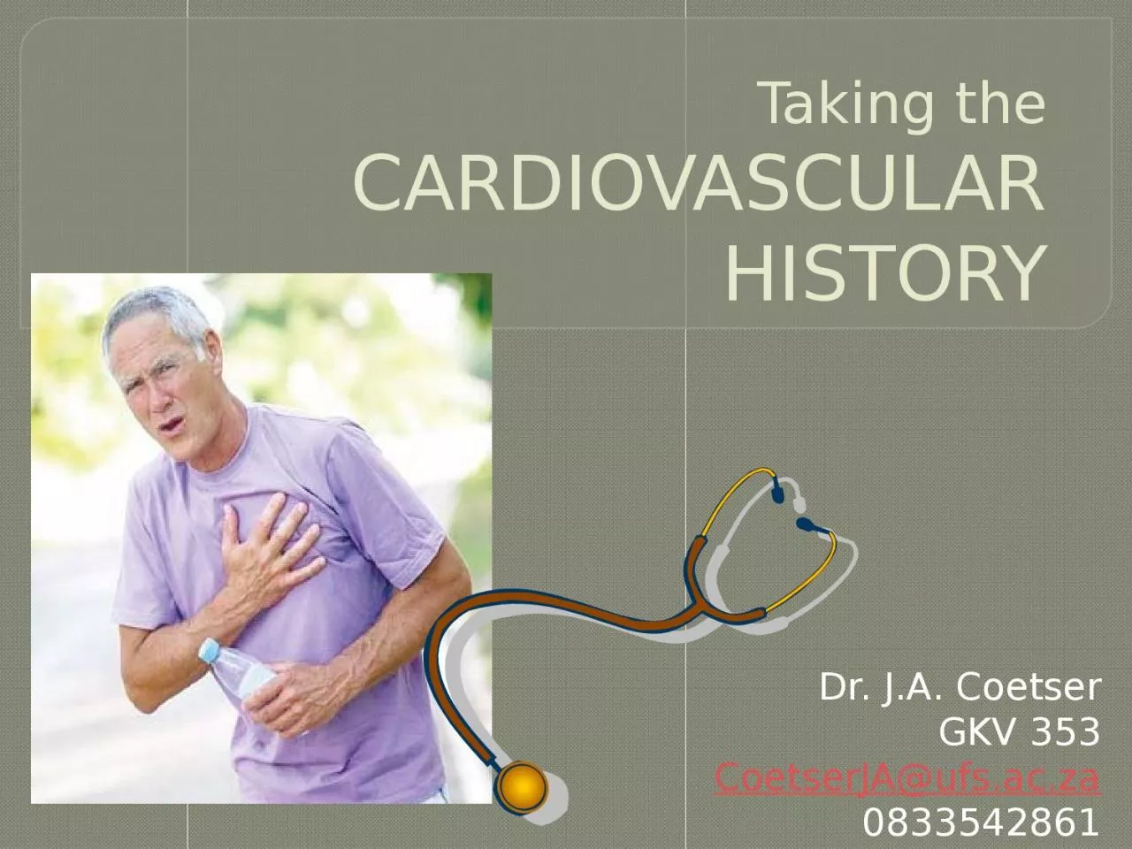
HISTORY Dr JA Coetser GKV 353 CoetserJAufsacza 0833542861 CASE STUDY A 56 year old white male presents to casualties at 3h40am complaining of severe chest pain that started 30min earlier ID: 931243

symptoms presenting chest pain presenting symptoms pain chest dyspnoea angina disease cardiac coronary syncope palpitations oedema claudication heart class

Share:

Link:

Embed:

Download Presentation from below link

Download Presentation The PPT/PDF document "Taking the CARDIOVASCULAR" is the property of its rightful owner. Permission is granted to download and print the materials on this web site for personal, non-commercial use only, and to display it on your personal computer provided you do not modify the materials and that you retain all copyright notices contained in the materials. By downloading content from our website, you accept the terms of this agreement.


Presentation Transcript

Slide1

Taking theCARDIOVASCULARHISTORY

Dr. J.A. CoetserGKV 353CoetserJA@ufs.ac.za0833542861

Slide2

CASE STUDYA 56 year old white male presents to casualties at 3h40am, complaining of severe chest pain that started 30min earlier.

WHICH IMPORTANT ASPECTS WOULD YOU ELICIT FROM THE HISTORY?

Slide3

Presenting symptoms:Chest pain

When evaluating symptomatic complaintsSiteOnsetCharacterR

adiationAlleviating factors

T

iming

E

xacerbating factors

S

everity

Slide4

Presenting symptoms:Chest pain

Determine the cause!4 cardinal featuresDuration (timing)Location (site)Quality (character)Precipitating and aggravating factors

Slide5

Presenting symptoms:Chest pain

AnginaCrushing pain, heaviness, discomfort or choking sensation in retrosternal areaCentral rather than left chestMay radiate to jaw and/or armsRarely below umbilicusTypical vs. atypical angina

Slide6

Typical vs. atypical angina

Typical anginaMeets all 3:Characteristic restrosternal chest discomfort – typical quality and duration

Provoked by exertion or emotionRelieved by rest or GTN or both

Atypical angina

Meets 2 of above

Non cardiac chest pain

Meets 1

or none of above

Slide7

Presenting symptoms:Chest pain

Pain from acute coronary syndromes (myocardial infarction and unstable angina)Often comes on at restPain present >30min

Slide8

Clot dissolves

Coronary blood flow returns No cardiac muscle damage

Clot persists Coronary blood flow cut off

Cardiac muscle dies

UNSTABLE ANGINA

MYOCARDIAL INFARCT

Angina

Acute coronary syndromes

Slide9

Slide10

Presenting symptoms:Chest pain

Pleuritic painDue to movement of pleural surfaces on one anotherInflammation of pleura or pericardiumViral infection of pleuraPneumoniaPulmonary embolismMade worse by inspirationOften relieved by sitting up and leaning forward

Slide11

Presenting symptoms:Chest pain

Dissecting aneurysm3 featuresSevere, tearing painRapid onsetRadiates to backProximal aorta dissection = anterior chest painDescending aorta dissection = interscapular painHx

of HPT, or connective tissue disorder e.g. Marfan’s syndrome

Slide12

Presenting symptoms:Chest pain

Massive pulmonary embolismSudden onsetMay be retrosternal/angina-likeCan be associated with dyspnoea, collapse and cyanosis

Slide13

Presenting symptoms:Chest pain

Spontaneous pneumothoraxSharp pain and severe dyspnoeaLocalized to one part of chest

Slide14

Presenting symptoms:Chest pain

Oesophageal disordersReflux disease can mimic anginaOesophageal spasmEspecially after drinking hot or cold fluidAssociated with dysphagiaRelieved by nitrates

Slide15

Presenting symptoms:Chest pain

Don’t forget:CholecystitisHerpes zoster

Slide16

Presenting symptoms:Dyspnoea

Dyspnoea definition: unexpected awareness of breathingSensation of increased force needed for work of breathingNeed to distinguish between cardiac and respiratory causes

Slide17

Presenting symptoms:Dyspnoea

Cardiac dyspnoeaLV output fails to rise during exerciseIncreased LV end-diastolic pressureRaised pressure in LARaised pressure in pulmonary venous systemLeakage of fluid into interstitial spaceDecreased lung compliance

Slide18

NYHA classification of dyspnoea

Class IDisease is present, but no dyspnoea ORDyspnoea only on heavy exertion

Class IIDyspnoea on moderate exertion (climbing stairs)

Class III

Dyspnoea on minimal exertion (getting dressed, washing)

Class IV

Dyspnoea

at rest

Slide19

Presenting symptoms:Dyspnoea

OrthopnoeaDyspnoea in the supine positionIn supine position, interstitial oedema distributes to all lung zones, decreasing overall oxygenationIn sitting position, oedema redistributes to lower zones, leaving upper zones free for oxygenationOther causes of orthopnoeaMassive

ascitesPregnancyBilateral diaphragmatic paralysisLarge pleural effusion

Severe pneumonia

Slide20

Presenting symptoms:Dyspnoea

Paroxysmal nocturnal dyspnoea (PND)Severe dyspnoea that wakes patient from sleepHas to sit up and gasps for breathMechanismSudden failure of LVReabsorption of peripheral oedema at night while supine with overload of LVDon’t forget

anxiety as cause of dyspnoeaInability to take deep enough breath to fill lungs in satisfying way

Slide21

Presenting symptoms:Ankle swelling

Ankle oedema of cardiac originUsually symmetricalWorst in evenings, improves during nightAs failure progresses, involves legs, thighs, genitalia and abdomenFind out if pt is on a calcium channel blocker, i.e. Adalat XL® (nifedipine

), amlodipine, etc., which can also cause ankle oedemaIf oedema also involves face, think of nephrotic

syndrome

Slide22

Presenting symptoms:Palpitations

Definition palpitations: unexpected awareness of the heartbeatAsk pt to tap out beat with fingerAsk if palpitations are slow or fast, regular or irregular, and what the duration isAny fast arrhythmia can produce angina if pt also has ischaemic heart disease

Slide23

Presenting symptoms:Palpitations

Atrial fibrillationCompletely irregular rhythmAtrial or ventricular ectopic beatSensation of skipped beat, followed by particularly heavy beatVentricular tachycardiaRapid palpitations followed by syncope

Slide24

Presenting symptoms:Syncope, presyncope and dizziness

Syncope = transient loss of consciousness resulting from cerebral anoxia, usually due to inadequate cerebral blood flowPresyncope = transient sensation of weakness without loss of consciousness (I’m about to faint)NB: ask about family history of sudden deathLong QT syndrome /

Brugada syndrome

Slide25

Presenting symptoms:Syncope, presyncope and dizziness

Postural syncopeLOC when standing for long periods or standing up suddenlyAsk about drugs that can cause postural hypotensionMicturition syncopeLOC when passing urineVasovagal syncope

LOC with emotional stressSyncope due to arrhythmiaLOC regardless of positionExertional

syncope

Aortic

stenosis

Hypertrophic

cardiomyopathy

Slide26

Presenting symptoms:Intermittent claudication and peripheral vascular disease

Claudication = pain in one or both calves (thighs or buttocks) on walking more than a certain distance (claudication distance)6 P’s of peripheral vascular diseasePain

PallorPulselessnessParasthesiae

Perishingly

cold

Paralysed

Lumbar spinal

stenosis

(pseudo

claudication

)

Pain relieved by flexing spine

Exacerbated by walking downhill

Slide27

Presenting symptoms:Fatigue

Common symptom of cardiac failureRemember other causesLack of sleepAnaemiaDepression

Slide28

Risk factors for coronary artery disease

Previous ischaemic heart diseaseHypercholesterolaemiaSmokingHypertensionFamily history1st degree relatives (parents of siblings)

Especially if <60yrsDiabetes mellitusDM is a coronary heart disease equivalent

Risk of diabetic for MI is the same as a non-diabetic who has had an infarct

Chronic kidney disease

Slide29

TreatmentWhich medications?Any side-effects?

Previous procedures, e.g. CABG, angioplastyAsk how many arteries were bypassed?How many stents were placed?

Slide30

Past historyPrevious MI or angina?

Increases risk for further eventsRheumatic feverHypertensionAlcohol useSalt intakeObesityLack of exerciseKidney diseaseNSAIDs

Slide31

Social historyIschaemic heart disease can interfere with daily functioning

Is patient still working?Has living arrangements changed?Enquire about rehabilitation programs

Slide32

QUESTIONS?