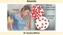
Anemia True or absolute anemia is defined as a decrease in erythrocyte mass within the body HCT hemoglobin and RBC count values are usually below their reference intervals Low erythrocyte parameters may also be present in blood when the totalbody erythrocyte mass is normal relative anemia ID: 932619
Download Presentation The PPT/PDF document "Anemia By Dr. Hussein AlNaji" is the property of its rightful owner. Permission is granted to download and print the materials on this web site for personal, non-commercial use only, and to display it on your personal computer provided you do not modify the materials and that you retain all copyright notices contained in the materials. By downloading content from our website, you accept the terms of this agreement.
Slide1
Anemia
By
Dr. Hussein
AlNaji
Slide2AnemiaTrue or absolute anemia is defined as a decrease in erythrocyte mass within the body. HCT, hemoglobin, and RBC count values are usually below their reference intervals.Low erythrocyte parameters may also be present in blood when the total-body erythrocyte mass is normal (relative anemia).
Slide3This can result from 1- overhydration resulting in erythrocyte dilution 2- splenic sequestration of erythrocytes as occurs with splenic relaxation during anesthesia,heparin-induced erythrocyte agglutination in horses.various causes of splenomegaly.Anemia may occur following blood loss, increased erythrocyte destruction, or decreased erythrocyte production.
Slide4Factors that can be useful in categorizing anemia into these broad causes (and often into more specific causes) include reticulocyte counts.erythrocyte indices.erythrocyte morphology on stained blood films. the appearance of the plasma.plasma protein concentration.serum iron measurements. bone marrow evaluation.
Slide5There are two types of anemia: regenerative and non-regenerative. In non-regenerative anemia, the cause is usually a decrease in erythropoietin (a hormone controlled by the kidneys that influences red blood cell production as a response to low oxygen in tissues) or bone marrow abnormalities. The bone marrow does not effectively respond to the decrease levels of red blood cells.Regenerative anemia, alternatively, the bone marrow responds to the anemia by increasing production of red blood cells and releasing reticulocytes (immature red blood cells that don’t have a nucleus). Regenerative anemia can be caused by a hemorrhage or hemolysis (the rupture or destruction of blood cells).
Slide6Causes of Regenerative Anemia poisoning, infection, trauma, or other conditions that may cause a loss of blood or hemolysis (shortened life span of erythrocytes).Oxidation Blood loss Immune-mediated hemolysis:Decreased marrow production Babesia spp infection Hemolysis caused by phosphorous deficiency, seen in dogs with diabetes, hepatic lipidosis, and refeeding syndrome Genetic
Slide7Symptoms of Regenerative Anemia in Dogs.Symptoms vary depending on the degree, duration, and cause of the anemia, but may include: Tachycardia (fast heart rate) Pale mucous membranes Hypotension (low blood pressure) Lethargy, Weakness and Anorexia Splenomegaly (enlarged spleen) and Abdominal distension Heart murmur 7. Jaundice8. Shock 9. Death
Slide8Classification of Anemia Using Erythrocyte IndicesAn anemia can also be classified using the MCV and MCHC values to assist in determining its cause. The terms used to indicate size are macrocytic (increased MCV), normocytic (normal MCV), and microcytic (decreased MCV). The terms used to describe MCHC values are normochromic (normal MCHC) and hypochromic (decreased MCHC).
Slide9Mean corpuscular volume ( MCV) : It is a measure of the average volume of a red blood corpuscle (or red blood cell). MCV 10 B. Mean corpuscular hemoglobin concentration (MCHC)a measure of the concentration of haemoglobin in a given volume of packed blood
MCHC
100 =
The anemia depended on MCV and MCHC include four types A- Normocytic Normochromic1. Hemolytic anemia. 2. Hemorrhage.3. Early iron-deficiency anemia before microcytes predominate4. Chronic inflammation and neoplasia (sometimes slightly microcytic)5. Chronic renal disease 6. Endocrine deficiencies8. Selective erythroid aplasia8. Aplastic and hypoplastic bone marrows9. Lead toxicity (may not be anemic
Slide11B- Macrocytic Hypochromic1. Regenerative anemias with marked reticulocytosis2. Spurious with prolonged storage of blood sampleC- Macrocytic Normochromic1. Regenerative anemias (decreased MCHC is not always present)2. FeLV infections with no reticulocytosis (common)3. Erythroleukemia (AML-M6) 5. Hyperthyroid cats (slight macrocytosis without anemia)6. Folate deficiency (rare)7. Congenital dyserythropoiesis of Hereford calves
Slide12- Microcytic Normochromic/Hypochromica1. Chronic iron deficiency (months in adults, weeks in nursing animals)2. Anemia of inflammatory disease (usually normocytic)3. Hepatic lipidosis in cats (usually normocytic)4. Prolonged recombinant erythropoietin treatment (mild)5. Copper deficiency (rare)6. Drugs or compounds that inhibit heme synthesis7. Pyridoxine deficiency (experimental)8. Spurious when platelets are included in erythrocyte histograms