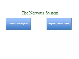
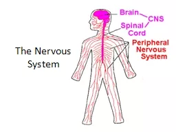
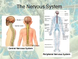
/
duced the nervous system which comprises the central nervous system duced the nervous system which comprises the central nervous system

duced the nervous system which comprises the central nervous system - PDF document

adah
adah . @adah
Follow
342 views
Uploaded On 2022-08-25

duced the nervous system which comprises the central nervous system - PPT Presentation

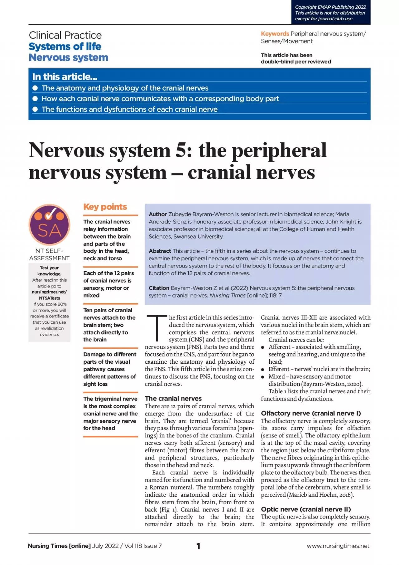
1 Key points The cranial nerves relay information between the brain and parts of the body in the head neck and torsoEach of the 12 pairs of cranial nerves is sensory motor or mixedTen pairs of crani ID: 941657

cranial nerve fibres visual nerve cranial visual fibres nerves muscle sensory motor 150 eye vagus facial 2021 optic 146

Share:

Link:

Embed:

Download Presentation from below link

Download Pdf The PPT/PDF document "duced the nervous system which comprises..." is the property of its rightful owner. Permission is granted to download and print the materials on this web site for personal, non-commercial use only, and to display it on your personal computer provided you do not modify the materials and that you retain all copyright notices contained in the materials. By downloading content from our website, you accept the terms of this agreement.


Presentation Transcript

1 duced the nervous system, which comprises the central nervous system (CNS) and the peripheral nervous system (PNS). Parts two and three focused on the CNS, and part four began to examine the anatomy and physiology of the PNS. This fifth article in the series continues to discuss the PNS, focusing on the cranial nerves.The cranial nervesThere are 12 pairs of cranial nerves, which emerge from the undersurface of the brain. They are termed ‘cranial’ because ings) in the bones of the cranium. Cranial nerves carry both afferent (sensory) and Key points The cranial nerves relay information between the brain and parts of the body in the head, neck and torsoEach of the 12 pairs of cranial nerves is sensory, motor or mixedTen pairs of cranial nerves attach to the brain stem; two attach directly to Author Zubeyde Bayram-Weston is senior lecturer in biomedical science; MariaAndrade-Sienz is honorary associate professor in biomedical science; John Knight isassociate professor in biomedical science; all at the College of Human and HealthSciences, Swansea University.Abstract This article – the fifth in a series about the nervous system – continues to examine the peripheral nervous system, which is made up of nerves that connect the In this article... The anatomy and physiology of the cranial nerves How each cranial nerve communicates with a corresponding body part The functions and dysfunctions of each cranial nerveClinical PracticeSystems of lifeNervous system Nursing Times [online] July 2022/ Vol 118 Issue 7www.nursingtimes.net Copyright EMAP Publishing 2022 This article is not for distributionexcept for journal club use ASSESSMENT Test your knowledge.After reading this article go to NTSATests If you score 80% or more, you will receive a certificate that you can use as revalidation evidence. 2 Some defects are obvious, such as a lesion to the optic nerve, which causes full blindness at that side (1, Fig 3), while others – such as a lesion to the optic nerve (2, Fig3) – may be hidden yet still result in some sight loss in both eyes. rupt the crossing-over of fibres. The most asma is a pituitary tumour, which compresses it from below. If the full thickness of the chiasma is damaged, this typically causes bitemporal hemianopia (tunnel vision) (3, Fig3). As the name suggests, this means blindness in the temporal halves of the visual field but normal vision in the nasal halves. mous hemianopia; this is sight loss on the same side of the visual field in both eyes (4-8, Fig 3). The central vision may remain intact, termed ‘macular sparing’. The visual cortex processes visual information and regulates involuntary eye movements that maintain fixation of gaze on a target in the visual field, so any damage to the visual cortex can interfere with vision; called cortical blindness, it demolishes visual awareness and leads to chronic Oculomotor nerve (cranial nerve III)The oculomotor nerve contains the bulk of somatic motor nerves that supply the extraocular muscles; these are responsible for eye movement. The oculomotor nerve innervates five of the seven extraocular muscles and, thereby, operates elevation of objects, and the temporal area

is involved with object identification (Mtui et al, 2015). Interruption to the visual pathway can cause various visual defects. Fig3 shows the visual defects that can arise from lesions in: Nearby structures that affect the visual pathway;The visual cortex. myelinated axons and is surrounded by an The partial crossing over of the optic nerves occurs in the optic chiasma (Fig2); chiasma means crossover. The optic chiasma is essential for binocular vision, three-dimensional vision and depth perception in the visual scene. Fibres from the medial half of each retina (the nasal retina) cross over in the optic chiasma and join uncrossed fibres from the lateral half of the retina (the temporal retina) to form the optic tract (Fig2). As a result, information carried to the right cerebral hemisphere represents the left half of the visual field, and information representing the right half of the visual field is relayed to the left hemisphere. After crossing the chiasma, most of the axons travel a short distance to terminate in lateral geniculate nuclei of the thalamus and are then conveyed to the visual cortex for processing. The human visual cortex is large and includes the: Posterior portion of the parietal lobe;Posterior region of the temporal lobe. This facilitates all aspects of vision, including the recognition of objects and the perception of colour, depth and motion. The occipital and posterior parietal areas of the visual cortex ascertain the position of Clinical PracticeSystems of life The cranial nerves Optic nerve (II)Olfactory nerve (I)Trochlear nerve (IV)Oculomotor nerve (III)Abducens nerve (VI)Facial nerve (VII)Vestibulocochlear nerve (VIII)Glossopharyngeal nerve (IX)Hypoglossal nerve (IX)Accessory nerve (XI)Vagus nerve (X) Trigeminal nerve (V) SuperiorInferiorRightLeft The visual pathway Visual cortex Optic nerveOptic chiasmOptic tractLateral geniculate body Temporal retinaLeft visual eldRight visual eldOptical len Eye Nasal retina JENNIFER N.R. SMITH 3 Clinical PracticeSystems of lifeMandibular branch (Fig4). It carries the sensations of touch, pressure, pain and temperature from the: Oral cavity (teeth, gums and paranasal sinuses). Additionally, the trigeminal nerve carries the nerve fibres from the muscles of mastication and the temporomandibular joint, which enable the opening and closing action of the jaw. Most of the afferent fibres are located in the trigeminal ganglion. This is in a cavity in the dura mater, concealing a depression on the anteporal bone. Herpes zoster infection of the trigeminal nerve (shingles) causes pain and eruption of vesicles in the dermatome innervated by a branch of the trigeminal nerve (Huff and Daly, 2021). Abducens nerve (cranial nerve VI)The abducens nerve contains only somatic motor neurons and innervates the lateral rectus muscle; this controls eye abduction (movement away from the midline). A lesion of the abducens nerve leads to an inability to move the eye outwards (Nguyen et al, 2021). Facial nerve (cranial nerve VII)The facial nerve has two roots: one contains sensory and parasympathetic fibres, and the other contains motor fibres. The sensory fibres of the facial nerve supply: Taste sensation from the

anterior two-thirds of the tongue; Touch sensation in part of the external ear. The facial nerve’s motor fibres control: The middle-ear muscle (stapedius).ical problem involving the facial nerve; it is generally a temporary condition without threat to overall heath. A lesion or impairment of the facial nerve leads to weakness of one side of the face and paralysis of the facial muscles, causing:Loss of taste sensation on the tongue. The symptoms are similar to those of more serious medical problems, such as Dilation of the pupil, initiated by unopposed action of the dilator pupillae muscle in the iris (sympathetic innervation). The first sign of compression of the oculomotor nerve is slowness of the ipsilateral pupillary response to light (Standring et al, 2008). Trochlear nerve (cranial nerve IV)The trochlear nerve is the smallest cranial nerve in terms of diameter, contains only somatic motor neurons and is the only cranial nerve that emerges from the dorsal region of the brain stem. It innervates only one muscle: the superior oblique muscle of means pulley, which describes the sling of connective tissue that contains the tendon of the superior oblique muscle. The action of this muscle is complex but the trochlear nerve controls the abduction and intorsion of the eye (Vilensky et al, 2015). Trigeminal nerve (cranial nerve V)The trigeminal nerve is the most complex cranial nerve, and the largest in diameter. nents. It is the major sensory nerve for the head, and its sensory fibres are distributed to various structures via three branches: the upper eyelid as well as controlling the following functions of the eyeball: Elevation (upward movement);Depression (downward movement);Adduction (inward movement). The oculomotor nerve also contains preganglionic parasympathetic neurons that innervate the smooth muscle in the pupillary sphincter (constrictor) muscle of the iris. The size of the pupil controls the amount of light entering the eye: light on the retina causes the iris’ sphincter muscles to contract, thereby constricting the pupil and reducing the amount of light reaching the retina. This is called the direct light reflex. In the absence of abnormality, even if only one retina is illuminated, the tion of the pupil of the non-illuminated eye is known as the consensual light reflex. Injury to the oculomotor nerve can cause third nerve palsy; this can cause paralysis of the muscles controlled by the oculomotor nerve while they are deprived of a nerve supply. The outcomes of such an injury are:Drooping of the upper eyelid (ptosis);An eye that is deviated downwards and laterally (lateral strabismus), initiated by unopposed action of the lateral Inability to direct the eye medially or vertically; Visual eld loss associated with lesions of the visual pathway 1234567812345678 Visual cortex Left eyeRight eyeOptic nerveOptic chiasmOptic tractLateral geniculate bodyOptic radiations JENNIFER N.R. SMITH 4 Clinical PracticeSystems of life Accessory nerve (cranial nerve XI)The accessory nerve only has motor function. It has two parts: Spinal. The cranial part originates from the medulla, joins the vagus nerve and is distributed to the muscle of the soft palat

e, pharynx and larynx. The spinal part originates from the upper part of the spinal cord and enters the cranial cavity via the foramen magnum, where it briefly joins the cranial portion. It then innervates two zius), which are involved in moving the head and shoulders (AlShareef and Newton, 2022). Hypoglossal nerve (cranial nerve XII)The hypoglossal nerve only has motor function. It innervates the muscles of the tongue, controlling its shape; it also receives fibres from the motor cortex that contribute to voluntary movements of the tongue, such as those that happen during speech. The hypoglossal nerve’s fibres are also involved in controlling the reflex movements of chewing, sucking and swallowing. ConclusionThis article has explained the anatomy and function of the cranial nerves. The next article in this series will examine the components of the autonomic thetic and parasympathetic divisions. ReferencesAlShareef S, Newton BW (2022) Accessory Nerve Injury. StatPearls Publishing.Banasik JL, Copstead L-EC (2019) Pathophysiology. Elsevier. Bayram-Weston Z (2020) Chapter 6: The nervous system. In: Knight J et al (eds) Understanding Anatomy and Physiology in Nursing. Sage. Bordoni B et al (2021) Neuroanatomy, Cranial Nerve 8 (Vestibulocochlear). StatPearls Publishing.Huff T, Daly DT (2021) Neuroanatomy, Cranial Nerve 5 (Trigeminal). StatPearls Publishing.Kenny BJ, Bordoni B (2021) Neuroanatomy, Cranial Nerve 10 (Vagus Nerve). StatPearls Publishing.(2016) Human Anatomy and Physiology. Pearson.(2015) Fitzgerald’s Clinical Neuroanatomy and Neuroscience. Elsevier.Nguyen V et al (2021) Neuroanatomy, Cranial Nerve 6 (Abducens). StatPearls Publishing. (2014) Identification of facial nerve during parotidectomy: a combined anatomical & surgical study. Indian Journal of Otolaryngology and Head and Neck Surgery; 66: 1, 63-68. Standring S et al Gray’s Anatomy: The Anatomical Basis of Clinical Practice. Churchill Livingstone/Elsevier.Thomas K et al (2021) Neuroanatomy, Cranial Nerve 9 (Glossopharyngeal). StatPearls Publishing.Vilensky JA et al (2015) The Clinical Anatomy of the Cranial Nerves: The Nerves of ‘On Old Olympus Towering Top’. Wiley-Blackwell.Warner MJ et al (2022) Bell Palsy (Nursing). StatPearls Publishing.one muscle, the stylopharyngeus muscle. This muscle is responsible for elevating the pharynx and larynx and is, therefore, involved in swallowing. The parasympathetic component is involved in innervation of the parotid salivary gland and initiates the production of saliva (Thomas et al, 2021). Vagus nerve (cranial nerve X) The vagus nerve contains sensory, motor and parasympathetic fibres. Vagus means wandering, as the vagus nerve branches and spreads extensively. The sensory fibres carry information from the:Pharynx, larynx, oesophagus, tympanic membrane, external auditory meatus and part of the external ear – for general sensation;Aortic bodies (which contain chemoreceptors) and aortic arch (which contain baroreceptors) – to monitor blood pressure and levels of carbon dioxide, oxygen and acidity in Receptors of the thoracic and abdominal organs – to convey sensory information. The motor component of the vagus nerve innervat

es the muscle of the soft palate, pharynx, larynx and upper part of the oesophagus. These motor fibres are vital for controlling speech and swallowing. The parasympathetic component of the vagus nerve is widely distributed through the cardiovascular, respiratory and gastrotions of the thoracic and abdominal organs, cles and glands in the stomach, intestine and bladder (Kenny and Bordoni, 2021).stroke and multiple sclerosis, so it needs urgent medical attention (Saha et al, 2014). The leading cause of Bell’s palsy remains unknown, but causes include viral infections such as herpes simplex virus, varicella-zoster virus and Epstein-Barr virus (Warner et al, 2022). Vestibulocochlear nerve (cranial nerve VIII)The vestibulocochlear nerve is a sensory nerve that carries impulses from the inner ear. It has two components: Vestibular nerve – this conveys information related to position and movement of the head;Cochlear nerve – this conveys auditory information to the auditory cortices (Bordoni et al, 2021). Glossopharyngeal nerve (cranial nerve IX)The glossopharyngeal nerve is mainly a sensory nerve, but it also contains parasympathetic fibres and some motor fibres. The sensory fibres convey information Pharynx (throat), posterior third of the tongue, Eustachian tube and middle ear – for general sensation;Pharynx and posterior third of the tongue – to supply the taste buds and facilitate the gag reflex with the vagus nerve (cranial nerve X);Carotid body (which contains chemoreceptors) and carotid sinus (which contains baroreceptors) – to continually monitor arterial blood geal nerve is very small and innervates only For more articles on neurology, go to nursingtimes.net/neurology Sensory distribution of the trigeminal nerve Ophthalmic branch Maxillary branchMandibular branch JENNIFER N.R. SMITH 5 Table 1. Functions and dysfunctions of the cranial nervesCranial nerve TypeAssociated dysfunction(s)Olfactory (I) Sense of smell Unilateral or bilateral loss of sense of smellLoss of taste Optic (II) Vision Loss of vision Oculomotor (III) MotorMovement of the eyeball and upper eyelid Eye-movement problemsParasympatheticPupil constrictionTrochlear (IV) MotorMovement of the eyeballEye-movement problemsTrigeminal (V) General sensation in face, scalp, corneas, and nasal and oral cavities Loss of facial sensationMotorChewingAbducens (VI)MotorMovement of the eyeballEye-movement problemsFacial (VII) TasteLoss of tasteInability to close eyeMotorFacial expressionParasympatheticSecretion of tears and salivaVestibulocochlear Hearing and balanceLoss of hearing and balance Glossopharyngeal Taste and sensation from back of tongue Inability to swallow Hoarse voiceMotorSwallowing and speechParasympatheticSecretion of salivaVagus (X)Taste and sensation from epiglottisInability to swallow Hoarse voice Delayed gastric emptyingMotorSwallowing and speechParasympatheticMuscle contraction of thoracic and abdominal organs and secretion of digestive fluidsAccessory (XI)MotorHead and shoulder movementInability to move head and raise shouldersHypoglossal (XII)MotorMovement of the tongue musclesInability to move tongueSource: Bayram-Weston (2020)Clinical PracticeSystems of l