/
Journal of Nutrition Metabolism and Health Science Journal of Nutrition Metabolism and Health Science

Journal of Nutrition Metabolism and Health Science - PDF document

adia
adia . @adia
Follow
342 views
Uploaded On 2022-10-11

Journal of Nutrition Metabolism and Health Science - PPT Presentation

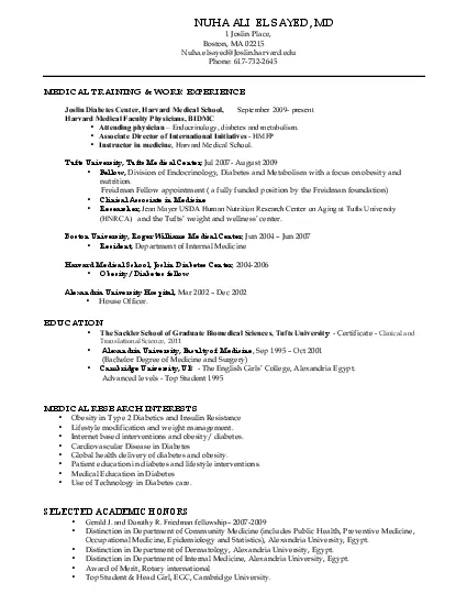
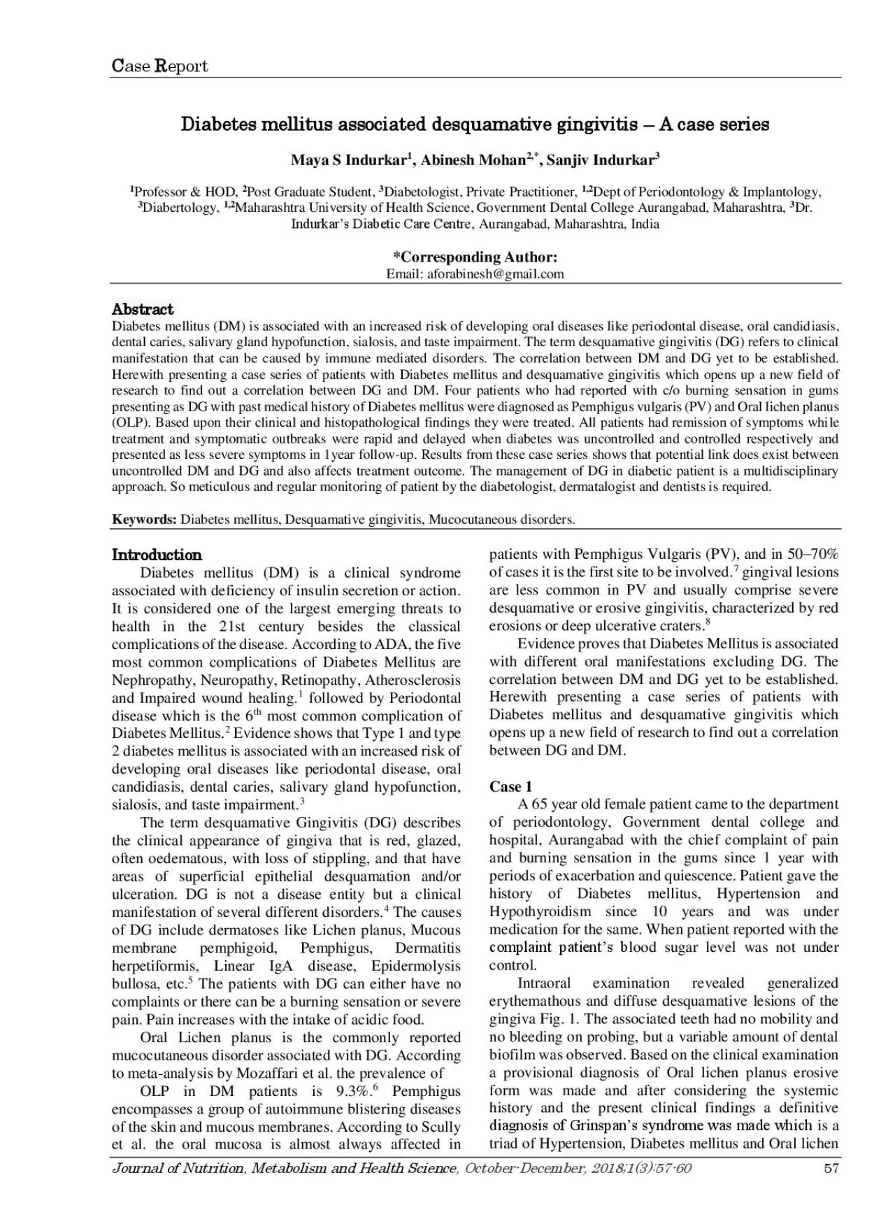
C ase R eport October December 20181 3 57 60 57 Diabetes mellitus associated desquamative gingivitis A case series Maya S Indurkar 1 Abinesh Mohan 2 Sanjiv Indurkar 3 1 Profe ID: 958712

patient oral mellitus diabetes oral patient diabetes mellitus lesions desquamative clinical planus days examination case lichen pemphigus patients gingivitis

Share:

Link:

Embed:

Download Presentation from below link

Download Pdf The PPT/PDF document "Journal of Nutrition Metabolism and Heal..." is the property of its rightful owner. Permission is granted to download and print the materials on this web site for personal, non-commercial use only, and to display it on your personal computer provided you do not modify the materials and that you retain all copyright notices contained in the materials. By downloading content from our website, you accept the terms of this agreement.


Presentation Transcript

C ase R eport Journal of Nutrition, Metabolism and Health Science , October - December, 2018;1( 3 ):57 - 60 57 Diabetes mellitus associated desquamative gingivitis – A case series Maya S Indurkar 1 , Abinesh Mohan 2,* , Sanjiv Indurkar 3 1 Professor & HOD, 2 Post Graduate Student, 3 Diabetologist, Private Practitioner, 1,2 Dept of Periodontology & Implantology, 3 Diabertology, 1,2 Maharashtra University of Health Science , Government Dental College Aurangabad, Maharashtra, 3 Dr. Indurkar’s Diabetic Care Centr e, Aurangabad, Maharashtra, India *Corresponding Author: Email: aforabinesh@gmail.com Abstract Diabetes mellitus (DM) is associated with an increased risk of developing oral diseases like periodontal disease, oral candid iasis, dental caries, salivary gland hypofunction, sialosis, and taste impairment. The term desquamative gingivitis (DG) refers to clinical manifestation that can be caused by immune mediated disorders. The correlation between DM and DG yet to be established. Herewith presenting a case series of patients with Diabetes mellitus and desquamative gingivitis which opens up a new field of research to find out a correlation between DG and DM. Four patients who had reported with c/o burning sensation in gums presenting as DG with past medical h istory of Diabetes mellitus were diagnosed as Pemphigus vulgaris (PV) and Oral lichen planus (OLP). Based upon their clinical and histopathological findings they were treated. All patients had remission of symptoms whi le treatment and symptomatic outbreaks were rapid and delayed when diabetes was uncontrolled and controlled respectively and presented as less severe symptoms in 1year follow - up. Results from these case series shows that potential link does exist between uncontrolled DM and DG and also affects treatment outcome. The management of DG in diabetic patient is a multidisciplinary approach. So meticulous and regular monitoring of patient by the diabetologist, dermatalogist and dentists is required. Keywords: Diabetes mellitus, Desquamative gingivit is, Mucocutaneous disorders. Introductio n Diabetes mellitus (DM) is a clinical syndrome associated with deficiency of insulin secretion or action. It is considered one of the largest emerging threats to health in the 21st century besides the classical c omplications of the disease. According to ADA, the five most common complications of Diabetes Mellitus are Nephropathy, Neuropathy, Retinopathy, Atherosclerosis and Impaired wound healing. 1 followed by Periodontal disease which is the 6 th most common compl ication of Diabetes Mellitus. 2 Evidence shows that Type 1 and type 2 diabetes mellitus is associated with an increased risk of developing oral diseases like periodontal disease, oral candidiasis, dental caries, salivary gland hypofunction, sialosis, and ta ste impairment. 3 The term desquamative Gingivitis (DG) describes the clinical appearance of gingiva that is red, glazed, often oedematous, with loss of stippling, and that have areas of superficial epithelial desquamation and/or ulceration. DG is not a disease entity but a clinical manifestation of several different disorders. 4 The causes of DG include dermatoses like Lichen planus, Mucous membrane pemphigoid, Pemphigus, Dermatitis herpetiformis, Linear IgA disease, Epidermolysis bullosa, etc. 5 The patie nts with DG can either have no complaints or there can be a burning sensation or severe pain. Pain increases with the intake of acidic food. Oral Lichen planus is the commonly reported mucocutaneous disorder associated with DG. According to meta - analysis by Mozaffari et al. the prevalence of OLP in DM patients is 9.3%. 6 Pemphigus encompasses a group of autoimmune blistering diseases of the skin and mucous membranes. According to Scully et al. the oral mucosa is almost always affected in patients with Pemp higus Vulgaris (PV), and in 50 – 70% of cases it is the first site to be involved. 7 gingival lesions are less common in PV and usually comprise severe desquamative or erosive gingivitis, characterized by red erosions or deep ulcerative craters. 8 Evidence pro ves that Diabetes Mellitus is associated with different oral manifestations excluding DG. The correlation between DM and DG yet to be established. Herewith presenting a case series of patients with Diabetes mellitus and desquamative gingivitis which opens up a new field of research to find out a correlation between DG and DM. Case 1 A 65 year old female patient came to the department of periodontology, Government dental college and hospital, Aurangabad with the chief complaint of pain and burning sensation in the gums since 1 year with periods of exacerbation and quiescence. Patient gave the history of Diabetes mellitus, Hyperten

sion and Hypothyroidism since 10 years and was under medication for the same. When patient reported with the complaint patient’s b lood sugar level was not under control. Intraoral examination revealed generalized erythemathous and diffuse desquamative lesions of the gingiva Fig. 1. The associated teeth had no mobility and no bleeding on probing, but a variable amount of dental biofil m was observed. Based on the clinical examination a provisional diagnosis of Oral lichen planus erosive form was made and after considering the systemic history and the present clinical findings a definitive diagnosis of Grinspan’s syndrome was made which is a triad of Hypertension, Diabetes mellitus and Oral lichen Mohan A. et al. Diabetes mellitus associated desquamative gingivitis… Journal of Nutrition, Metabolism and Health Science , October - December, 2018;1( 3 ):57 - 60 58 planus. It has been suggested that the antihypertensive medications that the patients take may cause a lichenoid mucosal reaction. Patient was prescribed with an oral gel containing 0.1% kenacor t thrice daily for 15 days multivitamin tablets Cap A to Z grid for 15 days and mucopain anesthetic gel along with 0.2% chlorhexidine mouthwash twice daily for 15 days. After 15 days patient reported back with the reduction of pain symptoms but with persis tent erythematous lesions over the gingiva. Oral prophylaxis was performed to remove plaque and calculus deposits. After which oral hygiene instructions were given and Patient was advised to continue the above prescribed medications for another 15 days and recalled for follow - up examination. In next visit, there was no remission of gingival lesions, so biopsy was taken for histopathological examination. Biopsy of the lesion along with some parts of connective tissue was taken from the mandibular molar regio n left side. Biopsy report was inconclusive. So patient was referred to the dermatologist for the further management. Systemic corticosteroids along with immunosuppressants were prescribed to the patient for one month. After one month patient reported with the reduction in the gingival lesions and reduction in the pain. Fig. 1 Case 2 A 55 year old female patient came to the department of periodontology, Government dental college and hospital, Aurangabad with the chief complaint of burning sensation of t he gums in the left and right lower back jaw region since 6 months. Patient gave the history of Diabetes mellitus for the last 10 years and was under medication. Her blood sugar levels were uncontrolled when she reported to the department. She experienced menopause at the age of 50. Examination revealed erythematous and desquamative lesions on the marginal gingiva involving maxillary and mandibular teeth and it was bilateral. Fig. 2 the associated teeth had no mobility and no bleeding on probing, but a vari able amount of dental biofilm was observed. Based on the clinical examination a diagnosis of Erosive Lichen planus was made. Patient was prescribed with topical corticosteroids Kenacort 0.1% oral paste for 7 days multivitamin tablets Cap A to Z grid and 0. 2% chlorhexidine mouthwash for 15 days. After 15 days patient reported with relief in pain symptoms. Oral prophylaxis was performed to remove plaque and calculus deposits. Oral hygiene instructions were given and patient was asked to continue topical appli cation of kenacort for next 1 month and kept on follow up examination once a month for 6 months. Fig. 2 Case 3 A 66 year old female patient came to the department of periodontology, Government dental college and hospital, Aurangabad with the chief complaint of pain and burning sensation in the right and left lower molar region. Patient gave the history of diabetes mellitus & was under the medication for the same since last 10 years. Intraoral examination revealed severe gingival & mucosal erythemato us desquamative lesions which were localized on the right and left mandibular distomolar region cheek mucosa and labial marginal gingiva. Fig. 3 There was also a presence of cheek bite in upper and lower second molar region left side. Patient had a general ized moderate chronic periodontitis. Patient was not able to maintain the oral hygiene properly due to pain and burning sensation. Extra oral examination revealed the small, round, brown to black coloured vesiculobullous lesions on the chest on right side & in umbilical regions suggestive of healing vesicles. Based on the clinical examination a diagnosis of Pemphigus vulgaris was made. Incisional biopsy was performed and superficial biopsy reveals strips of squamous epithelium with acute on chronic inflamma tory fibrinous exudate is also seen. Also biopsy revealed tissue showing absence of dysplasia or malignancy

was not suggestive of Pemphigus vulgaris. Patient was prescribed 0.1% kenacort oral paste for gingival lesions and 0.1% kenacort cream for extra ora l lesions for 7 days. Patient was also prescribed with Tantum aesthetic mouthwash and 0.2% chlorhexidine mouthwash for 7 days. In follow - up examination after 7 days there was reduction in pain and so she was referred to dermatologist for the further manage ment where patient was prescribed with systemic corticosteroids Mohan A. et al. Diabetes mellitus associated desquamative gingivitis… Journal of Nutrition, Metabolism and Health Science , October - December, 2018;1( 3 ):57 - 60 59 Zempred 16mg tablets for 15 days (2 tablets daily for first 5 days 1and half tablets daily for next 5 days and 1 tablet daily for next 5 days) and Kenacort cream. After 15days patient was advi sed change of drug to Methyl prednisolone tablets 4mg once daily for 30 days with Rabez tablet 20mg and Kenacort cream. Patient was examined every 15days for one and half months till the remission of the gingival lesions. During this period a good healing of gingival lesions were noted with reduction in erythema in some regions and complete resolution of pain. Fig. 3 Case 4 A 47 year old male patient came to the department of periodontology, Government dental college and hospital, Aurangabad with the ch ief complaint of pain and burning sensation in the gums. Patient gave the history of diabetes mellitus & was under the medication for the same since last 10 years. Intraoral examination revealed severe gingival erythemathous desquamative lesions which were localized on the right and left posterior jaw region. Patient was periodontally healthy. Fig. 4 Extra oral examination revealed the small, round, brown to black colored vesiculobullous lesions on the face upper and lower limbs suggestive of healing vesicl es. Based on the clinical examination a provisional diagnosis of Pemphigus vulgaris was made. Patient was prescribed 0.1% kenacort oral paste. Patient was also prescribed with tantum mouthwash and 0.2% chlorhexidine mouthwash for 7 days. In follow up examination there was no relief from pain so patient was referred to the dermatologist for further treatment. There the patient was prescribed systemic corticosteroids along with immunosuppressants Tablet Zempred for one month after which the patient got r elief of symptoms. Fig. 4 Discussion The present Case series describes the four cases of Diabetic patients who had reported with signs of desquamative gingivitis. Out of these four cases two cases were diagnosed with Erosive or ulcerative type of Oral L ichen Planus and two cases were diagnosed with Pemphigus vulgaris. In general clinical descriptions of oral lichen planus the gingival lesions could be classified into four general categories: (1) white popular keratotic lesions. (2) vesiculo bullous lesio ns. (3) Erosive or ulcerative lesions, and (4) atrophic lesions. Often there were combinations of the various clinical types of lesion, particularly that of keratotic and erosive or ulcerative patterns. 9 Since 1963 when David Grin span described a frequent association between lichen planus and diabetes mellitus where OLP lesions followed the clinical evolution of DM, many authors using heterogeneous diagnosis criteria have reported a strong association between lichen planus and DM. However this relationship could not always be proved and the association was qualified as coincidental or caused by anti - diabetic or other drugs. 10 The exact prevalence of the various causes related to DG are difficult to determine due to most evidence being (anecdotal) reported i n the form of case series. A recent study by Arduino et al. reported on the aetiology of DG cases reviewed in the Oral Medicine unit of the main hospital of Turin, Italy over a one - year period. A total of 382 cases were described. Oral lichen planus was s hown to be the most frequent condition, representing 58.4% of all diagnoses followed by mucous membrane pemphigoid (33.4%) pemphigus vulgaris (5.4%) and epidermolysis bullosa acquisita (2.8%). 11 Lo Russo et al. reviewed the aetiology of 125 patients with D G referred to the Oral Medicine Section in Palermo Italy. Similarly, oral lichen planus (75%) was the most common disease presenting as DG followed by mucous membrane pemphigoid 9%, and pemphigus vulgaris 4%. 12 The most widely accepted treatment for lesion s of DG involves topical or systemic corticosteroids to modulate the patient’s immune response which were prescribed to the patients presented in this case series Mohan A. et al. Diabetes mellitus associated desquamative gingivitis… Journal of Nutrition, Metabolism and Health Science , October - December, 2018;1( 3 ):57 - 60 60

after which there found to be remission of DG lesions. From this case series and it was also found that fluctuations in the blood sugar level is related to the exacerbation and remission of the lesions. Limitations The definitive diagnosis of DG is sometimes complex. It is often difficult to differentiate the causes of DG when there are few sympt oms and ⁄ or the associated disease is limited to the gingival tissues. Hence, there is a need for histopathological examination of lesional tissue, together mainly with direct, and perhaps indirect immunofluorescence to demonstrate lesional and circulatin g auto - antibodies respectively (Challacombe et al. 2001). The limitations in the case series is that the definitive diagnosis of the lesions was difficult as there was a scarcity of obtaining gingival tissue for direct immunofluorescence test. Conclusion Diabetes mellitus and desquamative gingivitis caused by Oral Lichen planus, Pemphigus vulgaris and Mucous membrane pemphigoid are disorders of humoral immunity which has been associated with reduced response of T cells and neutrophil function. It has been proposed that the endocrine dysfunction in diabetes mellitus may be related to immunologic defect or compromised immune system that may also contribute to the development of Lichen planus and Pemphigus vulgaris that eventually leads to development of desqu amative gingivitis. This case series highlights the combined immune - modulatory effect of both DM and DG. The correlation between DM and DG goes unnoticed by patient diabetologist and general dental practitioners in the general practice. Evidence shows that periodontitis is one of the common complications of Diabetes Mellitus. Hypothetically severe pain in DG restrain the patients from maintaining oral hygiene thereby leading to periodontal disease Various oral manifestations of DM which are mentioned in th e literature are periodontal disease, xerostomia, burning tongue etc. Further Cohort studies and research are needed to find out prevalence of DG in DM and whether DG can be one of the oral manifestation of DM. The management of DG in diabetic patient is a multidisciplinary approach. Systemic corticosteroids are used in the management of DG. Use of corticosteroids may cause increase in blood sugar level, susceptibility to infections. So meticulous and regular monitoring of patient by the diabetologist, derm atalogist and dentists is required. Patient education about the co - existence of the Diabetes Mellitus and desquamative Gingivitis & importance of good control of blood sugar need to be emphasized. Maintenance of good oral hygiene will help in preventing th e onset of periodontal disease in these patients. References 1. Fowler MJ, Microvascular and Macrovascular Complications of Diabetes. Clin Diabetes. 2011;29(3):116 - 22. 2. Harald Loe. Periodontal Disease - The sixth complication of diabetes mellitus. Diabetes Care . Supplement 993;16 3. Pedersen A M L. Diabetes Mellitus and Related Oral Manifestations. Oral Biosci Med 2004; 1: 229 - 248. 4. Robinson NA, Wray D. desquamative gingivitis: a sign of mucocutaneous disorders - a review. Aust Dent J 2003;48(4):206 – 11. 5. Al - Abeedi F. The Differential Diagnosis of desquamative Gingivitis: Review of the Literature and Clinical Guide. J Int Oral Health. 2015 ;7(1):88 – 92 6. Mozaffari H R. Prevalence of Oral Lichen Planus in Diabetes Mellitus: a Meta - Analysis Study. ACTA INFORM MED . 2016;24 (6):390 - 93 7. Scully C et al. Pemphigus vulgaris: The manifestations and long - term management of 55 patients with oral lesions. British Journal of Dermatol. 1999;140:84 – 9 8. Javali M A. Pemphigus vulgaris presenting as gingival involvement. Indian Dermatol Onli ne J . 2012;3(3): 202 – 4. 9. Dudhia B B. Oral lichen planus to oral lichenoid lesions: Evolution or revolution. J Oral Maxillofac Pathol. 2015;19(3):364 – 70 10. Romero MA. Prevalence of diabetes mellitus amongst oral lichen planus patients. Clinical and pathological characteristics . Med Ora.l 2002;7:121 - 29 11. Arduino PG, Broccoletti R, Sciannameo V, Scully C. A practical clinical recording system for cases with desquamative gingivitis. Br J Dermatol 2016. Doi: 10.1111/bjd.15075 12. Lo Russo L, Fierro G, Guiglia R, Compilato D, Testa NF, Lo Muzio L et al. Epidemiology of desquamative gingivitis: evaluation of 125 patients and review of the literature. Int J Dermatol . 2009; 48(10):1049 - 52. 13. Management of oral pemphigus vulgaris: A case report and a clinical update (PDF Download Available). Available from: https://www.researchgate.net/publication/282202979_ Management_of_oral_pemphigus_vulgaris_A_case_re port_and_a_clinical_updat e [accessed Feb 03 2018