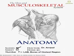
/
Bones of the gluteal region Bones of the gluteal region

Bones of the gluteal region - PowerPoint Presentation

alexa-scheidler
alexa-scheidler . @alexa-scheidler
Follow
380 views
Uploaded On 2016-05-06

Bones of the gluteal region - PPT Presentation

The Hip bone The hip bone is made of 1The ilium superior in position 2The ischiumposteroinferior in position 3The pubis antero inferior in position Anatomical position of the hip bone ID: 306961

hip surface bone important surface hip important bone medial gluteal iliac superior inferior pubis lateral part spine pubic upper

Share:

Link:

Embed:

Download Presentation from below link

Download Presentation The PPT/PDF document "Bones of the gluteal region" is the property of its rightful owner. Permission is granted to download and print the materials on this web site for personal, non-commercial use only, and to display it on your personal computer provided you do not modify the materials and that you retain all copyright notices contained in the materials. By downloading content from our website, you accept the terms of this agreement.


Presentation Transcript

Slide1
Slide2

Bones of the gluteal region Slide3

The Hip

bone

The hip bone is made of:

1-The

ilium: superior in position

2-The ischium:postero-inferior in position3-The pubis: antero-inferior in position

Anatomical

position

of the hip bone

It is very important to understand the anatomical position of the hip bone,

in anatomical position:1-The Anterior superior iliac spine and the pubic tubercle lie in the same vertical plane.2- The ischial spine and the upper border of the symphysis pubis lie in the same horizontal plane.

What does this mean?Slide4

The

ilium , ischium and pubis

meet one another

by means of triradiate (Y-shaped) cartilage

at the

Acetabulum.

While the inferior ramus of the pubis meets

with the ramus of the ischium by cartilaginous union

Ossifies near the age of 7 yearsat puberty the triradiate cartilage starts to ossify and near

the age of 17 the triradiate cartilage will be

replaced by bony

union

Remember that

X-ray?

What is the idea here?Slide5

The

hip bones articulate with the

sacrum

at the

sacroiliac joints

posteriorly while anteriorly they articulate with one another at the symphysis pubis

.

femur

hip bone

sacrum

symphysis pubis

sacroiliac joints

Thus the two hip bones form the

pelvic girdle

where the ilium corresponds to the scapula in the upper limb, the pubis corresponds to the clavicle while the ischium corresponds to the coracoid processSlide6

1-The Ilium

Two parts:

1- Ala

2- Body

Anterior border

Begins at the anterior superior iliac spine

(A.S.I.S)

and

ends at the

anterior Inferior iliac spine

(A.I.I.S)

Posterior borderBegins at the posterior superior iliac spine(P.S.I.S)And ends at the posterior inferior iliac spine(P.I.I.S)

Superior border

Is made by the iliac crest

Four borders:

1- superior

2-anteroir

3-posterior

4-medial

Three surfaces

1- gluteal surface

2- iliac fossa

3- sacropelvic surface

the

anterior superior spine of the ilium is easily felt and may be

visible in the thin subjectSlide7

Medial border

Forms the arcuate line

Which extends to the

ilio-pubic eminence

Iliac fossa

The sacropelvic surface presents:

1- Iliac tuberosity:

rough

area that gives attachment to the interosseous and dorsal sacroiliac ligaments

2- auricular surface:

Smooth

area

articulates with the sacrum to form the sacroiliac joint

3- pelvic surface

Smooth area Slide8

The gluteal surface is divided into 4 parts

by three lines:

1- Posterior gluteal line

2- Middle gluteal line

Or anterior

3- Inferior gluteal line

MAKE SURE you know the names of the muscles

that are attached to the areas between these lines

Which muscle is attached to the area between

The inferior and middle gluteal lines?Slide9

The body is flattened and has:

1- an upper border called

pubic crest

that ends laterally by

the pubic tubercle

2- symphyseal surface which articulates with

the opposite pubis to form the pubic symphysis

Pubic bone

Formed of a body and two rami:

superior and inferior

The inferior ramus of the pubic bone joins the ischial ramus

to form the conjoined tendon.

The superior pubic ramus has a pectineal line on

its medial surface

Pectineal lineSlide10

The ischium

Body

Ischial spine

Greater sciatic notch

lesser sciatic notch

Ischial tuberosity

Ischial

ramusSlide11

Ischial tuberosity

Divided by a transverse ridge into:

An upper quadrangular and a lower triangular parts

The upper quadrangular part is divided by an oblique ridge into:

1-Upper lateral part for the attachment of semimembranousus

2- lower Medial for the attachment of semitendinosus and long head of biceps

The lower triangular part is divided by a longitudinal ridge into: 1-lateral part that gives attachment to the adductor part of the adductor magnus muscle 2-medial part ( subcutaneous part)

The

ischial tuberosity is covered by gluteus maximus

when one stands

.

In the sitting position

the muscle slips away laterally .To palpate this bony point, therefore, feel for it uncovered by gluteus maximus in

the flexed position

of the hip.Slide12

The Acetabulum

It is a C-shaped cavity located on the lateral

aspect of the hip bone

directed

laterally, downwards and forwards

It is notched inferiorly by the acetabular notch which is bridged by the

transverse acetabular ligament

( part of the acetabular labrum)

anterior

Ilium forms the superior 2/5 of the lunate surface

The ischium forms the posterior 2/5

Of the lunate surfaceThe pubis forms the anterior 1/5 of the lunate surface

The

acetabular ligament converts the acetabular notch into foramen

Its cavity presents

a horse-shoe shaped articularsurface called Lunate surface

The Lunate surface surrounds a non articular depression

called acetabular fossa which is occupied by fat tissue in livingSlide13

Medial surface of the

right hip bone

OBTURATOR foramen

Covered by a membrane in living subjectsSlide14

Muscles and ligaments attached to the external surface of the right hip bone

Important

origin of glutei

muscles

ImportantSlide15

IMPORTANT

IMPORTANT

IMPORTANT

IMPORTANT

IMPORTANTSlide16

ligaments in the gluteal region

1- SACROTUBEROUS LIGAMENT

2- SACROSPINOUS LIGAMENTSlide17

B) The lesser sciatic foramen

is formed by

The lesser sciatic

notch of the hip bone and

the sacrotuberous

and the sacrospinous ligament Provides an entrance into the

perineum from the gluteal region

sacrotuberous ligament

sacrospinous ligament

A) The greater sciatic foramen

is formed by the greater sciatic notch of the hip bone and the sacrotuberous

and the sacrospinous ligament

Provides an exit

from the pelvis i

nto the gluteal regionSlide18

Bones the thighSlide19

Left femur (anterior view)

head of femur

fovea capitis

neck

Lesser trochanter

intertrochanteric line

greater trochanter

Shaft or body

Adductor tubercle

Medial epicondyle

Medial condyle

lateral epicondyle

lateral condyle

Pattelar surface

Long bone ?

Upper end

Lower end

And a shaft

The upper end or proximal end

The lower end or distal endSlide20

Left femur (posterior view)

intertrochanteric creast

Gluteal tuberosity

lateral lip of linea aspera

Medial lip of linea aspera

linea aspera

medial surface

lateral surface

medial supracondylar line

lateral supracondylar line

Adductor tubercle

lateral condyle

medial condyle

greater trochanter

Lesser trochanter

Spiral line

Intercondylar notchSlide21

The

greater trochanter of the femur lies a hand’s

breadth below the iliac crest; it is best palpated with the hip adducted so that the overlying hip abductors (tensor fasciae latae and gluteus medius and minimus) are relaxed. Slide22

importantSlide23

important

importantSlide24

importantSlide25

important