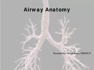
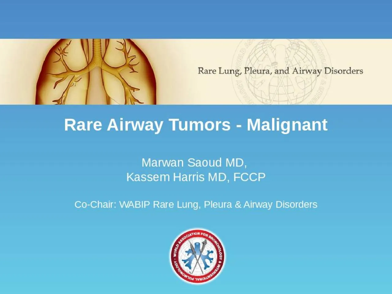
Marwan Saoud MD Kassem Harris MD FCCP CoChair WABIP Rare Lung Pleura amp Airway Disorders Background Rare Airway Tumors RATs Tracheobronchial tumors that have not been extensively studied in ID: 908211
Download Presentation The PPT/PDF document "Rare Airway Tumors - Malignant" is the property of its rightful owner. Permission is granted to download and print the materials on this web site for personal, non-commercial use only, and to display it on your personal computer provided you do not modify the materials and that you retain all copyright notices contained in the materials. By downloading content from our website, you accept the terms of this agreement.
Slide1
Rare
Airway
Tumors - Malignant
Marwan
Saoud
MD,
Kassem Harris MD, FCCP
Co-Chair
: WABIP Rare Lung, Pleura & Airway Disorders
Slide2BackgroundRare Airway Tumors (RATs)
Tracheobronchial tumors that have not been extensively studied in literature due to limited diagnostic feasibly
They represent 0.1% of all primary lung tumors
Occur anywhere from the subglottus to the segmental bronchioles
Often misdiagnosed in early stages as obstructive lung disease
Lancet Oncol
2006;
7
(1): 83-91
Slide3RATs Cell Type
Mesenchymal Cell Salivary Gland
Epithelial Cell
Miscellaneous
Slide4Mesenchymal Cell RATs
MalignantFibrosarcomaChondrosarcomaT-cell Lymphoma
Slide5Salivary Gland RATs
MalignantMyoepithelial CarcinomaMucoepidermoid Carcinoma
Adenoid
Cystic Carcinoma
Slide6Epithelial Cell RATs
MalignantCarcinoid Tumor
Slide7Fibrosarcoma
Malignant mesenchymal cell tumor Prevalent in children and young adults, males>females
Associated with exposure to radiation
Manifest as atelectasis or post-obstructive pneumonitis on x-ray and as smooth, lobular nodules or masses on CT scan
Appears as a multi-nodular mass on bronchoscopy
Biopsy is the definitive diagnosis and reveal spindle cells in herringbone pattern
Bronchoscopic resection is the preferred modality of treatment
Am J
Otolaryngol
2015;
36
(2): 287-9
Slide8Chondrosarcoma
Malignant mesenchymal cell tumor Mean age 30-60 years with male:female ratio of 1.3:1
Characteristic CT findings including bone and soft-tissue involvement with scattered areas of calcification
Appears as a polypoid mass on bronchoscopy
Biopsy is the definitive diagnosis and reveal cartilaginous and binuclear cells with open chromatin
Treatment options include:
Surgical resection
Adjuvant chemotherapy and/or radiation therapy for extensive tumors
Radiographics
2002;
22
(3): 621-37
Slide9T-cell Lymphoma
Malignant mesenchymal cell tumor Prevalent in adults age 40-60 years old, females>males
Associated with tobacco smoking
Variable size lesions on radiological imaging as well as bronchoscopy
Biopsy is the definitive diagnosis
Tissue stains positive for CD3, CD4, and CD5
Treatment options include:
Chemotherapy: pirarubicin, cyclophosphamide, vincristine and steroids
Surgical resection after chemotherapy
Respirol Case Rep
2015;
3
(2): 44-7
Slide10Computed tomography (CT) revealed chronic pyothorax with calcified foci on the right and a mass inside the bronchus intermedius.
Flexible bronchoscopy identified an endobronchial tumor obstructing the bronchus intermedius. Positron emission tomography with [18F] fluoro-2-deoxyglucose and CT revealed uptake at the endobronchial tumor. CT after the chemotherapy demonstrated that the endobronchial tumor markedly diminished.
Matsumoto et al.
Respirol
Case Rep
2015;
3
(2): 44-7
T-cell Lymphoma
Slide11Myoepithelial Carcinoma
Malignant salivary gland tumor 20 cases reported, Male:Female ratio of 1:1
Detected as opaque shadows with defined borders on x-ray and CT scans
Appears as a smooth, vascular mass with defined borders on bronchoscopy
Biopsy is the definitive diagnosis
Histology consistent with glandular differentiation with dual epithelial and myoepithelial cell population; occasional atypia and increased mitotic figures seen
Tissue stains positive for p-53 and c-Kit (CD117)
Surgical resection is the preferred treatment modality
Arch
Pathol
Lab Med
2004;
128
(1): 92-4
Slide12Mucoepidermoid Carcinoma
Malignant salivary gland tumor
Reported in younger population (<30 years of age), equal in males and females
Appears as ovoid or lobulated polypoid nodules on x-ray
Have the characteristic "
pneumonic consolidation
”
and "
punctuate calcifications
" on CT scan
Appears a glossy, non-vascular mass on bronchoscopy
Biopsy is the definitive diagnosis
Histology consistent with mucus-secreting cells, squamous cells and intermediate cells
Treatment options include:
Bronchoscopic resection
Surgical resection
Arch Pathol Lab Med
2007;
131
(9): 1400-4
Mod Pathol
2014;
27
(11): 1479-88
Slide13Mucoepidermoid Carcinoma
Left main stem completely occluded with mucoepidermoid tumor
Slide14Mucoepidermoid Carcinoma
Axial chest CT showing a highly vascularized left main stem occlusive mucoepidermoid tumor
Coronal chest CT showing a complete obstruction of the left main stem with mucoepidermoid tumor
Slide15Mucoepidermoid Carcinoma
Neoplastic tissue composed of round to oval epithelioid cells and occasional goblet cells punctuated by mucin containing cystic spaces
Slide16Adenoid Cystic Carcinoma
Malignant salivary gland tumor Equal prevalence in males and females, mean age of 46 years
Detectable on x-ray and CT as well as positive uptake on PET scan
Appears a nodular, vascular lesion with characteristic
“ice-berg
”
appearance on bronchoscopy
Biopsy is the definitive diagnosis with 3 histological cell subtypes:
Tubular , Cribriform
and
Solid
(most aggressive)
Tissue stains positive for keratin, CK7, CD117S‑100, and SMA
Treatment options include:
Surgical resection
Bronchoscopic resection
Pneumonectomy if there is extensive bronchial involvement
Oncol
Lett
2015;
9
(3): 1475-81
Clin Oncol (R Coll Radiol)
2015;
27
(12): 732-40
Slide17Carcinoid Tumor
Malignant epithelial cell tumor Prevalent in younger population (<35 years of age)
Appears as spherical or ovoid nodules on radiological imaging with vascular enhancement on CT scan
Appears as a large polypoid lesion with narrow stalk arising from the lumen on bronchoscopy
Biopsy is the definitive diagnosis with intra-cytoplasmic granules on electron microscope
Tissue stains positive for chromogranin and synaptophysin
Treatment options include:
Surgical resection
Bronchoscopic ablation
Mayo Clin Proc
1993;
68
(8): 795-803
Case Rep Pulmonol
2014;
2014
: 349707
Slide18Carcinoid Tumor
Carcinoid tumor of the right middle lobeRadial Endobronchial Ultrasound showing the highly vascularized tumor (arrows).
Slide19Outcomes
RATs prognosis depend on multiple factors:
Tumor malignant potential
Tumor location
Patient’
s co-morbidities
Risks of treatment modality
Benign tumors are usually localized and amendable to resection with no or minimal risk of recurrence
Outcome of malignant tumors depend mainly on lymph node and adjacent tissue metastasis
Tumors found on the carina have poor prognosis due to the high risk of surgical resection attributed to the anatomical feasibility
Intern Med
2013;
52
(18): 2113-6
Lancet Oncol
2006;
7
(1): 83-91
Slide20This presentation was prepared by
Marwan
Saoud
MD and
Kassem Harris MD, FCCP
and reviewed for accuracy and content by members of the
WABIP
Rare Lung, Pleura and Airway Disorders
section