/
Pancreatic Islets (Islets of Langerhans) Pancreatic Islets (Islets of Langerhans)

Pancreatic Islets (Islets of Langerhans) - PowerPoint Presentation

bety
bety . @bety
Follow
65 views
Uploaded On 2023-11-22

Pancreatic Islets (Islets of Langerhans) - PPT Presentation

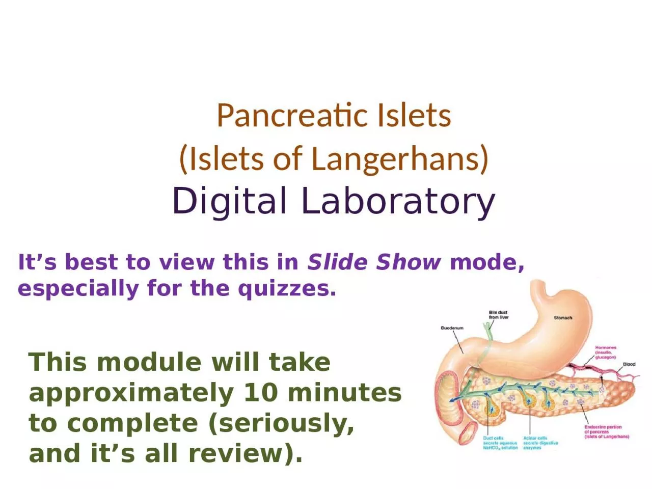
Digital Laboratory Its best to view this in Slide Show mode especially for the quizzes This module will take approximately 10 minutes to complete seriously and its all review After completing this exercise you should be able to ID: 1034281

islets cells exocrine pancreas cells islets pancreas exocrine endocrine pancreatic langerhans identify granules secretory secrete aciniexocrine stain side rer

Share:

Link:

Embed:

Download Presentation from below link

Download Presentation The PPT/PDF document "Pancreatic Islets (Islets of Langerhans)" is the property of its rightful owner. Permission is granted to download and print the materials on this web site for personal, non-commercial use only, and to display it on your personal computer provided you do not modify the materials and that you retain all copyright notices contained in the materials. By downloading content from our website, you accept the terms of this agreement.


Presentation Transcript

1. Pancreatic Islets(Islets of Langerhans)Digital LaboratoryIt’s best to view this in Slide Show mode, especially for the quizzes.This module will take approximately 10 minutes to complete (seriously, and it’s all review).

2. After completing this exercise, you should be able to: identify, at the light microscope level, each of the following: PancreasSerous aciniPancreatic islets (Islets of Langerhans)

3. GLANDSGlands in the body develop through an interaction of an epithelium and underlying connective tissue. The epithelial cells that line the surface of the body (e.g. epidermis of skin, inner lining of the gut) proliferate, and then invaginate into the underlying connective tissue. Note the basement membrane invaginates with the epithelium. From here, two things can happen:For exocrine gland formation, a lumen develops, and secretory cells secrete their substances out their apical side (arrows).For endocrine glands, the secretory cells lose their connection to the original epithelium, forming clusters of cells that secrete out their basal side, across the basement membrane, and into the bloodstream.In the pancreas, some cells become exocrine, while others become endocrine.

4. PANCREASThis image shows the stomach and the beginning of the small intestine, the duodenum. The pancreas (and liver) develop from the duodenum, and, therefore, are connected to it by ducts. The glandular cells in the pancreas are either exocrine (acinar) or endocrine (Islets of Langerhans).from:www.gopetsamerica.comseriously

5. The exocrine portion of the pancreas secretes a large amount of digestive enzymes. The details of these cells will be worked out in the Gastrointestinal Block, but you should recognize a cluster of serous acini when you see one.Because these cells synthesize a ton of digestive enzymes (proteins), they exhibit intense cytoplasmic basophilia on their basal side, and intense cytoplasmic eosinophilia on their apical side. I hope you remember why they stain this way. EXOCRINE PANCREAS

6. The endocrine portion of the pancreas forms clusters of cells called pancreatic islets (Islets of Langerhans - yellow outline). Although the cells within the islets secrete protein hormones, and, therefore, exhibit cytoplasmic basophilia and eosinophilia, their staining is not nearly as intense as that of the exocrine acinar cells. (FYI arrows are identifying possible α cells, really can’t say for sure)If you stop to think about it, this makes sense. Protein hormone production does require rER, Golgi, secretory vesicles; therefore, Islet cells will exhibit some staining characteristics. However, you don’t need much hormone to circulate in the body and elicit an appropriate response in target cells. After all, it’s only a message.The exocrine acinar cells, however, have to deal with large volumes of meals daily (especially on holidays). They need to secrete a ton of digestive enzymes, so they contain more rER, Golgi, and secretory vesicles than islet cells, and, therefore, stain much more intensely. ENDOCRINE PANCREASExocrine aciniExocrine aciniExocrine aciniExocrine acini

7. Video of pancreatic islets – SL108Link to SL 108 and SL 054Be able to identify:Exocrine pancreas (serous acini)Pancreatic Islets (Islets of Langerhans)Video of pancreatic islets – SL54Yeah, I know, I missed an obvious islet in the second video, but it wasn’t very nice, was it?ENDOCRINE PANCREAS

8. To underscore the difference in exocrine and endocrine pancreatic cells, this slide was specially prepared so that: DNA is stained red RNA is stained blueNote that, although the cytoplasm of the pancreatic islet cells do exhibit RNA staining, the exocrine acinar cell cytoplasm contains much more RNA PANCREAS – RNA STAIN

9. Video of pancreatic islets RNA – SL2ALink to SL 002A  Be able to identify:Exocrine pancreas (serous acini)Pancreatic Islets (Islets of Langerhans)MALE REPRODUCTIVE SYSTEM

10. Routine H&E cannot distinguish between the different cells within the islets of Langerhans. However, this specially-stained slide shows: α cells – stained red, secrete glucagon β cells – stain blue, secrete insulin δ cells (and other islet cells) - not specially stained here, release somatostatin ENDOCRINE PANCREAS – SPECIAL STAINExocrine aciniExocrine aciniExocrine aciniExocrine acini

11. Video of pancreatic islets special staining – SL109Link to SL 109 Be able to identify:Nothing specific, just realize cells in the pancreatic islets can be specifically identified with special stains, antibodies, etc.ENDOCRINE PANCREAS

12. This is another special stain for your viewing pleasure.ENDOCRINE PANCREAS – SPECIAL STAIN

13. Just FYI, this EM demonstrates that cells from the Islets of Langerhans have the protein machinery expected of cells with regulated secretion: rER (not easily seen), Golgi, secretory granules (arrows)ENDOCRINE PANCREAS

14. FYI again, for reference, this is a portion of the exocrine pancreas, with the lumen (L) of an acinus shown. Note that the acinar cells exhibit more elaborate rER, Golgi, and much larger secretory granules (Z, they are called zymogen granules).EXOCRINE PANCREASSimilar to what you encountered with the pituitary, etc., it’s often difficult to definitively identify similar cells on EM. In other words, rER, Golgi, secretory granules; the cell definitely is involved in regulated secretion, but it could be in a number of places. In this case, the presence of the lumen and the fact that the granules are toward the apical side of the cell is helpful in narrowing it down, but it does not completely rule out other tissues you will see, such as salivary glands.However, note that you certainly should be able to compare protein-secreting cells from steroid-secreting cells. In addition, once we learn about these exocrine cells in detail, if you were shown two EM images similar to the ones in this module, and you had to match them to exocrine or endocrine pancreas, we would hope you could do so. Or maybe that lumen and apical granules would help you when comparing to cells with granules on their basal side.

15. The next set of slides is a quiz for this module. You should review the structures covered in this module, and try to visualize each of these in light and electron micrographs. identify, at the light microscope level, each of the following: PancreasSerous aciniPancreatic islets (Islets of Langerhans)

16. Final quizSelf-check: Identify the organ and structure. (advance slide for answers). Do NOT let me down – this is for all the marbles.PancreasIslet of Langerhans