/
The Egyptian Journal of Hospital Medicine January 2014 Vol 54 Page The Egyptian Journal of Hospital Medicine January 2014 Vol 54 Page

The Egyptian Journal of Hospital Medicine January 2014 Vol 54 Page - PDF document

eliza
eliza . @eliza
Follow
345 views
Uploaded On 2022-10-26

The Egyptian Journal of Hospital Medicine January 2014 Vol 54 Page - PPT Presentation

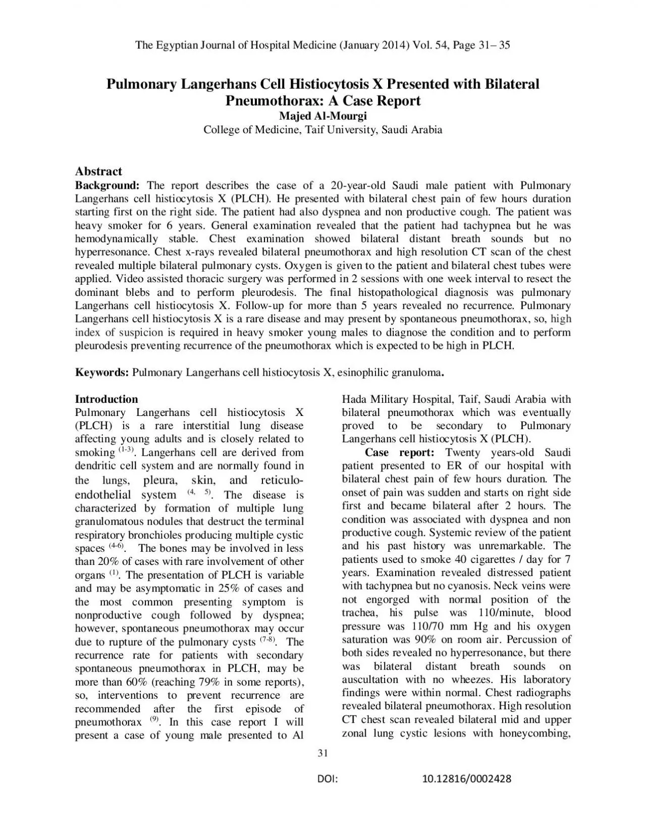
31 35 31 DOI 10128160002428 Pulmonary Langerhans Cell Hist i ocytosis X Presented with Bilateral Pneumothorax A Case Report Majed A l Mourgi College of Medicine Taif University Saud ID: 960399

langerhans pulmonary histiocytosis cell pulmonary langerhans cell histiocytosis pneumothorax disease bilateral patient chest lung plch revealed case ocytosis years

Share:

Link:

Embed:

Download Presentation from below link

Download Pdf The PPT/PDF document "The Egyptian Journal of Hospital Medicin..." is the property of its rightful owner. Permission is granted to download and print the materials on this web site for personal, non-commercial use only, and to display it on your personal computer provided you do not modify the materials and that you retain all copyright notices contained in the materials. By downloading content from our website, you accept the terms of this agreement.


Presentation Transcript

The Egyptian Journal of Hospital Medicine (January 2014) Vol. 54, Page 31 – 35 31 DOI: 10.12816/0002428 Pulmonary Langerhans Cell Hist i ocytosis X Presented with Bilateral Pneumothorax : A Case Report Majed A l - Mourgi College of Medicine, Taif University, Saudi Arabia Abstract Background: The report describes the case of a 20 - year - old Saudi male patient with Pulmonary Langerhans cell hist i ocytosis X (PLCH ) . He presented with bilateral chest pain of few hours duration starting first on the right side. The patient had also dyspnea and non productive cough. The patient was he a vy smoker for 6 years. General e xamination revealed that the patient had tachypnea but he was hemod ynamically stable . Chest exa mination showed bilateral distant breath sounds but no hyperresonance. Chest x - rays revealed bilateral pneumothorax and high resolution CT scan of the chest revealed multiple bilateral pulmonary cysts. Oxygen is given to the pat ient and bilateral chest tubes were applied. Video assisted thoracic surgery was performed in 2 sessions with one week interval to resect the dominant blebs and to perform pleurodesis. The final histopathological diagnosis was p ulmonary Langerhans cell his t i ocytosis X . Fol low - up for more than 5 years revealed no recurrence. Pulmonary Langerhans cell hist i ocytosis X is a rare disease and may present by s pontaneous pneumothorax , so, high index of suspicion is required in heavy smoker young males to diagnose the condition and to perform pleurodesis prevent ing recurrence of the pneumothorax which is expected to be high in PLCH. Keywords: Pulmonary Langerhans cell histiocytosis X, esinophilic granuloma . Introduction Pulmonary Langerhans cell hist i ocytosis X (PLCH) is a rare interstitial lung disease affecting young adults and is closely related to smoking ( 1 - 3) . Langerhans cell are derived from dendritic cell system and are normally fou nd in the lungs, pleura, skin, and reticulo - endothelial system (4, 5 ) . The disease is characterized by formation of multiple lung granulomatous nodules t hat destruct the terminal respiratory bronchioles producing multiple cystic spaces (4 - 6) . The bones may be involved in less than 20% of cases with rare involvement of other organs (1) . The presentation of PLCH is variable and may be asymptomatic in 25% of cases and the most common presenting symptom is nonproductive cough followed by dyspnea; however, spontaneous pneumothorax may occur due to rupture of the pulmonary cysts (7 - 8) . The recurrence rate for patients with secondary spontaneous pneumothorax in PLCH , may be more than 60% (reaching 79% in some reports) , so, interventions to prevent re currence are recommended after the first episode of pneumothorax (9) . In this case report I will present a case of young male presented to Al Hada Military Hospital, Taif, Saudi Arabia with bilateral pneumothorax which w as eventually proved to be secondary to Pulmonary Langerhans cell hist i ocytosis X (PLCH) . Case report: Twenty

years - old Saudi patient presented to ER of our hospital with bilateral chest pain of few hours duration. The onset of pain was sudden and starts on right side first and beca me bilateral after 2 hours. The condition was associated with dyspnea and non productive cough . Systemic review of th e patient and his past history was unremarkable. The patients used to smoke 40 cigarettes / day for 7 years. Examination revealed distressed patient with tachypnea but no cyanosis. Neck veins were not engorged with normal position of the trachea , his pulse was 110/minute , blood pressure was 110/70 mm Hg and his oxygen saturation was 90% on room air . Percussion of both sides revealed no hyperresonance , but there was bilateral distant breath sounds on auscultati on with no wheezes . His laboratory findings were within normal . Chest radiograph s revealed bilateral pneumothorax . High resolution CT chest scan revealed bilateral mid and upper zonal lung cystic lesion s with honeycombing , Pulmonary Langerhans Cell Histiocytosis X… 32 which was consistent with the diagnosis of PLCH , however, other cystic lung lesions were suspected as differential diagnos e s (Fig 1) . Oxygen was delivered to the patient through a mask with a rate of 5L/hr. Two intercost al tubes were inserted bilaterally and the patient was prep ared for Video - assisted thoracoscopic surgery (VATS) on the right side . Thoracoscopic examination showed diffused cystic lesions on the lungs, and then target areas (dominant blebs) were identified at the right upper lobe which was resected using a stapler device with enough amounts of surrounding tissues for histopathological examination . In the first session mechanical pleurodesis was added. The pathological report revealed presence of areas of scaring with nu merous Langerhans cells (positive for S - 100), esinophils, lymph ocytes and fibroblasts (Figure 2, 3 ) and a definitive diagnosis of pulmonary Langerhans cell histiocytosis X was established. A week later , v ideo - assisted thoracoscopic surgery was performed on the left side and the major blebs were also resected with talc pleurodesis as the talc was not available at the first session . The patient had uneventful postoperative course and he was discharged after 7 days . The p atient stopped smoking and follow up for more than 5 years revealed no recurrence and both l ung s were fully inflated with no difference between both sides , although different mechanism s of pleurodesis have been used . Discussion Pulmonary Langerhans cell hist i ocytosis X (PLCH) is a rare interstitial lung disease characterized by formation of multiple lung cysts with rare involvement of other organs (1) . A similar disease occurs in children but with multiple organ affection is known as pediatric histiocytic disorders (4) . The PLCH is also known as pulmonary histi ocytosis X or eosinophilic granuloma and it affects young adults with peak incidence around 2 0 - 40 years and males are more affected than

females (1 - 3) . In this case report the patient was 20 years old Saudi male . The actual prevalence of PLCH in Saudi Arabia and other developing countries is not known, however, some reports from developed countrie s estimated the prevalence of the disease to be 0.7 - 2.7 per million populations (3) . The disease is rel ated to tobacco smoking; and starts as peribronchiolar granulomatous infiltrates especially in the upper and middle lung zones (sites of smoking - related lung diseases) (1) , which is consistent with the finding of the present case. However, genetic, environmental, radiation, and chemot herapy are also encountered as susceptibility factors (4) . The PLCH granulomata are formed of activated Langerhans cells in addition to esinophils , macrophages, lymphocytes, plasma cells, and fibroblasts (5) . The same findings were detected in the current case. The granulomata destruct the terminal respiratory bronchioles and produce stellate scars ending in formation honeycombing and cystic spaces (5 - 6) . Presentation of PLCH is variable and the most common presenting symptom is non productive cough followed by dyspnea, easy fatigability, loss of weight, fever, chest pain, painful bone cysts which may lead to pathological fractures and about one quarter of patients are diagnosed incidentally during chest radiography (7) . Rupture of the pulmonary cysts will lead to spontaneous pneumothorax which is a recognizable presentation. The current patient presented with bilateral pneumothorax without any suggestive history preceding the presentation. There are no specific physical findings and are mostly related to the underlying present ation (7) . The pulmonary Langerhans cell histiocytosis X must be differentiated from; chronic obstructive pulmonary disease , cystic fibrosis, idiopathic or interstitial pulmonary fibros is, emphysema , and sarcoidosis ( 9) . Laboratory findings are non specific and pulmonary functions may be normal but in advanced disease it will show restrictive, obstructive, or mixed abnormalities (1 - 4) . The characteristic chest x - ray findings include; bilateral, symmetrical, ill - defined no dular lesions with ill defined outline which are mainly found in the upper or mid - pulmonary zones with sparing of the costophrenic angles (10) . The presence of cystic lesions of variable size and wall thickness or honeycomb Majed Al - Mourgi 33 appearance indicates advanced di sease (1,10. 11) . Lymphadenopathy and pleural effusion are rarely encountered (10) . Cystic bony lesions may be seen on the ribs or other sites ( 1, 11) . In the present case chest x - rays detected only bilateral pneumothorax with non identifiable other findin gs . H igh Resolution Computed Tomography (HRCT) of the chest may be diagnostic and the following findings are pathognomonic; upper or mid zonal nodules which are variable in size and may be cavitary in addition to presence of cysts of various wall thicknes ses and diameters (10, 11) . Consistent findings were detected in t he current patient ,

so , pulmonary histi ocytosis X, though it is rare , was the first differential diagnosis . The standard diagnostic modality is thoracoscopic or open lung biopsy and its sensitivity and specificity are better than analysis of the bronchoalveolar lavage fluid or transbronchial biopsy (10 - 12) . Langerhans cells are positive for S - 100, CD1 a, and mono clonal antibody MT - 1 (9 - 11 ) . Moreover , electron microscopy examination of the suspected PLCH lesion would show characteristic increased number of intra - cytoplasmic Birbeck granules (12) . In this case; the examination of the resected lung tissues taken by VATS confirmed the presence of typical Langerhans cells which was positive for S - 100 in addition to the presence of other granulomatous cells. As pulmonary Langerhans cell histiocytosis X is a rare disease, designed therapeutic protocols based on randomized prospective studies are lacking; however, abstinence of smoking is the mainstay in the management of PLCH and it often leads to disease regression or at least stabilizes the condition in addition to decreasing the risk of development of bronchogenic carcinoma (13 - 15) . Other therapeutic measures include Control of infection, bronchodilators if obst ructive ventilatory defect is found , in addition to oxygen supplementation if there is sign ificant hypoxemia ( 12) . Corticosteroids may be of value in progressive disease or persistent pulmonary symptoms (13) . Interleukin - 2 and antitumor necrosis factor alpha are still under investigation as a promising therapy ; however l ung transplantation may be an option in selected patients with advanced disease ( 13, 14, 16) . If spontaneous pneumothorax is developed insert ion of chest tube is indicated and prevention of recurrence, which is common, by talc insufflation, parietal pleurectomy, or mechanical pleurodesis is recommended by most of the experts (15) . In the current case, the major blebs were resected and pleurodesis was added and both lungs were fully inflated with no difference between both sides although different mechanisms of pleurodesis have been used. The course of the disease is unpredictable and some factors carr y the risk of a poorer prognosis as; recurrent pneumothorax, severe pulmonary hypertension, multiorgan affection other than the bones, especially, when the posterior pituitary is involved with diabetes insipidus, ( 4, 7 - 9 ) . However , most of the studies sho wed that the median survival is about 12 - 13 years after presentation (8) . Th e current patient stopped smoking postoperatively and follow up for more than 5 years revealed no recurrence . To conclude; Pulmonary Langerhans cell histiocytosis X is a rare disease and may present by spontaneous pneumothorax, so, high index of suspicion is required in heavy smoker young males to diagnose the condition and to perform pleurodesis prevent ing recurrence of the pneumothorax which is expected to be high in PL CH. Consent: Written informed consent was obtained from the patient for publication o

f this Case report . R eferences 1. Podbielski FJ, Worley TA, Korn JM, Connolly MM (2009) . Eosinophilic granuloma of the lung and rib. Asian Cardiovasc Thorac Ann ., 17 :194 - 5. 2. Thomeer M, Demedts M, Vandeurzen K (2001) . Registration of interstitial lung diseases by 20 centres of respiratory medicine in Flanders. Acta Clin Belg ., 56:163 - 72. 3. Watanabe R, Tatsumi K, Hashimoto S, Tamakoshi A, Kuriyama T (2001) . Clinico - epidemiological features of pulmonary histiocytosis X. Intern Med ., 40:998 - 1003. 4. Vassallo R, Ryu JH, Schroeder DR, Decker PA, Limper AH (2002) . Clinical outcomes of pulmonary Langerhans' - cell histiocytosis in adults. N Engl J Med ., 346:484 - 90. 5. Stach er E, Beham - Schmid C, Terpe HJ, Simiantonaki N, Popper HH (2009). Pulmonary Langerhans Cell Histiocytosis X… 34 Pulmonary histiocytic sarcoma mimicking pulmonary Langerhans cell histiocytosis in a young adult presenting with spontaneous pneumothorax: a potential diagnostic pitfall. Virchows Arch ., 455: 187 - 90. 6. Arico M (2004). Langerhans cell histiocytosis in adults: more questions than answers?. Eur J Cancer , 40: 1467 - 73. 7. Delobbe A, Durieu J, Duhamel A, Wallaert B (1996) . Determinants of survival in pulmonary Langerhans' cell granulomatosis (histioc ytosis X). Groupe d'Etude en Pathologie Interstitielle de la Société de Pathologie Thoracique du Nord. Eur Respir J ., 9 :2002 - 6. 8. El Fekih N, Inés K, Jones M, et al., (2013). Histiocytosis X Revealed by Diabetes Insipidus and Skin Lesions. Am J Derm., 35: 606 - 8 . 9. Howarth DM,Gilchrist GS,Mullan BP,Viseman GA ,Edmonson JH ,Schmberg PJ (1999) . Langerhans cell histiocytosis: diagnosis, natural history, management and outcome . Cancer, 85:2278 - 90. 10. Vassallor,Ryu JH,Golby TV. Et al . (2000). Pulmonary langerhans cell h istiocytosis.N Engl . J Med., 342:1969 - 78. 11. Dacic S, Trusky C, Bakker A, Finkelstein SD, Yousem SA (2003). Genotypic analysis of pulmonary Langerhans cell histiocytosis. Hum Pathol . , 34:1345 - 9. 12. King TE, Crausman RS (1998). Pulmonary histiocytosis X. In: Fishman AP, ed. Fishman's Pulmonary Diseases and Disorders . Vol 1. 3 rd ed. N ew York, NY: Mc - Graw Hill; 1163 - 70. 13. Sundar KM, Gosselin MV, Chung HL, Cahill BC (2003). Pulmonary Langerhans cell histiocytosis: emerging concepts in pathobiology, radiology, and clinical evolution of disease. Ches ., 123:1673 - 83. 14. Tazi A (2006) . Adult pulmonary Langerhans' cell histiocytosis. Eur Respir J . , 27(6):1272 - 85. 15. Vassallo R, Ryu JH (2004). Pulmonary Langerhans' cell histiocytosis . Clin Chest Med ., 25: 561 - 71. 16. Alalawi R, Whelan T, Bajwa RS, Hodges TN (2005) . Lung tran splantation and interstitial lung disease. Curr Opin Pulm Med ., 11 :461 - 6. Figure 1: S hows bilateral pneumothorax with multiple cysts and honeycombing Figure 2: Shows the histiocytic granuloma (Hx & E x40) Majed Al - Mourgi 35 Figure 3: Langerhans cells positive for S - 100 (x 40