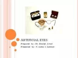
/
 Artificial eyes Prepared by : Dr.  Artificial eyes Prepared by : Dr.

Artificial eyes Prepared by : Dr. - PowerPoint Presentation

briana-ranney
briana-ranney . @briana-ranney
Follow
347 views
Uploaded On 2020-04-09

Artificial eyes Prepared by : Dr. - PPT Presentation

Khaled Awad Presented by T Aisha I Lubbad Introduction An artificial eye is a replacement for a natural eye lost because of injury disease trauma tumor or end stage ocular disease such as glaucoma or diabetes can be devastating at any age ID: 776539

eye implant prosthesis artificial eye implant prosthesis artificial socket cases enucleation muscles evisceration orbital motility iris size exposure ocular

Share:

Link:

Embed:

Download Presentation from below link

Download Presentation The PPT/PDF document " Artificial eyes Prepared by : Dr. " is the property of its rightful owner. Permission is granted to download and print the materials on this web site for personal, non-commercial use only, and to display it on your personal computer provided you do not modify the materials and that you retain all copyright notices contained in the materials. By downloading content from our website, you accept the terms of this agreement.


Presentation Transcript

Slide1

Artificial eyes

Prepared by : Dr. Khaled AwadPresented by : T. Aisha I. Lubbad

Slide2

Introduction

An artificial eye is a replacement for a natural eye lost because of injury ,

disease , trauma

, tumor, or end stage ocular disease such as glaucoma, or diabetes can be devastating at any age

.

Although the replacement cannot provide sight, it fills the cavity of the eye socket and serves as a cosmetic enhancement.

Slide3

It may have a major impact on one’s self-image, self-confidence, and self-esteem, not to mention the adjustment required in adapting to monocular (single-eye) vision.

There

may even be some job restrictions that apply for one-eyed patients (e.g., commercial drivers, airline pilots, policemen, firefighters, etc.).

Numerous

patients have been fitted with prosthetic eyes who are currently employed in the professions mentioned above

.

Slide4

Monocular patients should try to return to their work if possible and lead as normal a life as possible.

Although there is some loss of depth perception and peripheral vision, they are not as “handicapped” as many believe

.

Slide5

Before the availability of artificial eyes, a person who lost an eye usually wore a patch. An artificial eye can be attached to muscles in the socket to provide eye movement

Today, most artificial eyes are made of plastic, with an average life of about 10 years

.

Children require more frequent replacement of the prosthesis due to rapid growth changes. As many as four or five

prosthesis

may be required from infancy to adulthood.

Slide6

According to the Society for

the Prevention

of Blindness, between 10,000 and 12,000 people per year lose an eye.

Though

50% or more of these eye losses are caused by an accident (in one survey more males lost their eyes to accidents compared to females), there are a number of inherited conditions that can cause eye loss or require an artificial eye.

Slide7

Microphthalmia

is a birth defect where for some unknown reason the eye does not develop to its normal size. These eyes are totally blind, or at best might have some light

perception .

Slide8

Some people are also born without one or both eyeballs. Called

anophthalmia

, this presents one of the most difficult conditions for properly fitting an artificial eye. Sometimes the preparatory work can take a year or more. In some cases, surgical intervention is necessary.

Slide9

Retinoblastoma is a congenital (existing at birth) cancer or tumor, which is usually inherited. If a person has this condition in just one eye, the chances of passing it on are one in four, or 25%. When the tumors are in both eyes, the chances are 50%. Other congenital conditions that cause eye loss include cataracts and glaucoma. One survey showed that 63% of eye loss due to disease occurs before 50 years of age.

Slide10

There are two key steps in replacing a damaged or diseased eye. First, an ophthalmologist or eye surgeon must remove the natural eye. There are two types of operations. The

enucleation removes the eyeball by severing the muscles, which are connected to the sclera (white of eyeball). The surgeon then cuts the optic nerve and removes the eye from the socket. An implant is then placed into the socket to restore lost volume and to give the artificial eye some movement, and the wound is then closed.

14/2/2014

Slide11

With evisceration, the contents of the eyeball are removed. In this operation, the surgeon makes an incision around the iris and then removes the contents of the eyeball. A ball made of some inert material such as plastic, glass, or silicone is then placed inside the eyeball, and the wound is

closed.

Slide12

At the conclusion of the surgery, the surgeon will place a conformer (a plastic disc) into the socket. The conformer prevents shrinking of the socket and retains adequate pockets for the prosthesis. Conformers are made out of silicone or hard plastic. After the surgery, it takes the patient from four to six weeks to heal. The artificial eye is then made and fitted by a professional

ocularist

.

Slide13

Slide14

Primary socket procedure

There are three types of eye removal

Evisceration

- removal of the internal eye contents, but the

sclera

is left behind with the

extraocular

muscles still attached.

Enucleation

- removal of the

eyeball

, but the adjacent structures of the eye socket and eyelids remain. An intraocular tumor excision requires an

enucleation

, not an evisceration

.

Slide15

Exenteration

- removal of the contents of the eye socket (orbit) including the eyeball, fat, muscles and other adjacent structures of the eye. The eyelids may also be removed in cases of cutaneous cancers and unrelenting infection.

Exenteration

is sometimes done together with

Maxillectomy

which is removal of the maxilla or the upper jaw bone/cheekbone

Slide16

Reasons for eye removal

Cancer

of the eye (

retinoblastoma

, melanomas, any other cancers of the eye or orbit)

Severe injury of the eye when the eye cannot be saved or attempts to save the eye have failed

End stage

glaucoma

Painful

, blind

eye .

Slide17

In cases of

sympathetic

ophthalmia

(inflammation of the eye) to prevent travel to other eye, in which, if untreated can cause

blindness

Congenital cystic eye

In a deceased person, so the

cornea

can be used for a living person who needs a

corneal transplant

by a surgical operation called

keratoplasty

.

Constant infection in a blind, or otherwise useless eye.

Slide18

Enucleation

Definition Enucleation is the surgical removal of the eyeball that leaves the eye muscles and remaining orbital contents intact.

18/2/2014

Slide19

Purpose

Enucleation

is performed to remove large-sized eye tumors or as a result of traumatic injury when the eye cannot be preserved. In the case of tumors, the amount of radiation required to destroy a tumor of the eye may be too intense for the eye to bear. Within months to years, many patients who are treated with radiation for large ocular melanomas lose vision, develop glaucoma, and eventually have to undergo

enucleation

.

Slide20

The two types of eye tumors that may require

enucleation

are:

Intraocular eye melanoma. This is a rare form of cancer in which malignant cells are found in the part of the eye called the uvea, which contains cells called melanocytes that house pigments. When the melanocytes become cancerous, the cancer is called a melanoma. If the tumor reaches the iris and begins to grow, or if there are symptoms,

enucleation

may be indicated.

Slide21

Retinoblastoma. Retinoblastoma is a malignant tumor of the retina. The retina is the thin layer of tissue that lines the back of the eye; it senses light and forms images. If the cancer occurs in one eye, treatment may consist of

enucleation

for large tumors when there is no expectation that useful vision can be preserved. If there is cancer in both eyes, treatment may involve

enucleation

of the eye with the larger tumor, and radiation therapy for the other eye.

Slide22

Slide23

The conjunctiva (outer covering of eye) is removed with blunt scissors (A). The four rectus muscles are removed from their attachments to the eyeball (B). The optic nerve is severed (C), and the eyeball is removed. A synthetic globe replaces the eyeball in the socket, and the rectus muscles are sutured around it (D).

Slide24

Enucleation

Anesthesia The procedure may be performed under general or local anesthesia, according to the surgeon's preference. In either case, the injection is given in the retrobulbar space. An antibiotic (e.g., cefazolin) and an anti-inflammatory (e.g., dexamethasone) are also given intravenously.

23/2/2014

Slide25

Peritomy

, Muscle Identification, and

Neurectomy

A 360°

peritomy

is then performed, with care taken to preserve as much of the conjunctiva and

Tenon's

capsule as possible. The

extraorbital

muscles are then tagged with 5-0

Vicryl

sutures and are released by resection at their attachments to the globe. A

neurectomy

is then performed, according to the surgeon's preference, and the globe is delivered. Care should be taken to maintain hemostasis until the implant is placed in the orbit.

Slide26

Evaluation and Preparation of

Tenon's

capsule

Tenon's

capsule is then inspected for its readiness to accommodate the implant. In order to minimize the risk of implant exposure and subsequent infections, it is important to ensure that the tissues overlying the implant can be closed without tension.

Slide27

Traditionally, the orbital implant was placed within

Tenon's

capsule, as first described by Frost (

Frost 1887

), necessitating the use of a smaller implant. This technique usually required the use of implants with a diameter of 16 mm to 18 mm. However, due to the

enophthalmos

associated with these smaller

implants .

Slide28

Determination of Implant Size

Proper sizing of the orbit is essential to avoid complications and to achieve an optimum fit of the implant to both the orbit and the artificial eye. An undersized implant may result in

enophthalmos

or a deep superior sulcus, and may necessitate weight-increasing modifications to the artificial eye which, in turn, induce lower lid sag and restricted motility.

Slide29

Conversely, an oversized implant increases the risks of wound dehiscence, exposure, and infection, and may complicate the coupling of the artificial eye to the motility peg. An implant is of the proper size when its volume, added to the volume of the implant wrapping material and the volume of the artificial eye, equals the volume of the enucleated globe (

Perry 1983

).

Slide30

Refer to a

sizing table

for determining implant size from the size of the globe. A set of graduated sizing spheres should be used to properly size the orbit. A sizing sphere is of the proper size when it is the largest sphere that allows closure of the conjunctiva and anterior

Tenon's

capsule without tension.

Slide31

The diameter of the sizing sphere is noted from markings on its handle, although a calculation must be made to compensate for the thickness of the material used to wrap the implant. A scleral wrapping increases the diameter of the implant by an average of 1.5 mm. While it is known that the average orbit will accommodate an 18 mm or 20 mm implant wrapped in sclera, each orbit should nevertheless be individually sized.

Slide32

Wrapping the Implant

There are several benefits to wrapping the implant. These include: increased motility, ease of insertion, ease of muscle attachment, and decreased risk of exposure. The use of an unwrapped implant allows the rough surface of the hydroxyapatite material to snag on orbital tissues during insertion, and may also cause abrasion of the overlying tissues during movement, thereby increasing the risk of exposure.

Slide33

Prior to wrapping, the implant is sterilized and soaked in an antibiotic solution (80 mg gentamicin in 100 cc saline). The implant is placed in the scleral shell and the shell is sutured closed with 5-0

Vicryl

sutures. The excess tissue is trimmed and marks are made at the attachment sites of the four rectus muscles and at the anterior pole of the implant

Slide34

The oblique muscles can be attached, according to the surgeon's preference. Fornix-deepening sutures may be used at this point, as needed, to provide space for the artificial eye to move in concert with this highly motile implant.

Slide35

Closure

The wound should be closed with separate closures of

Tenon's

capsule and the conjunctiva in order to minimize the risk of exposure. Anterior

Tenon's

capsule is closed with interrupted 5-0

Vicryl

sutures; the conjunctiva is closed with a running 5-0

Vicryl

suture. Antibiotic ointment is applied to the socket and an acrylic conformer is placed over the conjunctiva, with care taken to avoid undue pressure between the conformer and the wound closure.

Slide36

Dressings and Postoperative Care

A firm pressure dressing is maintained for 4 to 6 days, oral antibiotics are given for 1 week, and steroids (prednisone 20 mg, adjusted according to patient status) are given 3 times daily for 4 days. Oral postoperative pain medication is also given as needed. The socket is evaluated after removal of the pressure dressing and, if the edema has subsided, the

tarsorrhaphy

sutures are removed.

Slide37

Topical antibiotics are applied 4 times daily for 4 weeks. The patient is generally ready for fitting with an artificial eye 6 to 8 weeks postoperatively.

Slide38

Enucleation Surgery

Enucleation refers to removal of the globe (eyeball). Surgery may be done under local stand-by anesthesia (twilight anesthesia) or general anesthesia (patient asleep). It is commonly done either as an outpatient or overnight stay patient. The surgical procedure generally takes about 1 hour. While under anesthesia, the lids are held open and the external coats of the eye (conjunctiva and Tenons) are trimmed away from the eyeball.

25/2/2014

Slide39

The

extraocular

muscles (responsible for moving the eye into different fields of gaze) are also trimmed away from the eye surface. Lastly the optic nerve is cut and the entire eyeball is removed. Bleeding is controlled by gentle cauterization.

Slide40

When someone loses an eye, two components are needed: an orbital implant to maintain the volume of the eye socket and an artificial eye or prosthesis.

Thus, following removal of the eye, an orbital implant is put into the socket and the tissues are closed over top of it. The

extraocular

muscles can be hooked up to the implant surface to help keep the implant from migrating and to help with socket movement.

Slide41

Initially, a temporary prosthetic conformer made of clear plastic is put in place over the orbital implant. This conformer maintains the tissue space behind the eyelids where the prosthetic eye will eventually sit. In approximately 6-8 weeks, the conformer is removed and a custom made artificial eye (prosthesis) is made for the socket. Eye drops or ointment are required during the first few weeks post-op. Pain is something patients worry about routinely with

enucleation

surgery

.

Slide42

There may be some discomfort post-operatively in the first few days but how much is variable, as everybody has a different pain threshold. What might be a lot of pain for one person may only be mild to moderate in another. Whatever pain is present (usually not severe) it subsides in the first 3 to 5 days. Pain killers are routinely prescribed after

enucleation

surgery along with antibiotics

Slide43

Indications

There

is a long and venerable debate about the relative merits of

enucleations

and eviscerations that began with the first medical report of an evisceration. While this debate is by no means concluded, some consider that an evisceration yields the best cosmetic result because it is less damaging to the structures of the orbit. However, there are many cases in which an

enucleation

may be indicated

Slide44

These include cases in which the majority of the contents of the eye must be removed, such as in cases of

uveal

melanoma,

choroidal

melanoma, or other malignant

intraorbital

tumors . Other indications may include objectionable appearance, chronic pain, atrophy

bulbi

, blindness with unilateral

iritis

, blindness with unilateral glaucoma, childhood hypoplasia, prophylaxis against sympathetic

ophthalmia

, and whenever a complete histological examination of the globe is required.

Slide45

Contraindications

An

enucleation

is most classically contraindicated after the onset of sympathetic

ophthalmia

, and in cases of virulent

endophthalmitis

. However, with improved diagnosis and technique, and considering the relatively rareness of sympathetic

ophthalmia

, there is some debate as to whether these diseases necessarily contraindicated an

enucleation

.

Slide46

An

enucleation

is also contraindicated in cases where it can be expected that the patient will experience increased psychological trauma from the thought of losing the "entire eye". In the latter case, an evisceration is indicated. Finally, an

enucleation

is relatively contraindicated in cases where excessive hemorrhaging is expected.

Slide47

Evisceration Surgery

Evisceration surgery refers to the removal of the inside contents of the eye (cornea, iris, lens, vitreous, and retina). The white shell of the eye (sclera) is left in place. The extraocular muscles are left attached to the eye surface and the optic nerve is not cut. Once the contents of the eye are removed, an implant is placed into the scleral shell. The sclera, Tenons, and conjunctiva are then closed over the implant and a temporary (clear plastic) conformer is put in place.

2/3/2014

Slide48

The real artificial eye (prosthesis) is made in about 6-8 weeks.

Evisceration, like

enucleation

, can be done under local stand-by (twilight

anaesthesia

) or general

anaesthesia

and may be performed as an outpatient or as an overnight stay visit. Like

enucleation

, pain is variable and

dependant

upon one's pain threshold. Generally, there is some pain but usually it is not severe. It settles in the first 3 to 5

days .

Slide49

Indications

An

evisceration is indicated in the cases listed above, where an

enucleation

is not specifically indicated. Some surgeons prefer an evisceration over an

enucleation

wherever possible. Classically, the clearest indication for an evisceration has been in cases of virulent

endophthalmitis

, to reduce the risk of the infection spreading via the optic nerve sheath. In addition, an evisceration is often less psychologically traumatic for the patient because it involves the loss of what is perceived as only a portion of the eye, as compared with the entire eye.

Slide50

Contraindications

An evisceration is contraindicated in cases where an

intraorbital

neoplasm is suspected, in cases where acute trauma may prevent complete removal of the

uveal

tissue, and in cases where a complete histological exam is needed.

Slide51

Exenteration Surgery

Exenteration is a more radical procedure than enucleation, evisceration or secondary orbital implantation. It involves removal of all the tissues within the entire eye socket. The conjunctiva, globe, extraocular muscles and orbital fat are all taken out. In these situations there is no place to put an orbital implant or artificial eye as described above. An oculo-facial prosthesis can be made however, to cover the eye socket opening. Although it does not move, it looks much better than an empty eye socket

4/3/2014

Slide52

Secondary Orbital Implant Surgery

In

some individuals who previously had an

enucleation

procedure years ago, the orbital implant may have shifted out of position, become exposed, infected, or is simply too small. In this situation a secondary surgery can be performed to remove the first implant (secondary orbital implant

surgery

Slide53

Secondary

orbital implant surgery can be more challenging than

enucleation

or evisceration especially if the surgeon tries to localize and reconnect the

extraocular

muscles.

It is therefore usually done under general

anesthesia

as an overnight stay patient. The post-operative healing is similar to

enucleation

and evisceration surgery.

Slide54

Secondary Implantation :

Anesthesia :

The procedure may be performed under general or local anesthesia .An antibiotic and an anti-inflammatory are also given intravenously.

Removing the implant .

Slide55

Dissection of the muscle .

Preparation of the orbit :

The socket is then inspected for scarring that may have contributed to the migration of the primary implant, and all such scars are remove to allow proper placement and to prevent migration of the new implant

Slide56

Determination of implant size :

Proper sizing of the orbit is essential to avoid complication and to achieve an optimum fit of the implant to both orbit and artificial eye.

Wrapping the implant .

Dressings and postoperative care .

Slide57

Indications

A secondary implant has been traditionally indicated in cases of chronic exposure, migration, or extrusion of the primary implant. In recent years, however, a rapidly growing number of patients are requesting secondary implants using the Bio-eye HA orbital implant in order to recover lost motility and to counteract lid sag via a motility/support peg.

Slide58

In most cases, the motility provided by the Bio-eye HA orbital implant is equal to or better than that provided by the primary implant. If the rectus muscles can be identified and attached directly to the implant, even greater motility can be expected. Fortunately, the rectus muscles can usually be identified because check ligaments attached to the

fornices

and

Tenon's

capsule prevent them from retracting too deeply after they are cut from the globe.

Slide59

If a socket containing a previously placed implant shows no motility, then damage to the rectus muscles or to the innervation to the rectus muscles is expected. There is no known implant design that will improve the motility of either the socket or the artificial eye in such cases. 

Slide60

Contraindications

A secondary implant is contraindicated in cases where there are no complications with the primary implant and where

cosmesis

and artificial eye motility is satisfactory

Slide61

General complication

Exposure

Early exposures :

if the exposure occurs within the first 6 months postoperatively, and if it is small (<= 3mm). If it widens, a graft should be considered. If the exposure does not widen and remains stable, continue to observe it, unless the patient complains of increased mucous discharge.

this is related to surgical technique and not related to the chemical properties of the implant.

Slide62

Late exposure :

If the exposure occurs later than 6 months postoperatively, and vascularization of the implant is confirmed, the exposure should simply be observed, unless the exposure widens or there is increased discharge. Most large exposure (> 3mm) will be accompanied by increased discharge. In some cases, even large exposure can remain stable for long periods of time if the implant is well vascularized .

Slide63

Indications

Enucleation

There is a long and venerable debate about the relative merits of

enucleations

and eviscerations that began with the first medical report of an evisceration. While this debate is by no means concluded, some consider that an evisceration yields the best cosmetic result because it is less damaging to the structures of the orbit. However, there are many cases in which an

enucleation

may be

indicated

Slide64

These

include cases in which the majority of the contents of the eye must be removed, such as in cases of

uveal

melanoma,

choroidal

melanoma, or other malignant

intraorbital

tumors . Other indications may include objectionable appearance, chronic pain, atrophy

bulbi

, blindness with unilateral

iritis

, blindness with unilateral glaucoma, childhood hypoplasia, prophylaxis against sympathetic

ophthalmia

, and whenever a complete histological examination of the globe is required.

Slide65

Evisceration

An evisceration is indicated in the cases listed above, where an

enucleation

is not specifically indicated. Some surgeons prefer an evisceration over an

enucleation

wherever possible. Classically, the clearest indication for an evisceration has been in cases of virulent

endophthalmitis

, to reduce the risk of the infection spreading via the optic nerve sheath. In addition, an evisceration is often less psychologically traumatic for the patient because it involves the loss of what is perceived as only a portion of the eye, as compared with the entire eye.

Slide66

Secondary Implantation

A secondary implant has been traditionally indicated in cases of chronic exposure, migration, or extrusion of the primary implant. In recent years, however, a rapidly growing number of patients are requesting secondary implants using the Bio-eye HA orbital implant in order to recover lost motility and to counteract lid sag via a motility/support peg.

Slide67

In most cases, the motility provided by the Bio-eye HA orbital implant

is

equal to or better than that provided by the primary implant. If the rectus muscles can be identified and attached directly to the implant, even greater motility can be expected. Fortunately, the rectus muscles can usually be identified because check ligaments attached to the

fornices

and

Tenon's

capsule prevent them from retracting too deeply after they are cut from the globe.

Slide68

If a socket containing a previously placed implant shows no motility, then damage to the rectus muscles or to the innervation to the rectus muscles is expected. There is no known implant design that will improve the motility of either the socket or the artificial eye in such cases. 

Slide69

Contraindications

EnucleationAn enucleation is most classically contraindicated after the onset of sympathetic ophthalmia, and in cases of virulent endophthalmitis. However, with improved diagnosis and technique, and considering the relatively rareness of sympathetic ophthalmia, there is some debate as to whether these diseases necessarily contraindicated an enucleation.

11/3/2014

Slide70

An

enucleation

is also contraindicated in cases where it can be expected that the patient will experience increased psychological trauma from the thought of losing the "entire eye". In the latter case, an evisceration is indicated. Finally, an

enucleation

is relatively contraindicated in cases where excessive hemorrhaging is expected.

Slide71

Evisceration

An evisceration is contraindicated in cases where an

intraorbital

neoplasm is suspected, in cases where acute trauma may prevent complete removal of the

uveal

tissue, and in cases where a complete histological exam is needed.

Slide72

Secondary Implantation

A secondary implant is contraindicated in cases where there are no complications with the primary implant and where

cosmesis

and artificial eye motility is satisfactory.

Slide73

Orbital implant

A variety of shapes and sizes have been tried in an attempt to promote some motility to the socket. In 1985 a new concept in eye socket implants began to evolve when a researcher (Dr. Arthur Perry, San Diego, CA) began to study sea coral as an ocular implant. Through a patented hydrothermal process the calcium carbonate component of sea coral was turned into calcium phosphate and a substance known as hydroxyapatite was made. Hydroxyapatite has the same chemical structure and porous structure as human bony tissue.

16/3/2014

Slide74

The implant material (

corralline

hydroxyapatite) is biocompatible, non-toxic and non-allergenic. The body’s tissue recognizes the material as similar and because of the porous nature, tissue will grow into it. The implant becomes more fixed and therefore resists migration. The implant allows attachment of the

extraocular

muscles which in turn leads to improved orbital implant

motility .

Slide75

The orbital implant can also be directly attached to the prosthesis through a peg, protruding from the implant

allowing

a wide range of prosthetic movement as well as the darting eye movements commonly seen when people are engaged in conversation. The increased range and fine darting movements allow a more life-like quality to the prosthetic

eye .

Slide76

The hydroxyapatite implant, also known as the Bio-Eye™ (integrated Orbital implants, San Diego) is called a “porous implant.” Since its introduction, a number of other “porous implants” have been introduced. One such implant (popularized by Dr. D.R. Jordan - University of Ottawa Eye Institute, Ottawa, Ontario, Canada) is a synthetic variety of hydroxyapatite (FCI3 HA implant) that has similar qualities to the original Bio-Eye™ but less expensive.

Slide77

A recent study has shown that HA has a more rapid rate of

fibrovascularization

than

Medpor

(porous polyethylene) .

It

is available in Canada and other parts of the world but due to patent restrictions is unavailable in the United States. Another synthetic porous implant that has become increasingly popular is a type of porous plastic known as Porous Polyethylene (

Medpor

™ -

Porex

Surgical

Inc

,

Cooledge

Park, GA).

Slide78

This material is a synthetic man-made material that previously has been used in a wide range of

cranio

-facial reconstructive procedures and facial fractures, with few problems. Its properties demonstrate high tensile strength, malleability, biocompatibility and

fibrovascular

ingrowth

.

The porous polyethylene orbital implants are also cheaper than the original Bio-Eye™, and are available in spherical, egg, conical or mounded shapes.

Slide79

Aluminum oxide (Al2O3) another man-made biomaterial that has been in use for more than 30 years as an implant in orthopedics and dentistry has also been studied extensively (Dr. D.R. Jordan - University of Ottawa Eye Institute, Ottawa, Ontario, Canada) and is emerging as an orbital implant in Canada, Europe and several other parts of the world. The aluminum oxide implant, also known as the

Bioceramic

Implant (FCI,

Issy

-Les-

Moulineaux

,

Cedex

, France) looks identical to the Bio-Eye™ hydroxyapatite with multiple interconnected pores. Like the synthetic HA and porous polyethylene, it is less expensive than the Bio-Eye™.

Slide80

There is evidence that human osteoblasts and fibroblasts appear to grow better on aluminum oxide than hydroxyapatite, suggesting it may be more biocompatible than hydroxyapatite and better tolerated in the eye socket.

Slide81

Implant types and chemical construction

There are many different types of implants, classification ranging from shape (spherical vs oval shaped), stock vs custom, porous non porous, and the presence of a peg or motility post. The most basic simplification can be to divide implant types into two main groups : non-integrated (non-porous) and integrated (porous)

18/3/2014

Slide82

Non-integrated implant

1- non-porous implants contain no unique apparatus for attachment to the

extraocular

muscles .

2- don’t allow in-growth of organic tissue into their inorganic substance .

3- no direct attachment to ocular prosthesis .

4- usually, these implants are covered with a material that permits fixation of the

extraocular

recti muscles, such as donor sclera which improves implant motility .

5- non-integrated implants include the acrylic (PMMA), glass, and silicon spheres

Slide83

Plymethyl

methacrylate (PMMMA) :

PMMA is a transparent thermoplastic available for use as ocular prosthesis, replacement intraocular lens when the original lens has been removed in the treatment of cataract and has historically been used as hard contact lenses.

PMMA has a good degree of compatibility with human tissue, much more than glasses.

Slide84

Integrated implant

1- the porous integrated implants allows

fibrovascular

ingrowth throughout the implant.

2- and thus also insertion of beg . Because direct mechanical coupling (PEG) is thought to improve artificial eye motility. Direct connected to the artificial eye.

3- implants that directly attached of the prosthesis were unsuccessful because of chronic inflammation or infection arising .

5-

integrated

implants include

the hydroxyapatite, aluminum oxide

and polyethylene .

Slide85

Bioceramic

:

1-

Bioceramic

prosthetics are made of

alminum

oxide (Al2O3) .

2- Low

friction, durability, stability, and inertness

.

3- more

biocompatible than HA .

Slide86

Slide87

Implant size

Proper implant sizing is crucial. Implant that provides about 65-70% of volume replacement is ideal, the remaining 35-30% being Contributed by the prosthesis. A smaller implant has a higher tendency to displace or migrate And develop superior sulcus deformity. A larger implant is known to improve both cosmoses and motility. However, an inappropriately large Implant may produce tension on the conjunctival wound and result in Wound gape and implant exposure.

23/3/2014

Slide88

Implant sizing has mostly been Empirical and is often decided in the operating room

.

Generally, a 16-18

Mm

implant is used in infants, 18-20 mm in older children, and 20-22 Mm in adults. There are implant

sizers

that may help gauge the Appropriate size. A recent trend is to use the axial length of the fellow Eye (axial length in mm - 2 = implant diameter in mm) to choose the Implant size.

Slide89

One should remember to deduct an additional 2-mm From the axial length if the implant is traditionally wrapped but not When the scleral cap technique is used.

Slide90

Ocular Prosthesis:

otherwise known as an artificial eye, is a plastic prosthesis used when an eye is surgically removed. During surgery, a ball implant is inserted into the eye socket to fill the area the eye once occupied. An ocular prosthesis is then molded to fit in front of the implant. It occupies the space between the eyelids and the skin (conjunctiva) covering the implant

Slide91

The ocular prosthesis is generally made four to six weeks after surgery in order to allow the socket tissues to heal adequately. Prior to that time, a thin plastic plate, called a conformer, is sometimes worn in place of the prosthesis. This prevents shrinkage of the tissue and helps to prepare the socket for ocular placement. Until the ocular prosthesis is fitted, the upper eyelid can be droopy in appearance. The prosthesis will help support the eyelid and allow the lids to open and close normally

.

Slide92

Scleral Cover Shell Prosthesis:

also known as a "cover shell", is a thin, plastic prosthesis used when an eye is malformed as a result of a birth defect, or becomes non-functioning due to subsequent illness or injury. In such cases, the non-functioning eye is left within the eye socket. A prosthesis can then be molded to fit between the lids and the blind eye

Slide93

Scleral cover shells fit directly over the malformed eye. Sometimes a "trial shell" is fashioned to allow the eye to grow accustomed to the direct contact of the prosthesis. Cover shells impart a better cosmetic appearance by closely resembling the remaining good eye, while leaving the affected eye in the socket.

Slide94

Types of Ocular Prosthesis

1- Artificial Eye :

-

patients who had one of their eyeballs enucleated or had grossly

phthisical

eyes, have been provided with artificial eyes. These are

Snellen

type of preformed eyes which vary in thickness from 2-10 mm and in size from 12 x 14 to 28 x 30 mm, the vertical side being the smaller one with a notch on the upper nasal

border

Slide95

2-

Moulded

Eye :-

In patients where a satisfactory fitting is not possible with the conventional type of artificial eyes described above, a

moulded

eye affords better results.

Moulded

eye is prepared from an impression of the socket .

Slide96

3- Cosmetic Contact Shell:-

This is an opaque contact lens painted for all the anterior segment details including conjunctiva and sclera. It requires all the fitting techniques and procedure of a scleral contact lens. This is given in cases who have normal or nearly normal shape of the eyeball having total corneal opacity or a disfigured eye where useful vision is not possible. These are extremely thin shells varying in thickness from 0.50-2.50 mm depending upon the contour and shape of the eyeball

Slide97

4- Cosmetic Contact Lens :-

This is akin to a cosmetic shell except that the pupillary area is left clear for visual purposes and is suitably powered to correct the refractive error. The scleral portion in such cases is also transparent

.

5- Spectacle Prosthesis :-

It is an ocular prosthesis attached to a spectacle frame and is indicated in cases whose sockets are markedly contracted beyond reasonable repair and cannot retain even the smallest eye or have been subjected to

exentration

Slide98

Before we deliver the prosthetic, we check seven basic considerations for assuring proper fit and life-like results:


1)  Size and lid contour

.


2)  Proper posterior fit of the prosthesis over the anterior tissues in the ocular cavity for comfort and motility

.


3)  Color of the sclera (white

).


4)  Position and plane of the iris

.


5)  Color of the iris and pupil size

.


6)  Movement of the artificial eye

.


7)  Patient comfort and a pleasing cosmetic result.

Slide99

Fabricating ocular prosthesis

Prosthetic eyes are made of an acrylic plastic material called methyl methacrylate. It can be formed into any size and shape, it's durable, and is well-tolerated by the surrounding eye tissues. The prosthesis is made through the following series of steps

Slide100

Sometimes the process begins by taking an impression of the eye socket to duplicate the exact nature of the space the prosthesis will fit into. The impression of the eye socket is used to make a wax model. Using the model, we determine the correct positioning of the iris (the colored part of the eye), the outer curvature, and the extent to which the eyelids will open

The iris is hand painted from direct observation of the unaffected eye.

Slide101

The wax model is then used to prepare a master mold which is filled with acrylic plastic to create a cast of the prosthesis.

The prosthesis is further colorized and vessels are added to simulate a natural appearance. It is then covered with a clear plastic protective coating and is cured once more The prosthesis is now ready for a polishing to give it a perfectly smooth surface and wet-looking appearance.

Slide102

Finally, the prosthesis is complete. Its appearance is deceivingly real, while the underside is an exact replica of the socket. It fits snugly and comfortably in

place .

Slide103

How prosthetic eyes are made

1- Clear Conformer The first step in fitting an artificial eye or scleral shell is to fit a clear acrylic conformer. This conformer fits inside the eye socket and opens the eyelids, so that both eyes are equal in shape and in overall appearance

Slide104

2- Alginate

ImpressionThe second step in the process is to take an impression of the eye socket, allowing a comfortable and acurate fit. The impression allows us to match the volume and lid contours of the fellow eye and also allows for excellent movement in the final prosthesis

Slide105

3- Cast of Impression

The impression is then duplicated within a material called castone, inside a stainless steel cast. This cast is used for making the white acrylic blank that will be the base of the prosthesis. The white blank is then processed using our specialized acrylic processo

Slide106

4- Iris

PaintingAll painting is done by hand with dry pigments in our specially designed paint room. The iris is painted on a flat acrylic disc, layer by layer. We use advanced techniques combined with careful attention to create a realistic and accurate match. This is one of the most important steps to matching a prosthesis to one's existing eye

Slide107

5- Lathe Cutting

A clear acrylic dome, similar to a cornea, is added over the painted surface. This dome brings up the depth and magnifies the detail of the iris. A specially designed lathe is used to cut a series of angles into the clear acrylic cap, allowing for a precise machined blend between the painted iris and the sclera portion of the prosthesis

Slide108

6- White Blank

This white blank is the exact size and shape of the molded clear conformer. It will be fit for comfort, lid size and shape, and movement. A well fit ocular prosthesis will move (track) right along with the orbital implant which is attached to the ocular muscles.

Slide109

7- Positioning Iris

While the patient is wearing the white blank, the iris position and plane are marked. This step ensures that the position of the completed prosthesis will match the natural eye. The completed iris and positioned blank are then prepared for attachment

Slide110

8- Attaching Iris

The iris is carefully attached with acrylic at the exact plane and position illustrated. This perfect line-up is very important for the finished overall cosmetic result

Slide111

9- Preparing for Veining

The excess acrylic and positioning lines are then trimmed away, leaving a white blank with attached iris. The prosthesis is now prepared for the veining

Slide112

10-

VeiningThe veining of the sclera (white of eye) is done with a frayed red silk thread. This thread is carefully applied to match the veins on the natural eye. This step is done in our color-corrected paint rooms

Slide113

11- Completed Veining

Once the veining is applied, the sclera is carefully tinted with our dry pigments to match the surface patterns and colors of one's existing eye. Notice how natural and realistic the eye looks at this stage

Slide114

12- Acrylic Capping

After the veined prosthesis is allowed to completely dry, a final cap of clear acrylic is applied. This final cap seals in all of the veining and pigments and also provides the prosthesis with its final polished surface. The prosthesis is then ground to the correct shape

Slide115

13- Final Adjustments

The prosthesis is polished with pumice, leaving a smooth durable finish for patient comfort. Following the polish with pumice, the prosthesis is finely polished with a high-speed wheel to remove all scratches and give it the final shiny finish

Slide116

14- Completed Prosthesis

The final prosthesis is then fit, adjusted and evaluated for size and contour, iris color and pattern, iris position and plane, scleral tinting and veining, movement, patient comfort and overall pleasing cosmetic result

Slide117

15- Completed

ProsthesisThe entire process of making a custom ocular prosthesis takes 3-4 appointments, which can be done over the course of 2-3 days depending on one's schedule.

Slide118

Artificial eye cleaning

Cleaning

Your artificial eye need only be cleaned occasionally. You may sleep with your artificial eye in, in fact it is advisable to do so. However, when it is removed, it should be cleaned by using hard contact lens cleaner, as the most common problem is irritation and watering caused by a protein build up.

Slide119

If not cleaned correctly this protein will collect upon the surface of the artificial eye as a crystalline coating which will eventually make the artificial eye feel gritty. The socket lining and the inner surface of the eyelids may become inflamed and sore with discharge in extreme cases

A HARD contact lens cleaner should be used to clean the artificial eye (it is not recommended that hand soap or washing-up liquid are used as cleaners).

Slide120

The cleaning procedure is as follows:

Remove artificial eye and place it on palm of hand.

Put a few drops of hard contact lens solution onto the artificial eye and gently rub over all surfaces.

Rinse the artificial eye thoroughly in sterile saline ensuring all the cleaner has been removed.

The artificial eye is now ready to be re-inserted.

Slide121

General Points

It is very important to wash hands thoroughly before inserting or removing the artificial eye for any reason.

The artificial eye should be removed and cleaned when it becomes sticky, dry and uncomfortable to wear. The frequency of this may vary from a few days to a couple of

months.If

dropped the artificial eye is unlikely to break, however, to

minimise

the risk of scratching or fracture great care must be taken when inserting and removing the artificial

eye.It

is recommended that the artificial eye should be checked and

polished

annually.