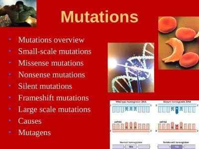
By Asha Kiran Akula Master of Research Gap Junctions Intercellular communication channels Gap junctions allow the selective permeability to ions and small molecules Movement through these channels is passive and non specific ID: 776542
Download Presentation The PPT/PDF document " Novel Mutations in the Connexin 43(GJA1..." is the property of its rightful owner. Permission is granted to download and print the materials on this web site for personal, non-commercial use only, and to display it on your personal computer provided you do not modify the materials and that you retain all copyright notices contained in the materials. By downloading content from our website, you accept the terms of this agreement.
Slide1
Novel Mutations in the Connexin 43(GJA1) may contribute to Nonsyndromic Hearing Loss.
By
Asha
Kiran Akula
Master of Research
Slide2Gap Junctions
Intercellular communication channels.
Gap junctions allow the selective permeability to ions and small molecules.
Movement through these channels is passive and non specific.
Gap junctions are made up of clusters of closely packed connexons
The structural unit of gap junction is Connexon.
Slide3Connexon
Consist of pairs of transmembrane channels.
The connexon hemi channel in one cell membrane docks with a connexon hemi channel in an adjacent cell.
Hexameric: they consist of arrays of 6 connexin protein subunits
Different connexin isoforms have been identified.
Homomeric or heteromeric
Slide4Slide5Functions
Gap junctions involve in regulation of
Tissue homeostasis
Regulation of cell growth
Embryonic development
Electrical and metabolic coupling
The loss of connexins, or the existence of mutations affecting their normal functions, has been implicated in a variety of diseases and disorders, including cancers.
Slide6Gap Junctions play a major role in intercellular calcium
signalling
.
Gap
junctional
opening is controlled by the intracellular concentration of calcium.
The low intracellular calcium concentration enables the gap junction channels to open and vice versa.
Controlled gating of gap junction channels may be responsible for the normal functioning of the cell.
Slide7Connexins
Show overlapping tissue expression patterns, most tissues expressing more than one connexin type.Each connexin contains 4 TM domains, with two extracellular and three cytoplasmic regions.
Slide8Both N- and C-termini –face the cytoplasm
The third TM domain -
amphipathic
in nature
forms the lining of the formed channel.
Amino acid sequence identity between the isoforms is ~50-80%, with the TM domains being well conserved.
Both extracellular loops – contain conserved
cysteine
residues, which likely form
intramolecular
disulphide bonds.
Single putative intracellular loop (between TM domains 2 and 3) and the
cytoplasmic
C terminus are highly variable among the family members.
Six connexins associate to form a hemi-channel, or connexon. Two connexons then interact (likely via the extracellular loops of their connexins) to form the complete gap junction channel.
Slide10Connexin Gene
Multigene
family comprising 20 in mouse and 21 genes in human genome (Cardiovascular Research, 2010).
α and
β
gene families.
The "
Gja
/
Gjb
" nomenclature-adopted by the NCBI data base.
Cells express multiple types of connexin-potentially associate to form gap junction channels containing more than one type of connexin.
Mutations
Alterations in the gap junction,
hemichannel
, or general functions of the connexins.
Cause various human diseases like skin diseases,
nonsyndormic
and
syndromic
deafness, cataracts,
Oculodentodigital
Dysplasia (ODDD), cancers etc.
Mutations in Cx32 - Charcot-Marie-Tooth disease. Cx26 -deafness and skin disease.
Cx30, Cx30.3, and Cx31- hearing loss and skin disorders.
Slide12Hearing Loss
Affects 1 in 1000 newborns.
Syndromic and nonsyndromic
70% of genetically related hearing loss-nonsyndromic.
Two types-
Conductive hearing loss
Sensorineural Hearing Loss
Mutations in Cx26- common cause of congenital bilateral non-syndromic sensorineural hearing loss.
Hearing
The ear is made up of three different sections:
1.the outer ear
External auditory canal
Tympanic
membrabe
2.the middle ear-bones
3.the inner ear
Cochlea
Vestibular system.
Cochlea
Main auditory portion of the inner ear.
Core component-organ of
corti
Organ of
corti
- sensory organ of hearing.
Organ of
corti
comprises - hair cells
supporting cells
Endolymph
Hair cells-two types
Inner hair cells
Outer hair cells
Connexins are present in supporting cells.
Slide16Slide17Sound waves
Tympanic membrane(Outer ear)
Bones maleus, incus and stapes(middle ear)
Movement of stapes
Pressure waves (fluid filled inner ear)
hairs to move in the inner ear
stimulate the auditory nerve
General mechanism of hearing
Slide18Organ of Corti
Outer hair cells
Inner hair cells
Tectorial
membrane
Basilar membrane
Supporting cells
Spiral
Limbus
Stria
Vascularis
Endolymph
Epithelial tissue
Slide19CX43- Four predicted membrane-spanning segments (M1–M4) linked by two extracellular (E1–E2)
and one cytoplasmic loop. Amino and carboxyl tails faced intracellularly.Expressed in non sensory epithelial cells of the inner ear.
CX43
Slide20Three novel missense mutations have been identified in the
GJA1 gene -
related to hearing loss.
The three missense mutations
c.205T>C (p.S69P) - Extracellular loop
c.932delC - C-terminal region
c.977C>T (p.T326I) - C-terminal
cytoplasmic
domain.
Studied the
intracellular distribution,
assembly and
the effects of the three Cx43 mutants with the wild type Cx43.
Slide21Plasmid construction with the mutationsPermanent transfected HeLa-CX43 cell lineImmunostainingDye Transfer
Slide22Plasmid
was constructed using CX43WT
cDNA
and cloned into pCDNA3.1 vector
Constructed plasmid-expression in HeLa cells via transfection-
lipofectamine
method.
Addition of G418- isolation of stable
transfectants
RT-PCR
– success of transfection and expression of transfected genes.
PCR products- Gel electrophoresis
CX43WT-Positive control.
HeLa and water-Negative control
β
-
actin
-internal control
Expression analysis of
GJA1 mRNA in four stable transfectedHeLa cells by RT-PCR. RT-PCR analysis of total RNA from HeLacells expressing CX43 WT, CX43 S69P, CX43 932delC and CX43T326I confirms expression of the corresponding mRNAs in stablytransfected HeLa cell lines (upper panel). -actin served as a reference for the loading amount of total RNA for each sample (lower panel).Mock HeLa and water were used as negative controls
Electrophoresis -Results
Slide24confirm the expressed CX43 mutant proteinsPrimary antibody- monoclonal anti-CX43 antibodySecondary antibody- HRP-conjugated anti-mouse IgGGAPDH-Internal ControlMock HeLa cells- Negative control
Western Blot-Results
Slide25Immunostaining
Transfected cells - washed and fixed.
Primary antibodies
Mouse anti-pan-
cadherin
antibody (anti-CH19) – cell membrane
Mouse anti-CX43-epitope of CX43 protein
Secondary Antibodies
Alexa
Fluor 488 and
Alexa
Flour 594
Nuclei-stained with DAPI
Slide26Immunostaining -Results
Localization analysis of CX43 WT in stably transfected HeLa cells by
immunocytochemistry
using anti-CX43 and pan-
cadherin
antibody. Analysis of fluorescence microscopy on HeLa cells expressing CX43 WT reveals localization of the CX43 protein in the
plasmamembranes
. CX43 proteins are indicated by
arrows. The cells were
counterstained with 4-6-diamidino-2-phenylindole, DAPI, to highlight the nuclei.
Scale bars 10 m
Slide27Slide28Dye Transfer
Functionality of gap junction formed.
Transfected HeLa cells-microinjected with Lucifer Yellow.
Slide29Cell line Dye-filled Number of Total number neighbor injections of cell (n)showing cell number (n) dye transfer (mean ± SE)HeLa-Cx43 WT 4.17 ±1.621 30 100HeLa-Cx43 S69P 0 30 0HeLa-Cx43 T326I 0 30 0HeLa-Cx43 A311V 0.54 ±1.471 50 86HeLa 0 30 0
Lucifer Yellow transfer stably expressed with WT or mutant
Cx43 HeLa cells
Slide30D
iscussion
With the above results, these three mutations-risk factor for the development of hearing.
Three mutations-loss of function of CX43 –hearing
loss.
CX43WT- found localized to the cell membranes at the point of contact between adjacent expressing cells.
Membrane localization-confirmed by
colocalization
with pan-
cadherin
.
Slide31THANK YOU