/
Body Systems – Part II Body Systems – Part II

Body Systems – Part II - PowerPoint Presentation

brianna
brianna . @brianna
Follow
0 views
Uploaded On 2024-02-16

Body Systems – Part II - PPT Presentation

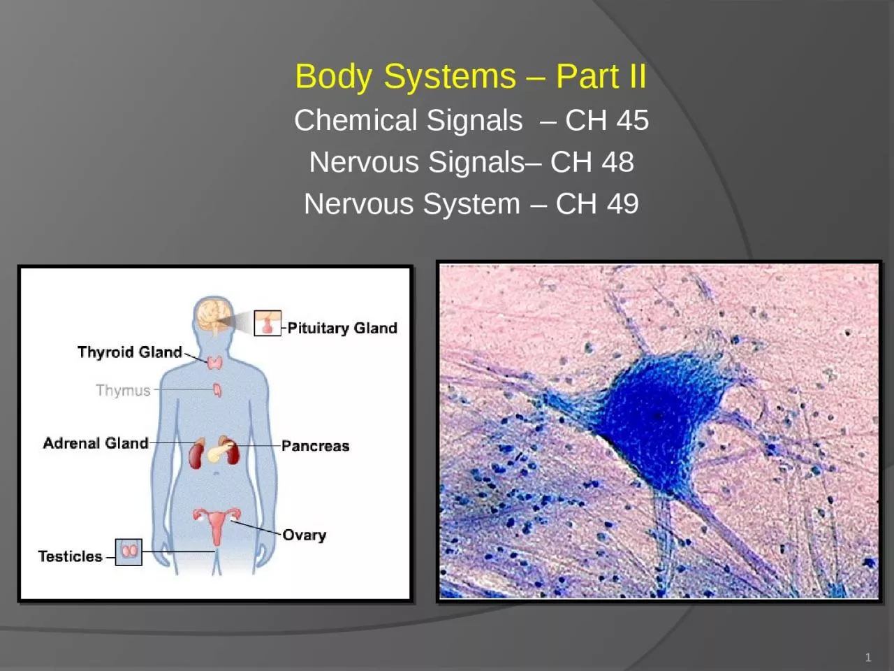
Chemical Signals CH 45 Nervous Signals CH 48 Nervous System CH 49 1 Chapter 45 Hormones and the Endocrine System 2 Hormone chemical excreted into body fluids used for communication within an organism ID: 1046384

cells cell potential system cell cells system potential nervous hormones blood signals brain action signal membrane neurons channels body

Share:

Link:

Embed:

Download Presentation from below link

Download Presentation The PPT/PDF document "Body Systems – Part II" is the property of its rightful owner. Permission is granted to download and print the materials on this web site for personal, non-commercial use only, and to display it on your personal computer provided you do not modify the materials and that you retain all copyright notices contained in the materials. By downloading content from our website, you accept the terms of this agreement.


Presentation Transcript

1. Body Systems – Part IIChemical Signals – CH 45Nervous Signals– CH 48Nervous System – CH 491

2. Chapter 45Hormones and the Endocrine System2

3. Hormone  chemical excreted into body fluids - used for communication within an organism - helps maintain homeostasis - modified amino acids and steroids - carried by the circulatory system to target cellsTarget Cells  equipped to respond to particular hormone; typically have membrane proteins that allow SPECIFIC hormones to bind**So this means that hormones in the blood can cause changes in SOME cells, and other cells will ignore them**3

4. Nervous vs. Endocrine SystemNervous System  high speed signals; Ex. Jerking your hand away from a flameEndocrine System  slower communication; Ex. Maturation of a butterfly; two parts: 4

5. Local RegulatorsAffects activity between neighbor cells; only uses LOCAL targets; there are 3 types: Growth Factors  peptides/proteins stimulate cell growth and development in target cells; includes nerve growth factorsNitric Oxide (NO)  a gas that has multiple functions including acting as a neurotransmitter (when secreted by neurons) and relaxing smooth muscle (when secreted by endothelial cells); it triggers a change in a target cell then breaks down quickly; it is also highly reactive and can be toxicProstaglandins  modified fatty acids first isolated in semen produced by prostrate; effect the female reproductive system (can cause smooth muscle contractions to help sperm reach egg; also induces uterine contraction in childbirth); aspirin and ibuprofen can inhibit the effects of PGs5

6. Local Regulators  divided into 2 groups: Paracrine  act on cells NEAR the secreting cellAutocrine  secreted regulators that act on the secreting cell itself!6

7. Synaptic Signaling vs. Neuroendocrine SignalingSynaptic Signaling  neurons form specialized junctions called synapses with target cells, such as other neurons and muscle cells; at synapses, neurons secrete molecules called neurotransmitters, which diffuse a very short distance to bind to receptors on the target cell7Neuroendocrine Signaling  specialized neurons called neurosecretory cells secrete chemical signals that diffuse from nerve cell endings into the bloodstream; these signals are a class of hormones called neurohormones (ex. ADH)- Endocrine – Ductless (secretes hormones into body fluids)- Exocrine – Uses ducts to send to specific locations (ex. sweat glands)

8. Signal Transduction Pathway3 main Processes: - Reception signal binds to a receptor protein- Signal Transduction  causes a change in the target cell- Response  causes a change in the receptor cell’s behaviorDifferent types of cells respond differently, so the SAME SIGNAL can bring about a DIFFERENT RESPONSE in various target cells.Only small amounts of regulators (ex. Hormones) are necessary because the pathway triggers enzyme cascades that can greatly amplify the signal.8

9. Several chemicals serve as both hormones of the endocrine system and signals in the nervous systemsEx. Epinephrine (This shows how the two systems are CHEMICALLY related)Nervous system = neurotransmitterEndocrine system = “fight or flight” hormoneEach system affects the output of the otherEx. Breast feeding – uses interdependent nervous and hormonal signals:Suckling = simulates sensory cells and nervous signals in the hypothalamus then trigger the release of oxytocin from the pituitary gland; this oxytocin cause the mammary cells to secrete milk The endocrine system and the nervous system are very closely related 9

10. Tropic HormonesTropic Hormone: have other endocrine glands as their targets10

11. Feedback Positive Feedback: causes a change in the same direction; ex. increasing milk release, labor during childbirth (oxytocin!) Negative Feedback: causes a change in the opposite direction; is responsible for the endocrine system’s ability to regulate homeostasis mechanisms; ex. sweating = cools the body; insulin and glucagon example (KNOW THIS ONE!)Feedback is COMMON TO BOTH THE ENDOCRINE AND THE NERVOUS SYSTEM!!11

12. PancreasSecretes bicarbonate ions to balance the pH from the acid chyme in the stomachAlpha cells secrete glucagon  signals the liver to release glucose back into blood stream (increases blood sugar levels); used when someone has not eaten in a while and blood sugar levels are lowBeta cells secrete insulin  signals body cells to take up glucose from the blood (decreases blood sugar levels); used when someone just ate and blood sugar levels are highExocrine  (secrete into ducts) bicarbonate ions and digestive enzymesEndocrine  (ductless) insulin/glucagon antagonists12

13. **Insulin and Glucagon are not steroids…they are PROTEIN hormones made of AA’s**Glucagon and Insulin are antagonistic hormones and work together to regulate the blood sugar levels in the body (recall our other antagonistic hormones are calcitonin and PTH for calcium)13If someone has fasted for 24 hours, what kinds of levels would we expect of these two hormones? HIGH levels of glucagon and LOW levels of insulin

14. DiabetesMost common endocrine disorder = Diabetes  caused by a deficiency in insulin or loss of response to insulin in target cellsType I diabetes  autoimmune disorder; immune system attacks pancreas; occurs in childhood and causes kid to not be able to make insulin; treated with insulin injections or a pumpType II diabetes  characterized by deficiency of insulin or reduced responsiveness in target cells; 90% of diabetics are type II; can usually be managed by diet and exercise; caused by genetics and obesity14

15. HypothalamusIntegrates the endocrine and nervous systems A good example of how they are structurally related is the NEUROSECRETORY CELLS located in the hypothalamusIt is part of the lower brain** It is important in homeostatic regulation (ex. body’s thermostat, regulates hunger/thirst, role in sexual/mating behaviors)** It regulates the Pituitary Gland** The neurosecretory cells of the hypothalamus secrete two hormones: oxytocin and ADH15

16. PituitaryLocated at base of the brainReferred to as “Master Gland” because it regulates so many other endocrine functionsIt obeys orders from the hypothalamusHas 2 discrete parts: Anterior Pituitary (front)Posterior Pituitary (back)16

17. Posterior Pituitary Posterior Pituitary Hormones  made by hypothalamus but secreted by posterior pituitary; these hormones act on specific structures rather than affecting other endocrine glandsStores and secretes two hormones: 1. Oxytocin  acts on muscles of uterus; induces contractions during childbirth and controls milk secretion during nursing2. Antidiuretic Hormone (ADH)  acts on the kidneys; causes kidneys to increase water retention thus decreasing urine volume; helps regulate the osmolarity of the blood17

18. Anterior Pituitary Secretes hormones directly into bloodHypothalamus secretes two kinds of hormones: Releasing hormones  makes anterior pituitary secrete its hormonesInhibiting hormones  makes anterior pituitary STOP secreting hormonesThe anterior pituitary secretes many hormones…18

19. Anterior Pituitary HormonesGrowth Hormone (GH)  promotes growth directly and also stimulates growth factorsHuman Growth disorders are related to GH production: Too much GH (hypersecretion) = gigantismToo little GH (hyposecretion) = pituitary dwarfism; this can be treated using growth hormones from cadaversInsulinlike Growth Factors (IGF)  stimulate bone and cartilage growthProlactin (PRL)  similar to GH; produces a variety of effects in different vertebrates (so scientists think this is an ANCIENT HORMONE); mammals = stimulates mammary gland growth and milk synthesis; freshwater fish = salt and water balance19

20. Anterior Pituitary HormonesFollicle-stimulating Hormone (FSH)  stimulates production of ova and spermLuteinizing Hormone (LH)  stimulates ovaries and testes** Gonadotropins = stimulate the activity of male and female gonads; FSH and LH are examples20

21. Anterior Pituitary HormonesThyroid Stimulating Hormone (TSH)  stimulates the thyroid gland (releases thyroid hormone which increases metabolic rate, raising body temp)Adrenocorticotropic Hormone (ACTH)  stimulates the production and secretion of steroid hormones by the adrenal cortex (part of the adrenal gland)Melanocyte-stimulating Hormone (MSH)  regulates the activity of pigment-containing cells in the skin; has a role in fat metabolismEndorphins  body’s natural opiates; inhibit the perception of pain21

22. Thyroid GlandConsists of 2 lobes on the tracheaThyroid hormone increases the metabolic rate, increasing the body temperatureThyroid has a critical role in vertebrate development & maturation (ex. Human development), homeostasis (blood pressure, heart rate, muscle tone, digestion)The term “thyroid hormone” refers to two closely related hormones: Triiodothyronine (T3)  causes changes to target cellsThyroxine (T4)  thyroid secretes mainly T4, but the target cells convert it to T3; this hormone stimulates and maintains metabolic processesBOTH T3 and T4 affect metabolic processes; important in bioenergetics Calcitonin  LOWERS calcium levels in blood (works antagonistically with PTH)22

23. 23

24. Thyroid Imbalances Include…Cretinism  deficiency in development, retarded skeletal/mental growth, develops in infants from hypothyroidismGoiter  deficiency of iodine in dietHyperthyroidism  Too much thyroid hormones; symptoms = weight loss, profuse sweating, high blood pressureHypothyroidism  Too little thyroid hormones; symptoms = weight gain, lethargyCretinismGoiter24

25. ParathyroidFound on the surface of the thyroid; function in homeostasis of calcium ions (very important to the normal functioning of all cells)Parathyroid Hormone (PTH)  raises calcium levels in blood; VERY important!Calcitonin  decreases calcium levels in the bloodPTH and Calcitonin are antagonistic hormones and work together to regulate the calcium levels in the blood (example of homeostasis)25

26. Antagonistic HormonesAntagonistic Hormones  hormones that work opposite each otherExamples: PTH (increases calcium in our blood) and calcitonin (decreases calcium in our blood) Insulin (decreases blood glucose levels) and glucagon (increases blood glucose levels)KNOW THESE TWO EXAMPLES!26

27. Adrenal GlandAdjacent to kidneysTwo parts: Adrenal cortex (outside)Adrenal medulla (inside)27

28. Adrenal Medulla (inside)Close ties to the nervous system – FIGHT OR FLIGHTCatecholamines – secreted in response to positive or negative stressNorepinephrine  sustaining blood pressure Epinephrine  heart and metabolic rates (Epi pen for anaphylactic shock); also acts as a neurotransmitter; GOOD EXAMPLE of how the endocrine and nervous systems are chemically related Mechanism:*Adrenal medulla under control of nerve cells from sympathetic division*Nerve cells excited by stressful stimulus*Acetylcholine released in adrenal medulla, and combines with receptors to release epinephrine28

29. Adrenal Cortex (outside)In contrast to the adrenal medulla, which reacts to nervous input, the adrenal cortex responds to endocrine signals. Corticosteroids include:Glucocorticoids  glucose metabolism; increases glucose in blood; secreted by the adrenal gland in response to stress and promotes the synthesis of glucose from non-carbohydrate substrates (ex. fats and/or proteins)  helps with long-term environmental issuesMineralocorticoids  helps inflammatory conditions; effects salt and water balance in kidneysBOTH help the body deal with LONG TERM stress (whereas epinephrine and norepinephrine deal with SHORT TERM stress)29

30. Adrenal Medulla vs. Adrenal Cortex30

31. Gonadal SteroidsControlled by gonadotropins from the anterior pituitary glandProduced in both males and females (in different proportions); produced in testes in males and ovaries in females; general function = affect growth and development and also regulate reproductive cycles and sexual behavior3 major types31

32. Types of Gonadal SteroidsAndrogens  ex. testosterone; development and maintenance of male reproductive system; produced in an embryo to turn the fetus into a male instead of a female; produced during puberty to stimulate secondary sex characteristics (hair growth, low voice)Estrogens  ex. estradiol; effects the female reproductive system and secondary sex characteristics in femalesProgestins  ex. progesterone; prepares and maintains the uterus which supports the growth and development of an embryo32

33. Pineal GlandSmall mass of tissue near the center of the brainSecretes the hormone melatonin, which regulates functions related to light/dark and seasons marked by changes in day length; related to biological clock rhythms33

34. Vertebrate Endocrine System34

35. 35

36. Chapter 48Neurons, Synapses, and Signaling36

37. Nervous SystemNeurons (functional unit of the nervous system) are nerve cells that transfer information within the body. Communication by neurons is based on two distinct types of signals: long-distance electrical signalsshort-distance chemical signals If an organism does NOT have an integration center, it would not be able to interpret stimuli. 37

38. Neurons (nerve cells)-Structural and functional unit of the nervous system-Has a cell body (contains the nucleus) and fiber-like extensions (dendrites and axons)-Dendrites-Short branched; many per cell body; receive INCOMING information and pass it to the cell body-Axons -Long; one per cell body; convey OUTGOING messages from the neuron to other cells-Axon hillock – part where the axon joins the cell body-Covered by myelin sheaths (insulated layer)-Synaptic terminals – specialized endings; relay signals from neuron to other cells by releasing chemical messengers called neurotransmitters-Site of contact between a synaptic terminal and a target cell is called a synapse-Presynaptic cell = transmitting cell-Postsynaptic cell = target cell38

39. 39

40. -Supporting Cells (called Glia)-Help support the nervous system and help it function properly-Originally thought to only have a structural role, but some synaptic interactions do occur between glia and neurons-In mature CNS, the glia are called astrocytes – they provide metabolic and structural support for neurons-Help form the blood-brain barrier  restricts passage of most substances into the brain which controls the extracellular chemical environment of the CNS (FORMED BY TIGHT JUNCTIONS)-Oligodendrocytes (in CNS) and Schwann cells (in PNS) are glia that form myelin sheaths around the axons of neurons-Necessary b/c can’t use regular cell membranes b/c they are made of lipids which are poor conductors of electrical currents; the myelin works better40

41. -Nervous system is made up of living neurons-Neurons are specialized for the fast transmission of impulses-Three major overlapping functions: -Sensory Input  sensory receptors take info from inside body and outside world and convey it to integrations centers-Integration  carried out in the CNS (central nervous system; brain and spinal cord); input is interpreted and body responds appropriately; if there was no integration center, organisms wouldn’t be able to interpret stimuli-Motor Output  conduction of signal from integration center to the effector cells (muscles or glands that carry out the signal)Overview -Signals are conducted by nerves-PNS (peripheral nervous system) = nerves that communicate motor and sensory signals between the CNS and the rest of the body-Information is communicated by both electrical and chemical signals 41

42. 42If Axon #1 was damaged, what would the result be? If Axon #2 was damaged, what would the result be?

43. 43If Axon #1 was damaged, what would the result be? The signal from the pinprick stimulus would not get to the brain If Axon #2 was damaged, what would the result be? The signal from the pinprick would get to the brain, be processed, but not get back to the finger, so the muscle will not respond

44. Cerebrospinal FluidMade in the brain by filtering the blood; fills the space in the brain and spinal cord44

45. The Nature of Nerve Signals-Nerve signals are changes in voltage across the membrane due to movement of ions -Membrane Potential  the potential charge difference between the cytoplasm and the extracellular fluid of a cell-Resting Potential  membrane potential of an unstimulated neuron-Can measure membrane potential as a voltage; typical animal cell is –50 to –100 mV (the negative means the inside of the cell is negative in charge w.r.t the outside)…in resting neurons, the membrane potential is more negative than the threshold potential45The inside of the cell is NEGATIVELY charged in comparison to the outside of the cell

46. Sodium Potassium Pump46-Differences in membrane potential are sustained by the actions of the sodium-potassium pump-Na+ pumped OUT (3 at a time)-K+ pumped IN (2 at a time)-THEREFORE…outside = positive; inside = negative-Goes against the gradient, so needs to use energy (ATP)  active transport-Ion channels are selective for specific ions; so a membrane can have different permeability's to different ions-They determine WHAT can pass through, but not the RATE

47. Gated Ion Channels47Gated ion channels are specialized proteins that span the membrane, and allow ions to diffuse back and forth across the membrane according to their respective gradients. Chemically-gated ion channels  respond to a chemical stimulus (ex. neurotransmitter)Voltage-gated ion channels  respond to a change in membrane potentialAllows only ONE kind of ion to pass through

48. -Graded Potentials  magnitude of change depends on the strength of the stimulus (larger stimulus will open more channels)-Hyperpolarization – increase in voltage across the membrane-Open K+ channel; K+ flows out and causes the inside of the cell to become more negative-Depolarization – reduction in the voltage across the membrane-Open a Na+ channel; Na+ flows in and causes the inside of the cell to become more positive48RESTING NEURONS: Resting membrane potential is more negative than the thresholdAt rest, the membrane is more permeable to sodiumThere is higher sodium concentrations OUTSIDE the cell (hence it diffuses IN during depolarization)

49. -Action Potentials  ALL OR NOTHING depolarization-DEPOLARIZATION causes an action potential-Triggered by graded potentials-When it reaches a certain point, the threshold potential (usually 15-20 mV more positive than the resting potential) it causes an action potential (NERVE IMPULSE!); all or none event-Occurs in the axons (not dendrites)-HYPERPOLARIZATION does NOT cause action potentials-Action Potential Mechanism: -Resting State – Na+ and K+ channels closed-Threshold – some stimulus opens Na+ channels; it reaches threshold potential, and therefore more Na+ channels open triggering an action potential-Depolarization - because Na+ channels are open and K+ channels are closed, the inside of the cell becomes more positive-Repolarization – Inactivation gates close the Na+ channels and the K+ channels open; K+ leaves the cell and the inside becomes more negative than the outside-Undershoot – K+ gates remain open because they are slow, but the Na+ gates are closed; resting state is restored very quickly (hyperpolarization happens for a millisecond)SEE Fig. 48.11 pg. 1068 explains everything perfectly…49Action potentials arise because some ion channels in neurons are voltage-gated ion channels, opening or closing when the membrane potential passes a particular level

50. 50KNOW THIS PICTURE!!WHEN do the voltage gated K+ channels open? WHEN do the Na+ inactivation gates close? When is the threshold reached? In this picture, what is the resting membrane potential? What happens first? (Na+ gates open)

51. -Because both gates of the Na+ channel are closed, if another stimulus arrives during this period, it is unable to trigger a change (inactivation gates had not had time to open back up yet)  called refractory period (neuron is insensitive to depolarization)-It is the number of action potentials per second, not their amplitude, that codes for a stimulus intensity in the nervous system-Action potentials propagate themselves along an axon (like tipping over the first of a long line of dominoes)-Factors that affect the speed of the action potentials (how fast they go along the axon):-Diameter of axon (larger diameter  faster transmission)-Saltatory conduction  action potentials that jump from node to node51

52. Think about…52If a toxin bound to the voltage gated sodium channels in an axon, what would the result be?

53. Think about…53If a toxin bound to the voltage gated sodium channels in an axon, what would the result be?The Na+ channel wouldn’t open and therefore the axon couldn’t reach the threshold, and therefore an action potential could NOT be obtained

54. -Communication between cells occurs at synapses-Synapses – unique cell junctions that control communication between a neuron and another cell; two types: electrical or chemical-Electrical Synapses-Allows action potentials to spread from presynaptic cell to postsynaptic cell via gap junctions-Not as common as chemical synapses-Chemical Synapses-Very common-Chemical synapses are called synaptic clefts; they separate presynaptic cell from postsynaptic cell-The cleft prevents an action potential from going directly from the pre to the postsynaptic cell-A series of events converts the electrical signal of the action potential arriving at the synaptic terminal into a chemical signal that travels across the synapse, where it is converted back into an electrical signal in the postsynaptic cell. (electrical signal  chemical signal  electrical signal)Fig. 48.16 pg. 1072…54Synapses

55. 551. An action potential DEPOLARIZES the PRESYNAPTIC membrane2. Na+ rush into the neuron’s cytoplasm3. Synaptic vesicles release neurotransmitters into the synaptic cleft4. Neurotransmitters bind with the receptors on the post-synaptic cell5. Ion channels open to allow specific ions into the target cellKNOW THESE STEPS!!!***A series of events converts the electrical signal of the action potential arriving at the synaptic terminal into a chemical signal that travels across the synapse, where it is converted back into an electrical signal in the postsynaptic cell. (electrical signal  chemical signal  electrical signal)Note: Calcium aids in the release of the neurotransmitters

56. When an action potential reaches the end of the chemical synapse, it causes a neurotransmitter to be released into the synapse-Neurotransmitters (intracellular messengers) are held in the tip of the pre-synaptic axon-Action potential releases these neurotransmitter molecules into the synapse (the action potential depolarizes the membrane)-The postsynaptic membrane has special receptors for neurotransmitters-Neurotransmitter binds  opens ion channels (chemically gated!) – postsynaptic membrane is either hyperpolarized or depolarized (depending on receptor)-Neurotransmitter is removed quickly (enzyme degradation)  therefore the effect is brief and precise-NOTE: nerve impulses can only transmit ONE way56Structure of a Chemical Synapse

57. -BOTH -Graded potentials-The electrical impact on the postsynaptic cell decreases with the distance away from the synapse-Excitatory  EPSP-Allows Na+ in and K+ out (more permeable to Na+, so more of that is allowed in)-Inside of cell becomes positive­ = DEPOLARIZES the plasma membrane (gets it closer to the action potential)-Excitatory Postsynaptic Potential (EPSP)  the name for the whole process that uses an excitatory synapse-Inhibitory  NO ACTION POTENTIAL! IPSP-K+ out and Cl- in (higher permeability to K+)-Inside of cell becomes negative = HYPERPOLARIZE the plasma membrane-Inhibitory Postsynaptic Potential (IPSP)  the name for the process that uses an inhibitory synapse57Excitatory Synapse vs. Inhibitory Synapse

58. 58

59. Summation-Several synaptic terminals working simultaneously on the same postsynaptic cell can have an additive effect  summation!-Temporal Summation  one or more synaptic terminals rapid fire with signals (voltage has no time to return to resting)-Spatial Summation  several synaptic terminals (on DIFFERENT cells) all send signals to the same postsynaptic cell (additive effect) 59

60. -Same neurotransmitter can produce different effects on different types of cells (depends on the receptors)-Major neurotransmitters -Acetylocholine – most common; vital for nervous system functions that include muscle stimulation, memory formation, and learning; can be either inhibitory or excitatory-Biogenic Amines – derived from amino acids-Epinephrine & Norepinephrine (excite and inhibit)  MORE ON THESE NEXT CHAPTER-Dopamine (generally excite)  affect sleep, mood, attention, and learning-Serotonin (generally inhibit)  affect sleep, mood, attention, and learningNeurotransmitters-Amino Acids – -GABA (gamma aminobutyric acid) – found at most inhibitory synapses; creates IPSP (increases Cl- permeability)  no action potential-Neuropeptides – short chains of AA’s-Substance P – excite Signal; mediates perception of pain-Endorphins – decreases perception of pain; emotional effects60

61. -Nitric Oxide (NO)  NO diffuses into neighboring target cells, produces a change, and is broken down within a few seconds; in its target cells, NO works like many hormones, stimulating an enzyme to synthesize a second messenger that directly affects cellular metabolism- Carbon Monoxide (CO)  In the brain, CO regulates the release of hypothalamic hormones; in the PNS, it acts as an inhibitory neurotransmitter that hyperpolarizes intestinal smooth muscle cells-Gasses not stored; cells make them on demand- They are used as LOCAL regulators61Gas signals of Nervous System

62. Chapter 49Nervous Systems62

63. Evolution and Diversity of Nervous Systems-Great Diversity in organization of new systems-Lack nerve systems (sponges)-Nerve nets (cnidarians)-Cephalization (neurons clustered in head)-Nerve cords (planarians)-Clearly defined CNS….etc.63

64. -The major types of glia (connective tissue of nervous system) nourish, support and regulate neurons. -Oligodendrocytes and Schwann cells function in axon myelination, a critical activity in the vertebrate nervous system. -Astrocytes induce cells that line the capillaries in the CNS to form tight junctions.The result is the blood-brain barrier, which controls the extracellular environment of the CNS by restricting the entry of substances from the blood.64Glia and Blood Brain Barrier

65. Central Nervous SystemThe brain integrates the complex behavior of vertebrates. The spinal cord conveys information to and from the brain and generates basic patterns of locomotion. The spinal cord acts independently as part of the simple nerve circuits that produce reflexes, the body’s automatic responses to stimuli (ex. pulling hand away from hot stove.)Cerebrospinal fluid is formed in the brain by filtering blood. In mammals, it fills the spaces in the brain and the spinal cord. The function is to act as a shock absorber. The brain and the spinal cord contain gray and white matter. Gray matter consists of cell bodies, dendrites, and unmyelinated axons. White matter contains axons with myelin sheaths. 65

66. Vertebrate Nervous SystemCNS  brain and spinal cordPNS  everything else!Parts of the peripheral nervous system: - Sensory (AFFERENT) division  incoming neurons - Motor (EFFERENT) division  outgoing neurons motor subdivides: - Somatic  carries signals to SKELETAL muscles - Autonomic  regulates internal environment autonomic subdivides: - Sympathetic  ACTIVE (fight/flight) - Heart beats faster, arousal/energy - Parasympathetic  INACTIVE (rest/digest) - Calming/conserving energy/ self-maintenance- Enteric  control the organs of the digestive, cardiovascular, excretory, and endocrine systemsCerebrospinal Fluid = made in the brain by filtering blood; fills the spaces in the brain and spinal cord66

67. Actions of two divisions of autonomic system67

68. The Brain68- The vertebrate brain has three major regions: the forebrain, midbrain, and hindbrain.- The FOREBRAIN activities include processing olfactory input (smell), regulation of sleep, learning, and any complex processing. Cerebrum, Diencephalons, Thalmus, Hypothalamus, Epithalmus- The MIDBRAIN coordinates routing of sensory input- The HINDBRAIN controls involuntary activities, such as blood circulation, and coordinates motor activities, such as locomotion. Pons, Medulla oblongata, Cerebellum

69. Brainstem (lower brain)- Major Functions: homeostasis, coordination of movement; conduction of information to higher brain centers- Consists of 3 parts - Medulla oblongata  Homeostatic control center; controls homeostatic functions (including respiration (breathing), swallowing, digestion, heart and circulation) - Pons  also helps control automatic functions (“assists” medulla) - Midbrain  acts as a projection center; sends coded sensory information to forebrain69

70. 70

71. CerebellumDevelops from the metencephalon (hindbrain)Functions in coordination; muscle actions; movement and balance; involved in learning and remembering motor responsesHand-eye coordination71

72. Thalamus/ HypothalamusDevelops from diencephalon (forebrain)Epithalamus – produces cerebrospinal fluidThalamus – main input center for sensory information going to cerebrum and main output center for motor information leaving the cerebrumHypothalamus – important in homeostatic regulationSource of hormonesRegulates the pituitary glandBody’s thermostat (regulates temperature)Regulates hunger/thirstRole in sexual/mating behaviors; fight or flight; pleasure/rageCircadian Rhythms Regular, repeated rhythmic behaviors (sleep/ wake)Hormone release; sex drive; sleep cycles; sensitivity to external stimuli72

73. Most HIGHLY DEVELOPED STRUCTURE of the mammalian brainDivided into left and right hemispheres; each hemisphere consists of: Gray matter (cerebral cortex) – coveringWhite matter – internal partBasal nuclei – found deep within the white matter; important in planning and learning movement sequencesCerebral Cortex (“gray matter”)LARGEST and MOST COMPLEX part of the mammalian brainEvolved a lot through evolutionNeocortex – 6 layers of tissue outside of the cortex; unique to mammalsRight half of brain controls the functions of the left side of the body; left half controls the right sideCorpus callosum – part of the brain that communicates between the left and right hemispheres73Cerebrum

74. Lobes of CerebrumFrontal, Parietal, Temporal, OccipitalPrimary motor cortex and primary somatosensory cortex form the boundary between the frontal lobe and parietal lobeMotor Cortex  sends commands to skeletal musclesSomatosensory Cortex  gets and integrates signals from touch, pain, pressure and temp receptors throughout the bodyFrontal Lobe  speech, motor cortex, emotionsParietal Lobe  somatosensory cortex, taste, speech, reading, touch, pain, pressure, tempTemporal Lobe  smell, hearingOccipital Lobe  visionSIDES OF THE CEREBRUMLeft Side  adept at language, math, logic, serial sequences of informationRight Side  pattern recognition, space recognition, spatial relations, nonverbal, emotions74

75. 75

76. Reticular FormationArousal and sleep are controlled in part by the reticular formation, a system of neurons that passes through the brainstem76

77. EmotionsDue to frontal lobe and limbic systemLimbic System  forms a ring around the brainstem; composed of hippocampus and the olfactory cortexResponsible for emotions (laughing, crying, feeding, aggression, sexuality)Amygdala  nucleus in the temporal lobe; recognizes emotional content of facial expressions and emotional memoriesInteresting note: frontal lobotomies, which disrupt the frontal lobes and limbic system, used to be performed to treat severe emotional disorders77

78. Language and SpeechProcessed by multiple areas of the cortex Broca’s area – frontal lobe  controls the muscles in the faceWernicke’s area – temporal lobe  controls the ability to comprehend speech but not the ability to speak78

79. Memory and LearningShort term memory (frontal lobe)  released if memory is irrelevantLong term memory (limbic system, hippocampus)  if info is pertinent; goes from short term to long term with repetition (“practice makes permanent”…not always perfect!)Skill memory  ex. walking, riding a bike, etc; once learned, it is hard to unlearn; learned by repetition (bad habits are hard to break!)  practice makes permanent79

80. Research for CNS InjuriesCNS can’t repair itselfNerve Cell DevelopmentNeurons develop by cell to cell communication, control of gene expression, and genetic basis HARD TO REPLICATE!!Axons grow to target cells and use molecular signals to direct them (don’t grow in a straight line)Sequence and time of development are important – therefore, again, it is HARD TO REPLICATEScientists are trying to get axons to regrow using different combinations of proteinsNeural Stem CellsIn adults, new cells are found in the hippocampus (memory/ learning)Function of the new stem cells = unclear!Issues with stem cell research  what can we use as a source?!?! LOTS OF DEBATES OVER THIS!!80

81. Relationship and similarities between nervous and endocrine systems * use chemical signals * response depends on action of receptor * chemical messengers produced by axons in parts of both81