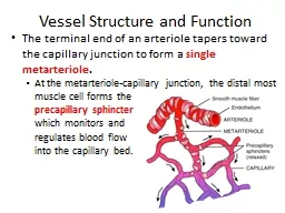
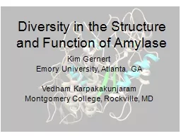
Kim Gernert Emory University Atlanta GA Vedham Karpakakunjaram Montgomery College Rockville MD Target Audience Students enrolled in Principles of Biology I BI 107 and II BI 108 Human salivary amylase used in one of our lab modules so students are familiar with this enzyme and i ID: 909741
Download Presentation The PPT/PDF document "Diversity in the Structure and Function ..." is the property of its rightful owner. Permission is granted to download and print the materials on this web site for personal, non-commercial use only, and to display it on your personal computer provided you do not modify the materials and that you retain all copyright notices contained in the materials. By downloading content from our website, you accept the terms of this agreement.
Slide1
Diversity in the Structure and Function of Amylase
Kim
Gernert
Emory University, Atlanta, GA
Vedham
Karpakakunjaram
Montgomery College, Rockville, MD
Slide2Target Audience
Students enrolled in Principles of Biology I (BI 107) and II (BI 108)
Human salivary amylase: used in one of our lab modules, so students are familiar with this enzyme and its function
Slide3Background
Alpha amylase:
http://www.rcsb.org/pdb/101/motm.do?momID=74
Alpha-amylase: begins starch breakdown
Starch chains broken into two or three glucose units
Most organisms synthesize amylase
Slide4Overview
Phylogenetic treeshttps://
sites.google.com/site/plasmodiumproblem/intro/about-phlyogenetic-trees
Blast
Protein sequence, amino acids
Protein structure, 3-D and secondary structural elements.
Slide5Big Picture: Concepts
Evolutionary relationships between organisms in
a molecular scale
Correlation between Structure and Function
Focus: modifications, if any, in amylase across organisms with unique lifestyles
Slide6Question # 1
What is the phylogenetic relationship between representative organisms from the three domains, in terms of evolution of amylase?
Slide7Study Organisms: a sample
Domain
Organism
Habitat/Lifestyle
Bacteria
E. coli
In
animal gut
Halothermothrix
orenii
Halothermophilic
Archaea
Pyrococcus
horishikii
HyperthermophilicP. woeseiEukaryaSaccharomycopsis fibuligeraUnicellularTenebrio molitorBeetle
6 out of 20 sequences archived for the
study
(1000’s of known amylase
sequences)
17 molecular structures are
presented
(100’s of solved
structures)
Slide8Data
The European Bioinformatics Institute sequences (
http://www.ebi.ac.uk/
)
Protein Data Bank Molecular Structures (
http://www.rcsb.org/pdb/home/home.do
)
>Aspergillus oryzae 2gvy
ATPADWRSQSIYFLLTDRFARTDGSTTATCNTADQKYCGGTWQGIIDKLDYIQGMGFTAI
WITPVTAQLPQTTAYGDAYHGYWQQDIYSLNENYGTADDLKALSSALHERGMYLMVDVVA
NHMGYDGAGSSVDYSVFKPFSSQDYFHPFCFIQNYEDQTQVEDCWLGDNTVSLPDLDTTK
DVVKNEWYDWVGSLVSNYSIDGLRIDTVKHVQKDFWPGYNKAAGVYCIGEVLDGDPAYTC
PYQNVMDGVLNYPIYYPLLNAFKSTSGSMDDLYNMINTVKSDCPDSTLLGTFVENHDNPR
FASYTNDIALAKNVAAFIILNDGIPIIYAGQEQHYAGGNDPANREATWLSGYPTDSELYK
LIASANAIRNYAISKDTGFVTYKNWPIYKDDTTIAMRKGTDGSQIVTILSNKGASGDSYT
LSLSGAGYTAGQQLTEVIGCTTVTVGSDGNVPVPMAGGLPRVLYPTEKLAGSKICSSS
Slide9Tools
Multiple alignments CLUSTALW
construct
phylograms
in:
www.phylogeny.fr
Sequence alignments Blast
http://blast.ncbi.nlm.nih.gov/Blast.cgi
Slide10Phylogeny based on Amylase
Slide11Discussion
Give a general set of observations on the tree. What clusters with the human sequences?
Identify mono-phyletic groups within the tree.
Why possibly
the
archaean
species are isolated in the tree?
Slide12Question
2
What is the percent similarity in structure of amylase based on the phylogenetic tree?
How does the sequence identity of the sequences match the clustering in the phylogenetic tree?
Blast for percent sequence identity and percent sequence similarity.
This will help students to quantitatively connect the information from phylogenetic tree to secondary structure/sequence
Slide13Sequence identity and
phylogeneticsby
Blast
Slide14Question # 3
Are the amino acid sequences (hence the structure) different across organisms?
Where are the conserved regions in the molecular structure?
Do they relate to the secondary structural elements
?
Slide15Conserved Sequences
Regions of the alignment that are highly conserved are highlighted in green and blue.
The same sequences are colored on the 3-dimensional structure in Chimera.
Slide16Tools
Use Chimera (
www.cgl.ucsf.edu/chimera
) to visualize and compare the amylase structure in various organisms
05-pdbs-061313-006.py
Structure file including human, porcine, beetle and bacterium
amylase
Slide17Discussion
What sections of the structure are colored green?What sections of the structure are colored blue?
Why are the sequence of these regions conserved?
What other regions do you think will be conserved?
Slide18Question # 4
Note that 4 or more conserved residues in a row highlight a critical region of the protein structure.
The
active site residues are in these regions
.
Slide19Tools
Resources:
Use NCBI (
http://www.ncbi.nlm.nih.gov/
)
for comparing the identity and percent similarities in the sequences across organisms
Slide20Tools
Resources:
Use Chimera (
www.cgl.ucsf.edu/chimera
) to visualize and compare the amylase structure in various organisms
Slide21How does the structure of the active site visually compare across different organisms?
Discussion
Slide22Multiple structure
overlay
Slide23Discussions
What are the critical active site residues?Are they present in all of the structures from different species?
Which structures have glucose or
another
starch bound?
Do the different species bind the starch differently?
Slide24Future plans
Study the binding of other molecules in the active site including inhibitors.
Study substrate analogs
.
Role of mutations in modifying the structure and function of amylase.
Slide25Known mutations
Mutations, structural
and
functional
effects.
1xgz MUTANT N298s
1nm9 MUTANT
W58A subsite 2. Critical for enzyme activity.
1q4n
MUTANT F256W salivary
1kgu
pancreatic MUTANT R377A (probing role of chloride ion).
1kgw
pancreatic MUTANT R337Q
1kgx pancreatic MUTANT R195Q (probing role of chloride ion).
Hordeum
vulgare (barley) 2xfr (2010, 0.97A) 2xff (2010, 1.31) H395a, (y105a, y308a), d180a (inactive)Glycine max (soybean) 1v3h, 1v3i soybean, E186, E380 (resolution 1.6 A) 1q6c, complex with maltose, M51T, E178Y, N340T (increased pH optimum)Bacillus cereus 1vem (2005, 1.85) Y164e, (t47m, Y164e, t328n)
Slide26Bibliography
http://www.ebi.ac.uk/
http://
www.rcsb.org/pdb
General textbook
1hny Protein
Sci.
1995 Sep;4(9):1730-42.
The
structure of human pancreatic alpha-amylase at 1.8 A resolution and comparisons with related
enzymes.
Brayer
GD
,
Luo
Y
, Withers SG.1smd Acta Crystallogr D Biol Crystallogr. 1996 May 1;52(Pt 3):435-46. Structure of human salivary alpha-amylase at 1.6 A resolution: implications for its role in the oral cavity. Ramasubbu N, Paloth V, Luo Y, Brayer GD,
Levine MJ
.