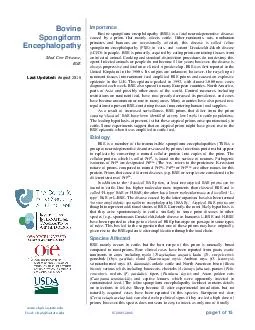
/
page of  Bovine Spongiform Encephalopathy Mad Cow Disease BSE Last Updated May  Importance page of  Bovine Spongiform Encephalopathy Mad Cow Disease BSE Last Updated May  Importance

page of Bovine Spongiform Encephalopathy Mad Cow Disease BSE Last Updated May Importance - PDF document

calandra-battersby
calandra-battersby . @calandra-battersby
Follow
555 views
Uploaded On 2015-02-26

page of Bovine Spongiform Encephalopathy Mad Cow Disease BSE Last Updated May Importance - PPT Presentation

Other ruminant species cats non human primates and humans are occasionally affected this disease is called feline spongiform encephalopathy FSE in cats and variant Creutzfeldt Jakob disease vCJD in people BSE is a relatively new disease that was fir ID: 39592

Other ruminant species cats

Share:

Link:

Embed:

Download Presentation from below link

Download Pdf The PPT/PDF document "page of Bovine Spongiform Encephalopath..." is the property of its rightful owner. Permission is granted to download and print the materials on this web site for personal, non-commercial use only, and to display it on your personal computer provided you do not modify the materials and that you retain all copyright notices contained in the materials. By downloading content from our website, you accept the terms of this agreement.


Presentation Transcript

www.cfsph.iastate.ed u Email: cfsph@iastate.edu © 2003 - 2016 page 1 of 15 Bovine Spongiform Encephalopathy Mad Cow Disease, BSE Last Updated: August 2016 Importance Bovine spongiform encephalopathy (BSE) is a fatal neurodegenerative disease, caused by a prion, that mainly affects cattle. Other ruminant s , cats, nonhuman spongiform encephalopat hy (FSE) in cats, and variant Creutzfeldt - Jakob disease (vCJD) in people. BSE is primarily acquired by eat ing prion - containing tissues from an infected animal. Cooking and standard disinfection procedures do not destroy this agent. Infected animals or peop le do not become ill for years; however, the disease is always progressive and fatal once clinical signs develop. BSE was first reported in the United Kingdom in the 1980s. Its origins are unknown; however, the recycling of ruminant tissues in to ruminant f eed amplified BSE prion s and caused an explosive diagnosed each week. BSE also spread to many European countries, North America, parts of Asia and possibly other areas of the wo rld. Control measures, including restrictions on ruminant feed, have now greatly decreased its prevalence, and cases have become uncommon or rare in many areas. Many countries have also passed new regulations to prevent BSE - containing tissues from entering human food supplies. As a result of increased surveillance, BSE prions that differ from the prion causing ‘classical’ BSE have been identified at very low levels in cattle populations. The leading hypothesis, at present, is that these atypical prions ari se spontaneous ly in cattle . Some experiments suggest that an atypical prion might have given rise to the BSE epizootic when it was amplified in cattle feed. Etiology BSE is a member of the transmissible spongiform encephalopathies (TSEs), a group of neurod egenerative disorders caused by prions, infectious proteins that appear to replicate by converting a normal cellular protein into copies of the prion. The cellular protein, which is called PrP c , is found on the surface of neurons. Pathogenic isoforms of Pr P c are designated PrP res (The ‘ res ’ refers to the proteinase K - resistant nature of prions, compared to normal PrP c ) . PrP Sc or PrP TSE are other names for this protein. Prions that cause different diseases (e.g. BSE or scrapie) are considered to be different strains of PrP res . In addition to the ‘classical’ BSE prion, at least two atypical BSE prions can be found in cattle. One has high er molecular mass fragments than classical BSE and is called ‘H - type’ BSE or H - BSE; the other has a lower molecular mass and is called ‘L - type’ BSE or L - BSE. T he disease caused by the latter organism ‘bovine amyloidotic spongiform ence phalopathy (BASE).’ Atypical BSE prions are thought to represent additional strains of BSE. Currently, the most likely hypothesis is that they arise spontaneously in cattle, similarly to some prion diseases in other species (e.g., spontaneous Creutzfeldt - J akob disease in humans). L - BSE and H - BSE have been reported to change to a classical BSE phenotype on passage in some types of mice. This has led to the suggestion that one of these prions may have originally given rise to the BSE epidemic after amplificat ion through the food chain. Species Affected compared to most prions. Rare clinical cases have been reported from goats ; exotic ruminants in zoos , includ ing nyala ( Tragelaphus angasi ), kudu ( Tr . strepsiceros ), gemsbok ( Oryx gazella ), eland ( Taurotragus oryx ), Arabian oryx ( O . leucoryx ), scimitar - horned oryx ( O . dammah ), ankole cattle and North American bison ( Bison bison ) ; various felids including housecats, cheetahs ( Acinonyx jubatus ), pumas ( concolor ), ocelots ( F. pardalis ), tigers ( Panthera tigris ) and Asian golden cats ( Catopuma temminckii ); and captive lemurs, which were apparently infected in contaminated feed. (The feline spongiform encephalopathy factsheet contains details on infections in felids.) Sheep become ill after experimental inoculation, but no naturally acquired cases have been reported in this species. European red deer ( Cervus elaphus elaphus ) can also develop clinical signs if they are fed a high dose o f prions; however, this species does not seem to easy to infect, as only one of 6 orally Bovine Spongiform Encephalopathy © 200 3 - 2016 www.cfsph.iastate.ed u  Email: cfsph@iastate.edu page 2 of 15 inoculated red deer became infected. M ink ( Mustela vison ) and cynomolgus macaque s ( Macaca fascicularis ) are also susceptible to oral inoculation. Common m armosets ( Callithrix jacchus ) and squirrel monkeys ( Saimiri sciureus ) have been infected by intracerebral inoculation; however, their natural susceptibility to BSE is unknown, as this method bypasses normal species barriers to prions. Pigs could be infecte d by simultaneous intracranial, intravenous and intraperitoneal routes or by intracerebral inoculation alone, but short - term feeding trials did not cause disease. One study reported that sea bream ( Sparus aurata) seemed to be susceptible to oral inoculatio n. L - BSE can infect sheep and cynomolgus macaques by intracerebral inoculation , but there are currently no reports of their susceptibility by ingestion . However, L - BSE has been transmitted to lemurs by the oral route, with the development of neurological s igns. Mice have been infected with L - BSE and H - BSE by intracerebral inoculation. Zoonotic potential Humans occasionally develop variant Creutzfeldt - Jakob disease after eating prion - containing tissues from an infected animal. To date , all known cases have been caused by the classical BSE prion . Whether H - BSE and L - BSE can cause disease in people is still uncertain . Some studies in laboratory models , but not others, have suggested that humans may be susceptible to L - type BSE. Geographic Distribution Cases o f classical BSE have been reported in indigenous cattle in some European countries, Canada , Israel and Japan. Some of these countries may have eradicated this disease, as it has not been reported in some time . Classical BSE was documented only in imported cattle in some nations , including the U.S., the F alkland Islands and Oman. Other countries , such as Iceland, Australia and New Zealand , seem to have remained completely free of classical BSE. The presence or absence of this disease cannot be determined in countries without adequate surveillance programs. Atypical BSE prions have been reported in Eur ope, the U.S., Canada, Japan and Brazil , as the result of surveillance programs for BSE. T hey are also likely to exist in other countries. Transmission BSE is usually transmitted when an animal or human ingests tissues containing the BSE prion. Young animals may be particularly susceptible : s ome studies suggest that most cattle become infected with BSE during the first six months of life. Sheep are , likewise, most susceptible to experimental (oral) inoculation during the first few months of life, especially during the first few weeks. In cattle, t he prions are thought to replicate initially in t he Peyer’s patches of the ileum, then are transported via the peripheral nerves to the central nervous system (CNS). P rions have been found in the brain of cattle as soon as 16 - 24 months after infection. T he highest prion concentration s occur in the CNS ( both the brain and spinal cord) and in the ileum . H owever, very sensitive detection methods have also found this agent in lymphoid tissues associated with the jejunum and colon , various nerve ganglia, peripheral nerves and adrenal glands , and in the optic nerve and retina . The accumulation of BSE in peripheral nerves , nerve ganglia and adrenal gland seems to coincide with or follow prion accumulation in the CNS. However, o ne group detected BSE in the jejunum as soon as 4 months after oral inocula tion . T here have been rare reports of BSE prions or infectivity in other locations, such as the tonsils ; bone marrow ; mesenteric lymph nodes; the esophagus, abomasum and rumen of one animal (possibly in nerve endings); sensory receptors (muscle spindles) o f muscles but not myofibrils; one muscle sample (probably associated with the endings of the sciatic nerve); the tongue and nasal mucosa of cattle in the terminal stages of the disease ; and even in concentrated saliva . These studies have generally used ver y sensitive techniques, found very small quantities of prions, and reported that these tissues contain prions only in animals with clinical signs. I n cattle, BSE prions do not seem to occur in the spleen or lymphatic tissues other than those associated wit h the gastrointestinal tract . Most studies have also not detected BSE in muscles. While one group reported evidence of its presence in a few plasma samples from cattle, others have not detected these prions in bovine blood. Epidemiological evidence and transmission studies suggest that BSE is not transmitted in milk, semen or embryos. There is no evidence that BSE is transmitted horizontally between cattle; however, there is an unexplained increase in the risk of BSE among the offspring of infected animals. In one study, calves seemed to be more likely to develop BSE when the dam was in the later stages of infection (i.e., nearer to the onset of clinical signs). These observations have led to speculation that vertical trans mission might be possible in cattle . If this occurs, it seems to be rare, and the route is unknown. I n experimentally infected sheep , BSE prions are more widely disseminated in the body than in cattle . They are readily found in many lymphoid tissues including the spleen, lymph nodes and gut - associated lymphoid tissue (GALT), as well as in the CNS. Blood - borne transmission has been demonstrated in this species. A number of ewes (18%) also transmitted BSE to t heir lambs in an experimental flock . The l ambs were more likely to become infected if the dam was in the later stages of the disease . P rions were not found in the placenta , except in one stillborn lamb , and the live lambs were thought to have be en infected shortly after birth. O ne lamb born to an BSE - negative sheep became infected; however, such horizontal transmission appears to be rare. In this experimental flock, a low transmission rate suggested that sheep would not maintain BSE long - term. Bovine Spongiform Encephalopathy © 200 3 - 2016 www.cfsph.iastate.ed u  Email: cfsph@iastate.edu page 3 of 15 Prions in the environment are not thought to be significant in the epidemiology of BSE. Nevertheless, there have been concerns about the ir possible longevity in sources such as buried carcasses. In one study, infectivity was reported to persist for at least 265 days in sewage or phosphate buffered saline , under laboratory conditions. BSE prions detected by immunoblot ting disappeared sooner than infectivity, and could not be found in sewage by 150 days. Other prions (e.g., the agents of scrapie and chronic wasting diseas e) can also persist in the environment for prolonged periods, and hamster - adapted scrapie prions have been shown to survive in the soil for at least 3 years. Prions are reported to remain infectious after passage through the digestive systems of birds (cro ws) and mammals (coyotes). Atypical BSE In cattle, some studies report that the tissue distribution of atypical L - BSE and H - BSE seems to resemble that of classical BSE, with prions detected mainly in the CNS. (There are, however, some differences in the pa ttern of distribution within the brain.) H - BSE and L - BSE have also been found in peripheral nerves , nerve ganglia and sensory receptors (muscle spindles) in some studies, and L - BSE was detected in the adrenal gland. In one study, prions were found in the m uscles of L - BSE infected cattle by immunostaining, and infectivity was detected in muscle homogenates with a highly sensitive mouse bioassay. Whether vertical transmission can occur is not known . One calf born to a cow in the late stages of infection with L - BSE was not infected . Transmission to humans including iatrogenic spread In humans, variant Creutzfeldt - Jakob disease usually results from eating BSE prions in contaminated animal tissues . S everal patients were infected via blood transfusions from asymp tomatically infected individuals , and highly sensitive prion detection techniques have found BSE prions in the blood of some symptomatic patients. There is also the potential for transmission by routes such as transplantation or the use of prion - contaminat ed equipment during surgeries. In humans, vCJD ( BSE ) p rions can be found in the CNS , the retina and optic nerves, various nerve ganglia and lymphoid tissues. P rions in lymphoid tissues are particularly common in the spleen, tonsils, appendix and other GALT ; however, they may also be found in other lymph nodes. Although very sensitive technique s have detected prions in the urine of some vCJD patients , there is no evidence that this disease can be transmitted during casual contact. Origins of the BSE epidemi c The origins of BSE are not well understood. This disease was first reported in the 1980s, but it was probably present in cattle since the 1970s or earlier. The two most popular hypotheses are that BSE originated as a spontaneous PrP c mutation in cattle, or that it came from a mutated scrapie prion that contaminated ruminant feed. Other sources suggest that BSE might have originated from a wildlife population or a human TSE agent. Once the BSE agent entered cattle populations, it was amplified by recycling tissues from infected cattle into ruminant feed supplements, mainly as meat - and - bone meal (MBM). MBM is a rendered concentrate derived from animal offal and carcasses. While r endering cannot completely inactivate prions even under optimal conditions , the epidemic may have been facilitated by changes in rendering practices that allowed more prions to survive. Disinfection Complete d econtamination of prion - contaminated tissues, surfaces and environments can be difficult. These agents are very resistant to most disinfectants , including formalin and alcohol. They are also resistant to heat, ultraviolet radiation, microwave irradiation and ionizing radiation, particularly when they are protected in organic material or preserved with aldehyde fixatives, or when the prion titer is high. Prions can bind tightly to some surfaces, including stainless steel and plastic, without losing infectivity. Prions bound to metal seem to be highly resistant to decontamination. Hamster - adapted scrapie prions are commonly used to assess prion disinfection methods; however, some studies have reported that BSE prions are more resistant to decontamination (e.g., to heat) than other prions . Few prion decontamination techniques have been published and confirmed to be effective for routine use . A 1 - 2 N sodium hydroxide solution, or a sodium hypochlorite solution containing 2% available chlorine (20,000 ppm) , has traditionally been recommended for equipment and surfaces. Surfaces should be treated for more than one hour at 20°C (68°F) . Overnight disinfection is recommended for equipment. Cleaning before disinfection removes organic material that may protect prions. Experimentally, some m ilder treatments have also been effective against certain prions, under some conditions. They includ e a specific phenolic disinfectant, various alkaline and enzymatic detergents (although the efficacy of specific agents within these classes varies), hydrogen peroxide gas plasma, r adiofrequenc y gas plasma, sodium dodecyl sulfate plus acetic acid , copper p lus hydrogen peroxide, and others . New commercial decontaminants have been developed for prions , though published tests of their efficacy vary . Some laboratories pre - treat tissues with formic acid (98%) to decrease infectivity before sectioning tissue bloc ks. Physical inactivation of prions (e.g., on surgical instruments) can be carried out by porous load autoclaving at 134°C (273°F) for 18 minutes at 30 lb/in 2 . Some reviews also recommend 132°C ( 269 °F) for 1 hour (gravity displacement sterilizer) . Tissue films containing prions are more difficult to decontaminate by steam after they have dried, and human guidelines for surgical instruments recommend that, after use, they be kept moist or wet until Bovine Spongiform Encephalopathy © 200 3 - 2016 www.cfsph.iastate.ed u  Email: cfsph@iastate.edu page 4 of 15 decontamination is performed. The cleaning agent use d before autoclaving should also be chosen with care, as certain agents (e.g., some enzymatic treatments) can increase the resistance of prions to steam sterilization. Some types of samples cannot be decontaminated effectively even at the recommended tempe ratures. For example, tissue macerates containing BSE were reported to require wet heat sterilization at ≥ 15 5 °C ( 311 °F) for 20 minutes , and resisted even these temperatures if the sample was dehydrated . Dry heat is less effective than moist heat ; hamster - adapted scrapie prions can survive dry heat at temperatures as high as 360°C (680°F) for an hour , and one group even reported that infectivity survive d incineration at 600°C ( 1112 °F) . A combination of chemical and physical decontamination can be more effec tive than either procedure alone , and effective combinations of chemical agents (e.g., NaOH) and autoclaving have been published . Even the harshest combination of chemical and physical disinfection is not guaranteed to destroy all prions in all types of sa mples. While the risk of transmitting vCJD on surgical instru ments decontaminated with prion - specific techniques is thought to be very low, disposable equipment and instruments may b e recommended during certain medical procedures. Anecdotal evidence and a recent study on scrapie suggest that decontaminat ing contaminated facilities , especially sites such as animal pens, can be very difficult. Incineration is commonly used for carcasses, but two studies found that composting may reduce or eliminate BSE and other prions in tissues, while another suggested that soil microorganisms might degrade prions in buried carcasses. In one of the two compost ing studies, BSE was found to be more resistant to decomposition than the prions that cause chronic wasting dis ease and scrapie. Infections in Animals Incubation Period The incubation period for classical BSE is estimated to be 2 to 8 years in cattle , and might be longer than a decade in a few instances . Published incubation periods in sheep fed BSE prions have ran ged from approximately 1.5 years to more than 6 years. Other reported incubation periods in animals , after oral inoculat ion, are 4 years, 9 months in one European red deer, 15 months in mink and several years in experimentally infected macaques. Clinical Signs Cattle with classical BSE Bovine spongiform encephalopathy is a neurological disease that usually has an insidious onset in cattle. The clinical signs may include gait abnormalities (particularly hindlimb ataxia) and difficulty negotiating obstacles , low carriage of the head, hyperresponsiveness to stimuli, tremors and behavioral changes such as aggression, nervousness or apprehension, changes in temperament, and even frenzy. A combination of behavioral changes, hyperreactivity to stimuli, and gait ab normalities is highly suggestive of BSE, but some animals exhibit only one category of neurological signs. Behavioral signs are often noted initially , and reluctance to be milked is reported to be a common early sign in dairy cattle. Pacing, a modified gait in which the legs move in lateral pairs, occurred in 25% of the cattle with BSE in one study, and may be suggestive of this disease. Intense pruritus is not usually seen in cattle, but some animals may lick or rub persistently. Nons pecific signs include loss of condition, weight loss, teeth grinding (possibly due to visceral pain or neurological disease) and decreased milk production. Decreased rumination, bradycardia and altered heart rhythms have also been reported. The signs of BS E usually worsen gradually over a few weeks to several months, but rare cases can develop acutely and progress rapidly. Rapid, acute onset neurological disease seems to be particularly common in exotic ruminants in zoos. Once clinical signs appear, BSE is always progressive and fatal. The final stages are characterized by recumbency, coma and death. Cattle with atypical BSE The features of atypical BSE in cattle are still incompletely understood. H - BSE and L - BSE have usually been found in asymptomatic catt le during routine surveillance, in fallen stock (‘downer’ cattle) or at emergency slaughter. H - BSE in one 13 - year - old cow was characterized by a change in behavior (unusual fear), while neurological signs were reported in a 19 - year - old zebu bull ( Bos indic us ) with H - BSE at a zoo. Experiments (all using intracerebrally inoculated cattle) have reported varying clinical signs, with some researchers concluding that L - BSE can be distinguished clinically from classical BSE, and others reporting that the spectrum of clinical signs overlaps. One group reported that Friesian and Alpine brown cattle infected with an Italian isolate of L - BSE developed an illness primarily characterized by inactivity, " mental dullness " (e.g., decreased alertness) , and muscle atrophy, w hich could be distinguished from classical BSE. The animals in this study were reported to be hyperresponsive to tactile facial stimuli, but not to light or sound. In this experiment, the same breeds inoculated with classical BSE prions developed behaviora l changes (e.g., aggressiveness, bellowing ) , as well as postural abnormalities and hyperresponsiveness to stimuli. Another group found that, in Holstein - Friesian cattle inoculated with German isolates of H - BSE and L - BSE, the initial signs seemed to be more nonspecific and subtle in atypical BSE (e.g., weight loss and loss of condition), but the differences were not sufficient to unambiguously distinguish these forms from classical BSE. T hese cattle were hyperresponsive to acoustic and visual stimuli as well as tactile facial stimuli . O ther clinical signs also appeared similar to classical BSE . A third experiment used Danish Bovine Spongiform Encephalopathy © 200 3 - 2016 www.cfsph.iastate.ed u  Email: cfsph@iastate.edu page 5 of 15 Holstein/ Aberdeen Angus crosses inoculated with an Italian L - BSE strain and an H - BSE strain. Both “dull” and “nervous” forms of the ill ness were reported in this study ; however, dullness was uncommon, and many cattle became hyperreactive to external stimuli, including tactile and facial stimuli . Behavioral, sensory and motor signs were all seen. In this study, the cattle tended to develop dysmetria and have difficulty in rising, but none progressed to permanent recumbency (unlike animals with classical BSE that develop ataxia). A study that used a Japanese L - BSE isolate in Holstein cattle reported decreased activity, hyperresponsiveness to stimuli, ataxia mainly of the hindlegs, difficulty rising and little aggression. Sheep with classical BSE Various neurological signs have been reported in experimentally infected sheep. In one study, Cheviot sheep mainly developed ataxia with minimal prur itus, and died in a few days to a week. In indigenous French breeds, clinical signs included ataxia and intense pruritus with loss of fleece. These animals deteriorated slowly and died in approximately 3 months. A third study mainly used Suffolk and Romney sheep, but also included a few individuals of other breeds, and reported that the clinical signs were similar in all animals. Pruritus was detected in all clinically affected sheep (however, it should be noted that this sign was also reported in 29% of th e sheep that did not have evidence of BSE at slaughter). Other signs in some animals included behavioral changes, teeth grinding, movement abnormalities including tremor and ataxia, hyperresponsiveness to auditory stimuli or decreased menace response in a few animals, and weight loss or loss of body condition. Altered behavior combined with ataxia and pruritus were detected in 40% of these sheep. Goats with classical BSE The few BSE cases documented in naturally infected goats were discovered during routine surveillance at slaughter. One goat was a scrapie suspect. Neurological signs have been reported in experimentally infected animals. In one study, the disease was characterized by ataxia and tremo rs, and progressed rapidly in intracerebrally inoculated goats; however, the signs in orally inoculated goats were mainly lethargy and weight loss, which progressed to recumbency over 3 weeks. Ataxia was not seen in orally inoculated goats, and neither int racerebrally nor orally inoculated goats had signs of pruritus. In another study, intracerebrally inoculated Saanen goats developed abnormalities in movement (e.g., ataxia, tremors, postural deficits and especially hypermetria) and hyperresponsiveness to s timuli. Over the course of the experiment, sniffing and nibbling of the animal handlers and instruments changed to aversive behavior, and these signs became more pronounced with time. One goat carried its head low when undisturbed and was inappetent. Other signs in some animals included pruritus, an absent menace response, teeth grinding and weight loss. Other species Neurological signs have also been reported in other species inoculated experimentally with BSE. W eight loss preceded behavioral signs (fear, restlessness) and other neurological signs (e.g., stereotypic head movements , abnormal flicking of the ears ) in one red deer . This animal also developed audible mouth breathing and ptyalism . In mink , the clinical signs included decreased appetite, l ethargy and neurological signs , which mainly consisted of hind limb ataxia , sometimes progress ing to hindleg paralysis . Hyperexcitability and hyperaggressiveness , which are common in transmissible mink encephalopathy , were not seen in these animals. Nonhum an primates developed neurological signs, and had gradual weight loss in the later stages of the disease. S ome orally inoculated , subclinical cynomolgus macaques gained weight, compared to uninoculatd animals, at a time when prion s were accumulati ng in the gastrointestinal tract . Post Mortem Lesions Click to view images Gross lesions are not found in BSE, with the exception of nonspecific signs, such as emaciation or wasting. The histopathologic lesions are confined to the CNS. Neuronal vacuolation and non - inflammatory spongiform changes in the gray matter are characteris tic of the disease in cattle. These lesions are usually but not always bilaterally symmetrical. Amyloid plaques are not typical of infections with the classical BSE prion or H - BSE, but are associated with L - BSE prions. Similar spongiform changes occur in e xperimentally infected sheep and macaques. Diagnostic Tests There is no live animal test for BSE. This disease is usually diagnosed by detecting prions (PrP res ) in the CNS. Sampling of the whole brain is mainly done at the level of the obex; however, the b rainstem can be sampled through the foramen magnum for some purposes (e.g., for surveillance with rapid tests). I mmunoblotting or immunohistochemistry are the most specific assays . A number of rapid diagnostic tests based on enzyme - linked immunosorbent ass ays (ELISAs), automated immunoblotting (Western blotting) and lateral flow assays are also available. Rapid tests allow large numbers of samples to be screened, and are often used in surveillance and slaughter testing. Positive samples in rapid tests are t raditionally confirmed with immunohistochemistry or immunoblotting. However, the World Organization for Animal Health (OIE) states that confirmation of positive results with a second BSE rapid test is acceptable under some circumstances ( details are availa ble in the OIE Manual of Diagnostic Tests and Vaccines and a t http://www.tse - lab - net.eu/documents/tse - oie - guide.pdf) . In autolyzed brains, BSE may also be diagnosed by finding characteristic prion fibrils called scrapie - associated fibrils (SAF) with electr on microscopy ; however, this test has low sensitivity. Histological examination of the brain can be Bovine Spongiform Encephalopathy © 200 3 - 2016 www.cfsph.iastate.ed u  Email: cfsph@iastate.edu page 6 of 15 very helpful in diagnosis (although it is not generally used as the sole confirmatory test), but some animals in the early stages of infection have few or no spongiform changes. Most tests used for BSE diagnosis are relatively insensitive and cannot detect prions in the brain until 3 - 6 months before the onset of clinical signs . Highly sensitive assays , includ ing protein misfolding cyclic amplification (PMCA) and quaking - induced conversion ( QuIC ) or real - time quaking - induced conversion , may be able to identify prions sooner . These techniques detect tiny amounts of prions by their ability to convert PrPc (the normal cellul ar protein) into prions in vitro . They are being investigated for diagnostic use, but have not yet been formally evaluated for surveillance programs. BSE can also be detected by inoculation into mice (rodent bioassays) ; however, an incubation period of sev eral months makes this technique impractical for routine diagnosis. Serology is not useful, as antibodies are not made against the BSE agent. Atypical prions (H - BSE or L - BSE) can be detected with the same tests, including rapid tests, as classical BSE . The distribution patterns of H - BSE and L - BSE in the brain differ somewhat from that of classical BSE, as well as from each other; however, the se prions can also be found in the obex. Atypical prions can be differentiated from classical BSE prions by their pro perties in tests such as immunoblotting. H - BSE has higher molecular mass fragments than classical BSE. It also reacts with a monoclonal antibody to an N - terminal epitope that is not found in classical BSE after proteinase K cleavage. L - BSE has a lower mole cular mass than classical BSE prions. Its glycosylation pattern differs from classical BSE, and it has an unusual deposition pattern in the brain characterized by amyloid plaques. BSE in small ruminants must be distinguished from scrapie, a far more common prion disease in these species. In most cases, t his can be accomplished with conventional prion tests . However, BSE is more difficult to distinguish from certain atypical (CH1641) scrapie prions. A limited number of assays such as PMCA, certain special types of immunoblots, PrPSc profiling or epitope mapping can differentiate the latter two agents . Treatment There is no treatment for BSE. Suspect animals are usually euthanized for testing. Control Disease reporting Veterinarians who encounter or suspect BSE should follow their national and/or local guidelines for disease reporting. This disease is reportable in most countries. In the U.S., state or federal veterinary authorities sh ould be informed immediately. Prevention Some countries test cattle , and sometimes sheep and goats, at slaughter to detect cases of BSE. In most cases, testing in healthy cattle and/or small ruminants intended for human consumption is targeted at animal s over a certain age. At one time, Japan tested all cattle, regardless of age. Testing requirements for high - risk animals (e.g., nonambulatory cattle or those with neurological signs) are usually more stringent. Some countries only (or mainly) test high ri sk animals . As the prevalence of BSE has decreased, even nations that once tested most or all cattle have scaled back their testing requirements. W hen an infected animal is identified, the affected herd is usually quarantined, and the source of the infect ion investigated. Cohorts of an infected animal (e.g., animals born or raised in the same herd for the first year of life ) are often euthanized and tested, as they are likel y to have been exposed to the same feed at the time of highest susceptibility . Due to the increased risk of BSE in the offspring of infected cattle, these animals may also be trace d and euthanize d . Classical BSE can be prevented by not feeding ruminant tissues that may contain prions to susceptible species. Complete avoidance is gen erally necessary, as cooking or rendering cannot completely inactivate prions. Many nations have now banned the use of either ruminant or mammalian proteins, with certain exceptions such as milk and blood, in livestock feed. The specific bans, and protein sources prohibited, vary with the country. In some countries, bans also apply to other animal feeds, or even to fertilizer. The latter measures can help prevent cross - contamination and accidental exposure of cattle to BSE prions. While feed bans can interr upt transmission and control BSE epidemics, the number of c ases may not decline for some time because the incubation period for this disease is so long . In addition, countries may place trade bans on the importation of live cattle and certain ruminant prot eins from affected countries. If atypical BSE represents sporadic (genetic) cases, this form of the disease cannot be eradicated. However, feed b ans may help prevent the se prions from being amplified in ruminant populations . Negli gi ble, controlled and unc ontrolled risk classification s for classical BSE The OIE recognizes countries as of “negligible risk” or “controlled risk” for classical BSE, if they conduct surveillance and traceback programs that meet OIE standards, and also fulfill certain other criter ia (e. g ., feed bans, laboratory support, BSE awareness programs for people who work with livestock). Countries with negligible risk have either had no cases of classical BSE in indigenous animals, or any infected animals were born more than 11 years ago, w hile those classified as controlled risk have had more recent BSE cases. Countries that do not meet the standards for either negligible risk or controlled risk are Bovine Spongiform Encephalopathy © 200 3 - 2016 www.cfsph.iastate.ed u  Email: cfsph@iastate.edu page 7 of 15 classified as “undetermined risk.” Aty p ical BSE is currently not considered in this system. Morbidity and Mortality Although some cases of classical BSE have been diagnosed in cattle as young as 22 months, the peak incidence occurs in 4 - 6 year - old - animals , and this disease is rare in animals less than 30 months of age . Dairy cattle have been predominantly affected. Nearly all L - BSE and H - BSE prions have been found in cattle older than 8 years. One exception was an L - BSE prion reported from a 23 - month old steer in Japan , and another was a case thought to be H - BSE in a 6.5 year - old animal in Switzerland. BSE is always fatal once the clinical signs appear. At one time, the estimated prevalence of classical BSE in various countries ranged from more than 100 cases per milli on cattle to fewer than 2 cases per million. BSE epidemics were reported in several European countries. The first outbreak occurred in the U.K., where more than 180,000 cases have been confirmed since the 1980s. The U.K. epidemic peaked in 1992, with nearl y 1,000 new cases confirmed each week , and an annual incidence of approximately 2 - 3% in affected herds. As a result of control measures (particularly feed bans), the incidence declined to approximately 5 - 10 new cases per week in 2004 , then to 7 to 11 cases each year between 2009 and 2011 , and 0 - 3 cases per year between 2012 and 2016 . The peak of the epidemic curve occurred later in countries where feed bans were established later ; however, classical BSE has now become rare or apparently absent in many natio ns. Some countries outside Europe also had cases of classical BSE . Japan, which tested all healthy cattle at slaughter until 2005, and cattle � 21 mont h s of after this time , found 3 6 cases between 2001 and 2009 , and none since that time . Canada and the U.S . have focused their test programs on high risk cattle (e.g., those with signs that could be consistent with BSE), although the U.S. conducted more extensive testing in 2004 - 2006 . Canada has detected approximately 20 BSE cases altogether , with the most recent classical BSE case i dentified in 2015 , and t he U.S. has reported one case of classical BSE in an animal imported from Canada . As of 2016 , approximately 100 animals infected with L - BSE or H - BSE have been identified worldwide . The major ity of these animals were asymptomatic, and identified mainly during surveillance programs for classical BSE. The incidence of atypical BSE appears to be much lower than classical BSE. Its prevalence in France and Germany may be as low as 1 case per 3 mill ion adult cattle. Small numbers of cases were also reported from Japan, Brazil and North America, including t hree cases in the U.S. Only two BSE cases have been published , to date, in goats , and no naturally acquired cases have been seen in sheep , despite their susceptibi li ty to experimental infection . S ome cases in small ruminants might have been misdiagnosed as scrapie, particularly before BSE surveillance was extended to these species. Surveillance conducted in Europe suggests that the prevalence of BSE is currently very low in sheep, if it occurs at all. Estimates of the maximum proportion of sheep TSE cases that could be BSE range from 0.7% to 5%. Experimentally infected sheep that are genetically resistant to scrapi e seem to have some resistance to BSE after intracerebral inoculation , but are not immune to infection or disease. However, some studies suggest that these sheep ( e.g., the ARR/ARR genotype ) might be completely resistant to oral inoculation. Infections in Humans Incubation Period The incubation period for vCJD is difficult to establish with certainty; however, the average incubation period is estimated to be 11 to 12 years, and some people have remained asymptomatic for up to 16 years before developing symp toms . In three cases transmitted in blood transfusions, the incubation period was 6 to 8.5 years. For comparison, some other human prion diseases have similar median incubation periods, but have been reported up to 40 years after exposure. Clinical Signs The symptoms of vCJD are broadly similar to the sporadic (genetic) form of CJD, but usually appear in younger patients. The first signs are usually psychiatric symptoms, such as anxiety, depression, insomnia, social withdrawal, delusions and/or persistent painful sensory symptoms. In most patients, frank neurological signs such as gait disturbances, ataxia, incoordination, memory loss, slurring of speech and tremor appear a few months later; however, neurological signs coincide with or precede psychiatric s ymptoms in a minority. Cognitive function gradually deteriorates. Involuntary movements (e.g., c horea, dystonia, myoclonus ) , visual disturbances and dementia typically develop late in the course of disease. Most patients die within two years. All of the k nown clinical cases, to date, have occurred in people with a certain genotype (see Morbidity and Mortality, below). Some authors have noted that, if other genotypes are more resistant but not actually immune to vCJD, their symptoms and/or the progression o f the disease might differ from the syndrome described so far . Diagnostic Tests A tentative diagnosis may be made before death by the history, clinical signs and cortical atrophy on magnetic resonance imaging (MRI) of the brain. The electroencephalogram ( EEG) is usually normal, and only rarely develops the characteristic abnormalities seen in sporadic CJD (f it occurs at all , this is seen late in the disease . ) A definitive diagnosis can be made by finding p rion s in tonsil biopsies with immunoblot ting (Western blot ting ) or immunohistochemistry. In other cases, the Bovine Spongiform Encephalopathy © 200 3 - 2016 www.cfsph.iastate.ed u  Email: cfsph@iastate.edu page 8 of 15 diagnosis is made by microscopic examination of brain tissue, usually at necropsy. Numerous amyloid plaques surrounded by vacuoles are found in vCJD; such plaques are seen in only 5 - 10% of ca ses of sporadic (genetic) CJD. Large amounts of prion protein can be found around the plaques by immunohistochemistry. Highly sensitive prion techniques (e.g., PMCA , QuIC ) can detect BSE prions in the blood of some vCJD patients and are being examined as p otential antemortem and blood screening tests . P rion s have al s o been found in the urine of some patient s . Treatment No treatment is available, other than supportive care . While various d rugs have been tried, none have been demonstrated to be effective , to date. Control Variant Creutzfeldt - Jakob disease can usually be avoided by not eating tissues from BSE - infected or potentially BSE - infected animals . Animals with clinical signs consistent with BSE (e.g., neurological signs, downer cattle) cannot be used as human food in many countries. Tissues that have a high risk of transmitting BSE in cattle (specified risk materials or SRM) are no longer allowed to enter the human food chain in many countries. They generally include the brain, spinal cord, associated bon es and some associated nerve ganglia; tonsils; and various portions of the spinal cord (e.g., currently, the distal ileum in the U.S., and the last 4 meters of the small intestine, the cecum and mesentery in the E.U.) . S urveillance of animals at slaughter , using rapid tests, can help prevent meat from infected animals from reaching the human food supply. (While most sources do not consider meat alone to be of risk, it could become contaminated with prions from neurological or GI lym phoid tissues during proc essing . ) Slaughter and processing techniques that have a high risk of contaminating muscle tissues with CNS (e.g., mechanically separated meat) have been prohibited in many countries. In the E.U., some slaughter regulations now apply to sheep and goats, as well as cattle. Special prion decontamination techniques, or the use of disposable surgical instruments, can reduce the risk of p erson - to - person transmission during surgeries . In addition to the risks from neurological tissues in vCJD suspects, there are concerns that some asymptomatic people may have BSE prions in tissues such as the appendix or tonsils. Transmission in blood cannot be completely prevented with the current techniques; however, many countries restrict blood donation s from people with a sig nificant risk of having been infected during the BSE epidemics . Some countries have also taken other measures, such as universal leucodepletion of blood, to reduce the risk of vCJD. Prion filters have been developed to reduce infectivity in plasma, but are still being evaluated and are not in wide use. Some countries import fresh frozen plasma from low - risk countries for patients without dietary exposure to BSE (e.g., patients born after 1996 in the U.K.). Although laboratory or abattoir - related cases have not been reported, veterinarians and laboratory workers should always take precautions when conducting necropsies on BSE suspects or handling tissues; BSL - 3 is the recommended level of protection. Because prions can survive in the environment for years and are difficult to disinfect, contamination of surfaces and equipment should be avoided as much as possible . Disposable plastic - coated paper sheets can be used to protect tables and other surfaces. Disposable instruments and work clothing may also be used. Morbidity and Mortality To date, vCJD has usually been seen in young patients. The reason is unknown, but it is possible that children and adolescents are more susceptible to infection and/ or have a more rapid progression of disease than adults. The media n age of onset is 26 years ( range 12 to 74 years); in contrast, it is 65 years (range 15 to 94 years) in the sporadic (genetic) form of Creutzfeldt - Jakob disease. Once symptoms develop, vCJD is always fatal. As of May 2016 , 228 cases of vCJD have been reported worldwide. This includes 178 cases in the U.K. , 2 7 cases in France, and 5 cases each in various Europe an , North Am erica n , Asia n and the Middle East ern countries . With the exception of the French cases, t he vast majority (185 people ) h ad resided in the U.K. for more than 6 months during the peak of the BSE epidemic, and are likely to have been infected there . The number of vCJD cases diagnosed in recent years has been falling. The incidence in the U.K. peaked in 2000, when 28 cases were diagnosed, and gradually fell to 5 cases per year in 2005. There were 2 - 5 cases per year between 2006 and 2011, and only two additional cases were diagnosed between 2012 and 2016. To date, all people with confirmed clinical cases have been homozygous for methionine at codon 129 in the PrP C protein (M/M). One person who was heterozygous for methionine/valine at this codon (M/V) was infected in a blood transfusion, but did not develop vCJD symptoms, and died of unrelated causes after 5 years. A possible, but unconfirmed, clinical case occurred in a M/V individual in 2009. It is not yet known whether people with resistant genotypes (V/V or M/V) are unlikely to develop vCJD, or simply have a longer incubation period. The number of people who have been infected asymptomatically, and the percentage of those likely to develop vCJD, are still unclear. Based on the pattern of infection in the U.K, some sources suggest that, at most, 70 additional cases can be expected . However, some studies tha t have examined lymphoid tissues, such as the tonsils or appendix, suggest that from 1 in 2000 to 1 in 10,000 people in the U.K. may be infected subclinically . This includes a recent, large scale survey, which found BSE prions in 0.0 5 % of appendix samples archived between 2000 and Bovine Spongiform Encephalopathy © 200 3 - 2016 www.cfsph.iastate.ed u  Email: cfsph@iastate.edu page 9 of 15 2012 . This study found that V/V individuals seemed to be overrepresented among those with BSE prions , and the M/V genotype was also seen. Whether these people will ever develop vCJD is not known. Internet Resources Canadian Food I nspection Agency http://www.inspection.gc.ca/animals/terrestrial - animals/diseases/reportable/bse/eng/1323991831668/13239 91912972 Centers for Disease Control and Prevention http://www.cdc.gov/prions/bse/index.html European Commission. TSE/BSE http://ec.europa.eu/food/food/biosafety/tse_bse/index_en.htm The Merck Veterinary Manual http://www.merckvetmanual.com/mvm/index.html The National Creutzfeldt - Jakob Disease Surveillance Unit, United Kingdom. www.cjd.ed.ac.uk United Kingdom. Department f or Environment Food and Rural Affairs. Bovine Spongiform Encephalopathy https://www.gov.uk/guidance/bse United States Department of Agriculture (USDA), Animal and Plant Health Inspection Service. Bovine Spong iform Encephalopathy http://www.usda.gov/wps/portal/usda/usdahome?navtype=S U&navid=BSE United States Food and Drug Administration. Bovine Spongiform Encephalopathy http://www.fda.gov/animalveterinary/guidancecompliancee nforcement/complianceenforcement/bovinespongiformence phalopathy /default.htm World Health Organization. Bovine Spongiform Encephalopathy http://www.oie.int/animal - health - in - the - world/official - disease - status/bse/list - of - bse - risk - status/ World Organization for Animal Health (OIE) http ://www.oie.int OIE Manual of Diagnostic Tests and Vaccines for Terrestrial Animals http://www.oie.int/international - standard - setting/terrestrial - code/access - online/ OIE Terrestrial Animal Health Code http://www.oie.int/international - standard - setting/terrestrial - manual/access - online/ Acknowledgements This factsheet was written by Anna Rovid Spickler, DVM, PhD, Veterinary Specialist from the Center for Food Security and Public Health. The U.S. Department of Agriculture Animal and Plant Health Inspection Service (USDA APHIS) provided funding for this fac tsheet through a series of cooperative agreements related to the development of resources for initial accreditation training. The following format can be used to cite this factsheet. Spickler, Anna Rovid. 2016 . Bovine Spongiform Encephalopathy . Retrieved from http://www.cfsph.iastate.edu/DiseaseInfo/factsheets.php . References Aguzzi A, Heikenwalder M, Miele G. Progress and problems in the biology, diagnostics, and therapeutics of prio n diseases. J Clin Invest. 2004;114:153 - 60. Andréoletti O, Morel N, Lacroux C, Rouillon V, Barc C, et al. Bovine spongiform encephalopathy agent in spleen from an ARR/ARR orally exposed sheep. J Gen Virol. 2006;87:1043 - 6. Andrews NJ. Incidence of variant Creutzfeldt - Jakob disease diagnoses and deaths in the UK January 1994 – December 2011 The National Creutzfeldt - Jakob Disease Surveillance Unit [CJD Unit], United Kingdom. .Available at: http://www.cjd.ed.ac.uk/data.html . Accessed 28 Aug 2016. Animal Health Australia. The National Animal Health Information System (NAHIS). Bovine spongiform encephalopathy. Available at: http://www.brs.gov.au/usr – bin/aphb/ahsq?dislist=alpha.* Accessed 7 Nov 2001. Anonymous. Japan to end mandatory testing for mad cow disease. Japan Times; 2016 Jul. Available at: http: //www.japantimes.co.jp/news/2016/07/14/world/science - health - world/japan - end - mandatory - testing - bse - next - year/ . Accessed 28 Aug 2016. Arnold ME, Wilesmith JW. Estimation of the age - dependent risk of infec tion to BSE of dairy cattle in Great Britain. Prev Vet Med. 2004;66:35 - 47. Baker HF, Ridley RM, Wells GAH. Experimental transmission of BSE and scrapie to the common marmoset. Vet Rec. 1993;132:403 - 6. Balkema - Buschmann A, Eiden M, Hoffmann C, Kaatz M, Ziegler U, Keller M, Groschup MH. BSE infectivity in the a bsence of detectable PrP(Sc) accumulation in the tongue and nasal mucosa of terminally diseased cattle. J Gen Virol. 2011;92(Pt 2):467 - 76. Balkema - Buschmann A, Fast C, Kaatz M, Eiden M, Ziegler U, McIntyre L, Keller M, Hills B, Groschup MH. Pathogenesis of classical and atypical BSE in cattle. Prev Vet Med. 2011;102(2):112 - 7. Balkema - Buschmann A, Ziegler U, McIntyre L, Keller M, Hoffmann C, Rogers R, Hills B, Groschup MH. Experimental challenge of cattle with German atypical bovine spongiform encephalopath y (BSE) isolates. J Toxicol Environ Health A. 2011;74(2 - 4):103 - 9. Bannach O, Reinartz E, Henke F, Dreßen F, Oelschlegel A, Kaatz M, Groschup MH, Willbold D, Riesner D, Birkmann E. Analysis of prion protein aggregates in blood and brain from pre - clinical an d clinical BSE cases. Vet Microbiol. 2013;166(1 - 2):102 - 8. Barlow RM, Middleton DJ. Dietary transmission of bovine spongiform encephalopathy to mice. Vet Rec. 1990;126:111 - 2. Bovine Spongiform Encephalopathy © 200 3 - 2016 www.cfsph.iastate.ed u  Email: cfsph@iastate.edu page 10 of 15 Baron T, Biacabe AG, Arsac JN, Benestad S, Groschup MH. Atypical transmissible spo ngiform encephalopathies (TSEs) in ruminants. Vaccine. 2007;25:5625 - 30. Baron T, Vulin J, Biacabe AG, Lakhdar L, Verchere J, Torres JM, Bencsik A. Emergence of classical BSE strain properties during serial passages of H - BSE in wild - type mice. PLoS One. 201 1 Jan 14;6(1):e15839. Barria MA, Ironside JW, Head MW. Exploring the zoonotic potential of animal prion diseases: in vivo and in vitro approaches. Prion. 2014;8(1):85 - 91. Beekes M, McBride PA. The spread of prions through the body in naturally acquired tra nsmissible spongiform encephalopathies. FEBS J. 2007;274:588 - 605. Belay ED, Blase J, Sehulster LM, Maddox RA, Schonberger LB.Management of neurosurgical instruments and patients exposed to Creutzfeldt - Jakob disease. Infect Control Hosp Epidemiol. 2013;34(1 2):1272 - 80. Bellworthy SJ, Dexter G, Stack M, Chaplin M, Hawkins SA, Simmons MM, Jeffrey M, Martin S, Gonzalez L, Hill P. Natural transmission of BSE between sheep within an experimental flock. Vet Rec. 2005;157:206. Belondrade M, Nicot S, Béringue V, Cos te J, Lehmann S, Bougard D. Rapid and highly sensitive detection of variant Creutzfeldt - Jakob disease abnormal prion protein on steel surfaces by protein misfolding cyclic amplification: application to prion decontamination Studies. PLoS One. 2016;11(1):e0 146833. Béringue V, Andréoletti O, Le Dur A, Essalmani R, Vilotte JL, Lacroux C, Reine F, Herzog L, Biacabé AG, Baron T, Caramelli M, Casalone C, Laude H. A bovine prion acquires an epidemic bovine spongiform encephalopathy strain - like phenotype on inters pecies transmission. J Neurosci. 2007;27:6965 - 71. Boujon C , Serra F , Seuberlich T. Atypical variants of bovine spongiform encephalopathy: rare diseases with consequences for BSE surveillance and control. Schweiz Arch Tierheilkd. 2016;158(3):171 - 7. Braun U, Gerspach C, Ryhner T, Hauri S. Pacing as a clinical sign in cattle with bovine spongiform encephalopathy. Vet Rec. 2004;155:420 - 2. Brown P, Abee CR. Working with transmissible spongiform encephalopathy agents. ILAR J. 2005;46:44 - 52. Brown P, McShane LM, Z anusso G, Detwile L. On the question of sporadic or atypical bovine spongiform encephalopathy and Creutzfeldt - Jakob disease. Emerg Infect Dis. 2006;12:1816 - 21. Brown P, Rau EH, Johnson BK, Bacote AE, Gibbs CJ, Jr., Gajdusek DC . New studies on the heat resistance of hamster - adapted scrapie agent: threshold survival after ashing at 600 degrees C suggests an inorganic template of replication. Proc Natl Acad Sci USA. 2000; 97 :3418 - 21. Canadian Food Inspection Agency (CFIA). Enhanced Animal h ealth p rotection from BSE - s pecified r isk m aterial (SRM) . CFIA; 201 5 Apr . Available at: http://www.inspection.gc.c a/animals/terrestrial - animals/diseases/reportable/bse/srm/eng/1299870250278/1334 278201780 . Accessed 28 Aug 2016 . Centers for Disease Control and Prevention [CDC]. Variant Creutzfeldt - Jakob disease [ website online]. CDC; 201 5 Feb . Available at: http://www.cdc.gov/prions/vcjd/index.html . Accessed 28 Aug 2016 . Centers for Disease Control and Prevention [CDC]. Bovine spongiform encephalopathy (BSE) or mad cow disease [ website online] . CDC; 2015 Feb. Available at: http://www.cdc.gov/prions/bse/index.html . Accessed 26 Aug 2016. Colchester AC, Colchester NT. The origin of bovine spongiform encephalopathy: the human prion disease hyp othesis. Lancet. 2005;366:856 - 61. Comoy EE, Casalone C, Lescoutra - Etchegaray N, Zanusso G, Freire S, Marcé D, Auvré F, Ruchoux MM, Ferrari S, Monaco S, Salès N, Caramelli M, Leboulch P, Brown P, Lasmézas CI, Deslys JP. Atypical BSE (BASE) transmitted from asymptomatic aging cattle to a primate. PLoS One. 2008;3(8):e3017. Cunningham AA, Kirkwood JK, Dawson M, Spencer YI, Green RB, Wells GA. Bovine spongiform encephalopathy infectivity in greater kudu ( Tragelaphus strepsiceros ). Emerg Infect Dis. 2004;10:1044 - 9. Dagleish MP, Martin S, Steele P, Finlayson J, Eaton SL, Sisó S, et al . Susceptibility of European red deer ( Cervus elaphus elaphus ) to alimentary challenge with bovine spongiform encephalopathy. PLoS One. 2015;10(1):e0116094. Dagleish MP, Martin S, Ste ele P, Finlayson J, Sisó S, Hamilton S, Chianini F, Reid HW, González L, Jeffrey M. Experimental transmission of bovine spongiform encephalopathy to European red deer ( Cervus elaphus elaphus ). BMC Vet Res. 2008;4:17. Diack AB , Head MW, McCutcheon S, Boyle A, Knight R, Ironside JW, Manson JC, Will RG. Variant CJD. 18 years of research and surveillance. Prion. 2014;8(4):286 - 95. Doherr MG. Brief review on the epidemiology of transmissible spongiform encephalopathies (TSE). Vaccine. 2007;25:5619 - 24. Editorial t eam. Fourth case of transfusion - associated vCJD infection in the United Kingdom. Euro Surveill. 2007;12:E070118.4. Edgeworth JA, Sicilia A, Linehan J, Brandner S, Jackson GS, Collinge J.A standardized comparison of commercially available prion decontaminat ion reagents using the s tandard s teel - b inding a ssay. J Gen Virol. 2011;92(Pt 3):718 - 26. Eloit M, Adjou K, Coulpier M, Fontaine JJ, Hamel R, et al . BSE agent signatures in a goat. Vet Rec. 2005;156:523 - 4. Espinosa JC, Andréoletti O, Castilla J, Herva ME, Mo rales M, Alamillo E, San - Segundo FD, Lacroux C, Lugan S, Salguero FJ, Langeveld J, Torres JM. Sheep - passaged bovine spongiform encephalopathy agent exhibits altered pathobiological properties in bovine - PrP transgenic mice. Virol. 2007;81:835 - 43. Espinosa J C, Morales M, Castilla J, Rogers M, Torres JM. Progression of prion infectivity in asymptomatic cattle after oral bovine spongiform encephalopathy challenge. J Gen Virol. 2007;88:1379 - 83. European Commission . Control of TSEs (including BSE and scrapie) . 2016. Available at: http://ec.europa.eu/food/safety/biosafety/food_borne_diseases/ tse_bse/index_en.htm# . Accessed 26 Aug 2016. European Food Safety Authority [EFSA]. EFSA opinion on the likelihood of BSE infectivity in specified risk material. EFSA; 2007 Jul. Available at: http://www.efsa.europa.eu/en/press/news/biohaz070511.htm. Acce ssed 25 Aug 2007. Bovine Spongiform Encephalopathy © 200 3 - 2016 www.cfsph.iastate.ed u  Email: cfsph@iastate.edu page 11 of 15 Everest SJ, Thorne LT, Hawthorn JA, Jenkins R, Hammersley C, Ramsay AM, Hawkins SA, Venables L, Flynn L, Sayers R, Kilpatrick J, Sach A, Hope J, Jackman R. No abnormal prion protein detected in the milk of cattle infected with the bovine spongiform encephalopathy agent. J Gen Virol. 2006;87:2433 - 41. Fast C, Keller M, Balkema - Buschmann A, Hills B, Groschup MH. Complementary studies detecting classical bovine spongiform encephalopathy infectivity in jejunum, ileum and ileocaecal junction in incubating cattle. Vet Res. 2013;44:123. Fichet G, Comoy E, Dehen C, Challier L, Antloga K, Deslys JP, McDonnell G. Investigations of a prion infectivity assay to evaluate methods of decontamination. J Microbiol Methods. 2007;70(3):511 - 8. Fichet G, Comoy E, Duval C, Antloga K, Dehen C, Charbonnier A, McDonnell G, Brown P, Lasmézas CI, Deslys JP. Novel methods for disinfection of prion - contaminated medical devices. Lancet. 2004;364:521 - 6. Franz M , Eiden M, Balkema - Buschmann A, Greenlee J, Schatzl H, Fast C, Richt J, Hildebrandt JP, Groschup MH. Detection of PrP(Sc) in peripheral tissues of clinically affected cattle after oral challenge with bovine spongiform encephalopathy. J Gen Virol. 2012;93(Pt 12):2740 - 8. Fukuda S, Iwamaru Y, Imamura M, Masujin K, Shimi zu Y, et al. Intraspecies transmission of L - type - like bovine spongiform encephalopathy detected in Japan. Microbiol Immunol. 2009;53(12):704 - 7. Giles K, Glidden DV, Beckwith R, Seoanes R, Peretz D, DeArmond SJ, Prusiner SB. Resistance of bovine spongiform encephalopathy (BSE) prions to inactivation. PLoS Pathog. 2008 Nov;4(11):e1000206. Gill ON , Spencer Y, Richard - Loendt A, Kelly C, Dabaghian R, et al. Prevalent abnormal prion protein in human appendixes after bovine spongiform encephalopathy epizootic: large scale survey. BMJ. 2013 Oct 15;347:f5675. Giovannini A, Savini L, Conte A, Fiore GL. Comparison of BSE prevalence estimates from EU countries for the period July to December 2001 to the OIE and EU GBR classifications. J Vet Med B Infect Dis Vet Publ ic Health. 2005;52:262 - 71. González L, Chianini F, Martin S, Sisó S, Gibbard L, Reid HW, Jeffrey M. Comparative titration of experimental ovine BSE infectivity in sheep and mice. J Gen Virol. 2007;88:714 - 7. Gough KC , Bishop K , Maddison BC. Highly sensitiv e detection of small ruminant bovine spongiform encephalopathy within transmissible spongiform encephalopathy mixes by serial protein misfolding cyclic amplification. J Clin Microbiol. 2014;52(11):3863 - 8. Gough KC , Rees HC , Ives SE , Maddison BC. Methods fo r d ifferentiating p rion t ypes in f ood - p roducing a nimals. Biology (Basel). 2015;4(4):785 - 813. Gray JG , Dudas S, Graham C, Czub S. Performance analysis of rapid diagnostic tests on atypical bovine spongiform encephalopathy. J Vet Diagn Invest. 2012;24(5):976 - 80. Guldimann C., Gsponer M., Drögemüller C., Oevermann A., Seuberlich T. Atypical H - type bovine spongiform encephalopathy in a cow born after the reinforced feed ban on meat - and - bone meal in E urope. J Clin Microb iol 2012; 50(12): 41 71 - 4 . Hall V , Brookes D , Nacul L , Gill ON , Connor N; CJD Incidents Panel. Managing the risk of iatrogenic transmission of Creutzfeldt - Jakob disease in the UK. J Hosp Infect. 2014;88(1):22 - 7. Hawkins SA, Simmons HA, Gough KC, Maddison BC. Persistence of ov ine scrapie infectivity in a farm environment following cleaning and decontamination. Vet Rec. 2015;176(4):99. Hedman C, Bolea R, Marín B, Cobrière F, Filali H, Vazquez F, Pitarch JL, Vargas A, Acín C, Moreno B, Pumarola M, Andreoletti O, Badiola JJ.Trans mission of sheep - bovine spongiform encephalopathy to pigs. Vet Res. 2016;47:14. Heim D, Mumford E. The future of BSE from the global perspective. Meat Sci. 2005;70:555 - 62. Henry C, Knight R. Clinical features of variant Creutzfeldt - Jakob disease. Rev Med V irol. 2002;12:143 - 50. Hill AF, Collinge J. Subclinical prion infection in humans and animals. Br Med Bull. 2003;66:161 - 70. Hilton DA. Pathogenesis and prevalence of variant Creutzfeldt - Jakob disease. J Pathol. 2006;208:134 - 41. Hirata Y, Ito H, Furuta T, I kuta K, Sakudo A. Degradation and destabilization of abnormal prion protein using alkaline detergents and proteases. Int J Mol Med. 2010;25(2):267 - 70. Hoffmann C, Eiden M, Kaatz M, Keller M, Ziegler U, Rogers R, Hills B, Balkema - Buschmann A, van Keulen L, Jacobs JG, Groschup MH. BSE infectivity in jejunum, ileum and ileocaecal junction of incubating cattle.Vet Res. 2011;42(1):21. Hoffmann C, Ziegler U, Buschmann A, Weber A, Kupfer L, Oelschlegel A, Hammerschmidt B, Groschup MH. Prions spread via the autonom ic nervous system from the gut to the central nervous system in cattle incubating bovine spongiform encephalopathy. J Gen Virol. 2007;88:1048 - 55. Holznagel E , Yutzy B , Kruip C , Bierke P , Schulz - Schaeffer W , Löwer J. Foodborne - t ransmitted p rions f rom the b rain of c ows w ith b ovine s pongiform e ncephalopathy a scend in a fferent n eurons to the s imian c entral n ervous s ystem and s pread to t onsils and s pleen at a l ate s tage of the i ncubation p eriod. J Infect Dis. 2015;212(9):1459 - 68. Horby P. Variant Creutzfeldt - J akob disease: an unfolding epidemic of misfolded proteins. J Paediatr Child Health. 2002;38:539 - 42. Houston F , Goldmann W , Foster J , González L , Jeffrey M , Hunter. Comparative s usceptibility of s heep of d ifferent o rigins, b reeds and PRNP g enotypes to c hall enge with b ovine s pongiform e ncephalopathy and s crapie. PLoS One. 2015;10(11):e0143251. Hunter N. Scrapie and experimental BSE in sheep. Br Med Bull. 2003;66:171 - 83. Hunter N , Houston F, Foster J, Goldmann W, Drummond D, Parnham D, Kennedy I, Green A, Stewart P, Chong A. Susceptibility of young sheep to oral infection with bovine spongiform encephalopathy decreases significantly after weaning. J Virol. 2012;86(21):11856 - 62. Irani DN. Johns Hopkins Department of Neurology. Resource on prion diseases [online]. Bovine spongiform encephalopathy. Available at: http://www.jhu – prion.org/animal/ani – bse – hist.shtml.* Accessed 7 Nov 2001. Bovine Spongiform Encephalopathy © 200 3 - 2016 www.cfsph.iastate.ed u  Email: cfsph@iastate.edu page 12 of 15 Iwamaru Y, Imamura M, Matsuura Y, Masujin K, Shimizu Y, Shu Y, Kurachi M, Kasai K, Murayama Y, Fukuda S, Onoe S, Hagiwara K, Yamakawa Y, Sata T, Mohri S, Okada H, Yokoyama T. Accumulation of L - type bovine prions in peripheral nerve tissues. Emerg Infect Dis. 2010;16(7):1151 - 4. Jeffrey M , Witz JP , Martin S , H awkins SA , Bellworthy SJ , Dexter GE , Thurston L , González L. Dynamics of the natural transmission of bovine spongiform encephalopathy within an intensively managed sheep flock. Vet Res. 2015;46:126. Kimura K, Haritani M. Distribution of accumulated prion protein in a cow with bovine spongiform encephalopathy. Vet Rec. 2008;162(25):822 - 5. Konold T, Bone GE, Clifford D, Chaplin MJ, Cawthraw S, Stack MJ, Simmons MM. Experimental H - type and L - type bovine spongiform encephalopathy in cattle: observation of two clinical syndromes and diagnostic challenges. BMC Vet Res. 2012 Mar 8;8(1):22. Konold T, Bone GE, Phelan LJ, Simmons MM, González L, Sisó S, Goldmann W, Cawthraw S, Hawkins SA. Monitoring of clinical signs in goats with transmissible spongiform encephalop athies. BMC Vet Res. 2010 4;6:13. Konold T, Bone G, Ryder S, Hawkins SA, Courtin F, Berthelin - Baker C. Clinical findings in 78 suspected cases of bovine spongiform encephalopathy in Great Britain. Vet Rec. 2004;155:659 - 66. Konold T, Bone G, Vidal - Diez A, Tortosa R, Davis A, Dexter G, Hill P, Jeffrey M, Simmons MM, Chaplin MJ, Bellworthy SJ, Berthelin - Baker C. Pruritus is a common feature in sheep infected with the BSE agent. BMC Vet Res. 2008;4:16. Kubler E, Oesch B, Raeber AJ. Diagnosis of prion diseases. Br Med Bull. 2003;66:267 - 79. Lacroux C, Comoy E, Moudjou M, Perret - Liaudet A, Lugan S, et al. Preclinical detection of variant CJD and BSE prions in blood. PLoS Pathog. 2014;10(6):e1004202. Langeveld JP , Jacobs JG, Erkens JH, Baron T, Andréoletti O, Yokoy ama T, van Keulen LJ, van Zijderveld FG, Davidse A, Hope J, Tang Y, Bossers A. Sheep prions with molecular properties intermediate between classical scrapie, BSE and CH1641 - scrapie. Prion. 2014;8(4):296 - 305. Lasmézas CI, Comoy E, Hawkins S, Herzog C, Mout hon F, Konold T, Auvré F, Correia E, Lescoutra - Etchegaray N, Salès N, Wells G, Brown P, Deslys JP. Risk of oral infection with bovine spongiform encephalopathy agent in primates. Lancet. 2005;365:781 - 3. Lehmann S, Pastore M, Rogez - Kreuz C, Richard M, Belon drade M, Rauwel G, Durand F, Yousfi R, Criquelion J, Clayette P, Perret - Liaudet A. New hospital disinfection processes for both conventional and prion infectious agents compatible with thermosensitive medical equipment. J Hosp Infect. 2009;72(4):342 - 50. L ombardi G, Casalone C, D' Angelo A, Gelmetti D, Torcoli G, et al. Intraspecies transmission of BASE induces clinical dullness and amyotrophic changes.PLoS Pathog. 2008;4(5):e1000075. Lord Phillips, chair. The BSE inquiry: The report. A report to the Minist er of Agriculture, Fisheries and Food, the Secretary of State for Health and the Secretaries of State for Scotland, Wales and Northern Ireland. Report no. HC 887 - 1. London: Her Majesty’s Stationery Office; 2000. Available at: http://www.bseinquiry.gov.uk/report/ . Accessed 2006 Jan. Ludlam CA, Turner ML. Managing the risk of transmission of variant Creutzfeldt Jakob disease by blood products. Br J Haematol. 2006;132:13 - 24. Maheshwari A, Fischer M, Gambetti P, Parker A, Ram A, Soto C, Concha - Marambio L, Cohen Y, Belay ED, Maddox RA, Mead S, Goodman C, Kass JS, Schonberger LB, Hussein HM. Recent US c ase of v ariant Creutzfeldt - Jakob d isease - g lobal i mplications. Emerg Infect Dis. 2015;21(5):750 - 9. Maluquer de Motes C, Espinosa JC, Esteban A, Calvo M, Girones R, Torres JM.Persistence of the bovine spongiform encephalopathy infectious agent in sewage. Environ Res. 2012;117:1 - 7. Martin S, Jeffrey M, González L, Sisó S, Reid HW, Steele P, Dagleish MP, Stack MJ, Chaplin MJ, Balachandran A. Immunohistochemical and biochemical characteristics of BSE and CWD in experimentally infected European red deer ( Cervus elaphus elaphus ). BMC Vet Res. 2009;5:26. Masujin K, Matthews D, Wells GA, Mohri S, Yokoyama T. Prions in t he peripheral nerves of bovine spongiform encephalopathy - affected cattle. J Gen Virol. 2007;88:1850 - 8. Masujin K , Orrú CD , Miyazawa K , Groveman BR , Raymond LD , Hughson AG , Caughey B. Detection of a typical H - t ype b ovine s pongiform e ncephalopathy and d iscrim ination of b ovine p rion s trains by r eal - t ime q uaking - i nduced c onversion. J Clin Microbiol. 2016;54(3):676 - 86. Masujin K, Shu Y, Yamakawa Y, Hagiwara K, Sata T, Matsuura Y, Iwamaru Y, Imamura M, Okada H, Mohri S, Yokoyama T. Biological and biochemical char acterization of L - type - like bovine spongiform encephalopathy (BSE) detected in Japanese black beef cattle. Prion. 2008;2(3):123 - 8. Matsuura Y, Ishikawa Y, Bo X, Murayama Y, Yokoyama T, Somerville RA, Kitamoto T, Mohri S. Quantitative analysis of wet - heat i nactivation in bovine spongiform encephalopathy. Biochem Biophys Res Commun. 2013;432(1):86 - 91. Matsuura Y , Iwamaru Y, Masujin K, Imamura M, Mohri S, Yokoyama T, Okada H. Distribution of abnormal prion protein in a sheep affected with L - type bovine spongiform encephalopathy. J Comp Pathol. 2013;149(1):113 - 8. McDonnell G, Dehen C, Perrin A, Thomas V, Igel - Egalon A, Burke PA, Deslys JP, Comoy E. Cleaning, disinfection and sterilization of surface prion contamination. J Hosp Infect. 2013;85(4):268 - 73. McGovern G , Martin S , Jeffrey M , Bellworthy SJ , Spiropoulos J , Green R , Lockey R , Vickery CM , Thurston L , Dexter G , Hawkins SA , González L. Influence of breed and genotype on the onset and distribution of infectivity and disease - associated prion protein in sheep following oral infection with the bovine spongiform encephalopathy agent. J Comp Pathol. 2015;152(1):28 - 40. McGovern G , Martin S , Jeffrey M , Dexter G , Hawkins SA , Bellworthy SJ , Thurston L , Algar L , González L. Minimum e ffective d ose of c attle and S heep BSE for o ral s heep i nfection. PLoS One. 2016;11(3):e0151440. Meloni D , Davidse A, Langeveld JP, Varello K, Casalone C, Corona C, Balkema - Buschmann A, Groschup MH, Ingravalle F, Bozzetta E.EU - approved rapid tests for bovine spongiform encephalopathy de tect atypical forms: a study for their sensitivities. PLoS One. 2012;7(9):e43133. Bovine Spongiform Encephalopathy © 200 3 - 2016 www.cfsph.iastate.ed u  Email: cfsph@iastate.edu page 13 of 15 Mestre - Francés N, Nicot S, Rouland S, Biacabe AG, Quadrio I, Perret - Liaudet A, Baron T, Verdier JM. Oral transmission of L - type bovine spongiform encephalopathy in primate m odel. Emerg Infect Dis. 2012;18(1):142 - 5. Moda F, Gambetti P, Notari S, Concha - Marambio L, Catania M, Park KW, et al. Prions in the urine of patients with variant Creutzfeldt - Jakob disease. N Engl J Med. 2014;371: 530 - 9. Nichols TA, Fischer JW, Spraker TR, Kong Q, VerCauteren KC. CWD prions remain infectious after passage through the digestive system of coyotes ( Canis latrans ). Prion. 2015;9(5):367 - 75. Novakofski J, Brewer MS, Mateus - Pinilla N, Killefer J, McCusker RH. Prion biology relevant to bovine s pongiform encephalopathy. J Anim Sci. 2005;83:1455 - 76. Okada H, Iwamaru Y, Fukuda S, Yokoyama T, Mohri S. Detection of disease - associated prion protein in the optic nerve and the adrenal gland of cattle with bovine spongiform encephalopathy by using highly sensitive immunolabeling procedures. J Histochem Cytochem. 2012;60(4):290 - 300. Okada H, Iwamaru Y, Imamura M, Masujin K, Matsuura Y, Shimizu Y, Kasai K, Mohri S, Yokoyama T, Czub S. Experimental H - type bovine spongiform encephalopathy characterized by pla ques and glial - and stellate - type prion protein deposits. Vet Res. 2011;42(1):79. Okada H , Miyazawa K, Fukuda S, Iwamaru Y, Imamura M, Masujin K, Matsuura Y, Fujii T, Fujii K, Kageyama S, Yoshioka M, Murayama Y, Yokoyama T.The presence of disease - associate d prion protein in skeletal muscle of cattle infected with classical bovine spongiform encephalopathy. J Vet Med Sci. 2014;76(1):103 - 7. Okada H, Murayama Y, Shimozaki N, Yoshioka M, Masujin K, Imamura M, Iwamaru Y, Matsuura Y, Miyazawa K, Fukuda S, Yokoya ma T, Mohri S. Prion in saliva of bovine spongiform encephalopathy - infected cattle. Emerg Infect Dis. 2012;18(12):2091 - 2. Ono F, Tase N, Kurosawa A, Hiyaoka A, Ohyama A, Tezuka Y, Wada N, Sato Y, Tobiume M, Hagiwara K, Yamakawa Y, Terao K, Sata T. Atypica l L - type bovine spongiform encephalopathy (L - BSE) transmission to cynomolgus macaques, a non - human primate. Jpn J Infect Dis. 2011;64(1):81 - 4. Orge L , Machado CG, Ramalho L, Carvalho R, Silva J, Almeida P, Tavares P, Ochoa C, Lima C, Pinto MJ, Simas JP. I dentification of H - type BSE in Portugal.Prion. 2015;9(1): 22 - 8. Orrú CD , Favole A , Corona C , Mazza M , Manca M , Groveman BR , Hughson AG , Acutis PL , Caramelli M , Zanusso G , Casalone C , Caughey B. Detection and discrimination of classical and atypical L - type bovine spongiform encephalopathy by real - time quaking - induced conversion. Clin Microbiol. 2015;53(4):1115 - 20. Ortiz - Pelaez A, Stevenson MA, Wilesmith JW, Ryan JB, Cook AJ. Case - control study of cases of bovine spongiform encephalopathy born after July 31, 1996 (BARB cases) in Great Britain. Vet Rec. 2012;170(15):389. Padilla D, Béringue V, Espinosa JC, Andreoletti O, Jaumain E, Reine F, Herzog L, Gutierrez - Adan A, Pintado B, Laude H, Torres JM. Sheep and goat BSE propagate more efficiently than cattle BSE i n human PrP transgenic mice. PLoS Pathog. 2011;7(3):e1001319. Piccardo P, Cervenak J, Yakovleva O, Gregori L, Pomeroy K, Cook A, Muhammad FS, Seuberlich T, Cervenakova L, Asher DM. Squirrel monkeys ( Saimiri sciureus ) infected with the agent of bovine spong iform encephalopathy develop tau pathology. J Comp Pathol. 2011 ;147(1):84 - 93. Pirisinu L , Migliore S, Di Bari MA, Esposito E, Baron T, D'Agostino C, De Grossi L, Vaccari G, Agrimi U, Nonno R. Molecular discrimination of sheep bovine spongiform encephalopa thy from scrapie. Emerg Infect Dis. 2011;17(4):695 - 8. Plinston C, Hart P, Chong A, Hunter N, Foster J, Piccardo P, Manson JC, Barron RM Increased susceptibility of human - PrP transgenic mice to bovine spongiform encephalopathy infection following passage i n sheep. J Virol. 2011;85(3):1174 - 81. Prince MJ, Bailey JA., Barrowman PR, Bishop KJ, Campbell GR, Wood JM. Bovine spongiform encephalopathy. Rev Sci Tech. 2003; 22:37 - 60. Quimby AE , Shamy MC. The Canadian m anagement of b ovine s pongiform e ncephalopathy in h istorical and s cientific p erspective, 1990 - 2014. Can J Neurol Sci. 2015;42(6):472 - 81. Richt JA, Kunkle RA, Alt D, Nicholson EM, Hamir AN, Czub S, Kluge J, Davis AJ, Hall SM. Identification and characterization of two bovine spongiform enc ephalopathy cases diagnosed in the United States. J Vet Diagn Invest. 2007;19:142 - 54. Robinson MM, Hadlow WJ, Huff TM, Wells GAH, Dawson M, Marsh RF, Gorham JR. Experimental infection of mink with bovine spongiform encephalopathy. J Gen Virol. 1994;75:2151 - 5. Rogez - Kreuz C, Yousfi R, Soufflet C, Quadrio I, Yan ZX, Huyot V, Aubenque C, Destrez P, Roth K, Roberts C, Favero M, Clayette P. Inactivation of animal and human prions by hydrogen peroxide gas plasma sterilization. Infect Control Hosp Epid emiol. 2009;30(8):769 - 77. Ronzon F, Bencsik A, Lezmi S, Vulin J, Kodjo A, Baron T. BSE inoculation to prion diseases - resistant sheep reveals tricky silent carriers. Biochem Biophys Res Commun. 2006;350:872 - 7. Rutala WA, Weber DJ; Society for Healthcare Ep idemiology of America. Guideline for disinfection and sterilization of prion - contaminated medical instruments. Infect Control Hosp Epidemiol. 2010;31(2):107 - 17. Saba R , Booth SA.The genetics of susceptibility to variant Creutzfeldt - Jakob disease. Public H ealth Genomics. 2013;16(1 - 2):17 - 24. Salta E, Panagiotidis C, Teliousis K, Petrakis S, Eleftheriadis E, Arapoglou F, Grigoriadis N, Nicolaou A, Kaldrymidou E, Krey G, Sklaviadis T. Evaluation of the possible transmission of BSE and scrapie to gilthead sea bream ( Sparus aurata ). PLoS One. 2009 Jul 28;4(7):e6175. Saunders SE, Bartelt - Hunt SL, Bartz JC.Prions in the environment: occurrence, fate and mitigation. Prion. 2008;2(4):162 - 9. Bovine Spongiform Encephalopathy © 200 3 - 2016 www.cfsph.iastate.ed u  Email: cfsph@iastate.edu page 14 of 15 Seuberlich T. Overview of bovine encephalopathy. Kahn CM, Line S, Aiello SE , editors. The Merck veterinary manual [online]. Whitehouse Station, NJ: Merck and Co; 2014. Bovine spongiform encephalopathy. Available at: http://www.merckvetmanual.com/mvm/nervous_system/bovin e_spongiform_encephalopathy/overview_of_bovine_spongifo rm_encephalopathy.html . Accessed 26 Aug 2016. Seuberlich T, Heim D, Zurbriggen A. Atypical transmissible spongifo rm encephalopathies in ruminants: a challenge for disease surveillance and control. J Vet Diagn Invest. 2010;22(6):823 - 42. Smith JD, Nicholson EM, Greenlee JJ. Evaluation of a combinatorial approach to prion inactivation using an oxidizing agent, SDS, and proteinase K. BMC Vet Res. 2013;9:151. Smith PG, Bradley R. Bovine spongiform encephalopathy (BSE) and its epidemiology. Br Med Bull. 2003;66:185 - 98. Sohn HJ, Lee YH, Green RB, Spencer YI, Hawkins SA, Stack MJ, Konold T, Wells GA, Matthews D, Cho IS, Joo YS. Bone marrow infectivity in cattle exposed to the bovine spongiform encephalopathy agent.Vet Rec. 2009;164(9):272 - 3. Somerville RA, Gentles N. Characterization of the effect of heat on agent strains of the transmissible spongiform encephalopathies. J G en Virol. 2011;92(Pt 7):1738 - 48. Spencer MD, Knight RS, Will RG. First hundred cases of variant Creutzfeldt - Jakob disease: retrospective case note review of early psychiatric and neurological features. BMJ. 2002;324:1479 - 82. Spiropoulos J, Lockey R, Sallis RE, Terry LA, Thorne L, Holder TM, Beck KE, Simmons MM. Isolation of prion with BSE properties from farmed goat. Emerg Infect Dis. 2011;17(12):2253 - 61. Stack M, Jeffrey M, Gubbins S, Grimmer S, González L, Martin S, Chaplin M, Webb P, Simmons M, Spencer Y , Bellerby P, Hope J, Wilesmith J, Matthews D. Monitoring for bovine spongiform encephalopathy in sheep in Great Britain, 1998 - 2004. J Gen Virol. 2006;87:2099 - 107. Strom A , Yutzy B , Kruip C , Ooms M , Schloot NC , Roden M , Scott FW , Loewer J , Holznagel E. Foo dborne transmission of bovine spongiform encephalopathy to non - human primates results in preclinical rapid - onset obesity. PLOS One. 2014;9(8):e104343. Suardi S, Vimercati C, Casalone C, Gelmetti D, Corona C, et al. Infectivity in skeletal muscle of cattle with atypical bovine spongiform encephalopathy. PLoS One. 2012;7(2):e31449. Terry LA, Howells L, Hawthorn J, Edwards JC, Moore SJ, Bellworthy SJ, Simmons H, Lizano S, Estey L, Leathers V, Everest SJ. Detection of PrPsc in blood from sheep infected with th e scrapie and bovine spongiform encephalopathy agents. J Virol. 2009;83(23):12552 - 8. Terry LA, Jenkins R, Thorne L, Everest SJ, Chaplin MJ, Davis LA, Stack MJ. First case of H - type bovine spongiform encephalopathy identified in Great Britain. Vet Rec. 2007 ;160:873 - 4. The National Creutzfeldt - Jakob Disease Surveillance Unit [CJD Unit], United Kingdom. Variant CJD cases worldwide .. CJD Unit, U.K.; 2016 May . Available at: http://www.cjd.ed.ac.uk/d ocuments/worldfigs.pdf . Accessed 28 Aug 2016 . Thomas JG, Chenoweth CE, Sullivan SE. Iatrogenic Creutzfeldt - Jakob disease via surgical instruments. J Clin Neurosci. 2013;20(9):1207 - 12. Torres JM, Andréoletti O, Lacroux C, Prieto I, Lorenzo P, Larska M, Baron T, Espinosa JC. Classical bovine spongiform encephalopathy by transmission of H - type prion in homologous prion protein context. Emerg Infect Dis. 2011;17(9):1636 - 44. Trieschmann L, Navarrete Santos A, Kaschig K, Torkler S, Maas E, Schatzl H, Bohm G, Ultra - sensitive detection of prion protein fibrils by flow cytometry in blood from cattle affected with bovine spongiform encephalopathy. BMC Biotechnol. 2005;5:26. Tyshenko MG. Bovine spongiform encephalopathy and the safety of milk from Canadia n dairy cattle. Vet Rec. 2007;160:215 - 8. United Kingdom Department for Environment Food and Rural Affairs (DEFRA). General statistics on BSE cases. DEFRA; 2012. Available at: http://vla.defra.gov.uk/science/docs/sci_tse_stats_gen.pdf .* Accessed 30 Apr 2012 . U.K. Food Standards Agency. BSE and beef: new controls explained. Available at: www.food.gov.uk/sites/default/files/multimedia/pdfs/publicati on/bsebookl et.pdf . Accessed 29 Aug 2016 . U.K. Veterinary Laboratories Agency (VLA). Ca ttle TSE surveillance statistics. VLA; 5 Apr 2012. Available at: http://vla.defra.gov.uk/science/sci_tse_stats_catt.htm .* Accessed 30 Apr 2012. U.S. Department of Agriculture, Anim al and Plant Health Inspection Service [USDA APHIS]. Bovine s pongiform e ncephalopathy (BSE): o ngoing s urveillance p rogram . Available at: https://www.aphis.usda.gov/aphis/ourfocus/animalhealth/anim al - disease - information/cattle - disease - information/sa_bse/ct_monthly_surv_test/ . Accessed 29 Aug 2016 . U.S. Department of Agriculture, Animal and Plant Health Inspection Service [USDA APHIS]. Bovine spongiform encephalopathy (BSE) response plan summary. USDA APHIS; 1998 Oct. Available at: http://permanent.access.gpo.gov/lps3025/bsesum.pdf.* Accessed 15 Aug 2007. U.S. Department of Agricu lture, Animal and Plant Health Inspection Service [USDA APHIS]. BSE s urveillance in the United States. USDA APHIS; 2016 Jun . Available at: https://www.aphis.usda.gov/aphis/ourfocus/animalhealth/anim al - disease - information/cattle - disease - information/sa_bse/ct_surv_in_usa . Accessed 29 Aug 2016 . U.S. Department of Health and Human Services [USDHHS] Federal agencies t ake special precautions to keep “mad cow disease” out of the United States [online]. USDHHS; 2001 Aug. Available at: http://www.cfsan.fda.gov/~lrd/hhsbse2.html.* Accessed 15 Aug 2007. U.S. Food and Drug Administration [FDA]. Bovine spongiform encephalopathy . FDA; 20 16 Mar . Available at: http://www.fda.gov/animalveterinary/guidancecomplianceenfo rcement/complianceen forcement/bovinespongiformencephalo pathy/default.htm . Accessed 30 Aug 2016 . Bovine Spongiform Encephalopathy © 200 3 - 2016 www.cfsph.iastate.ed u  Email: cfsph@iastate.edu page 15 of 15 Vamvakas EC. Universal white blood cell reduction in Europe: has transmission of variant Creutzfeldt - Jakob disease been prevented? Transfus Med Rev. 2011;25(2):133 - 44. VerCauteren KC, Pilon JL, Nash PB, Phillips GE, Fischer JW. Prion remains infectious after passage through digestive system of American crows ( Corvus brachyrhynchos ). PLoS One. 2012;7(10):e45774. Wells GA, Hawkins SA, Austin AR, Ryder SJ, Done SH, Green RB, Dexter I, Dawson M, Kimberlin RH. Studies of the transmissibility of the agent of bovine spongiform encephalopathy to pigs. J Gen Virol. 2003;84:1021 - 31. Wilesmith JW. Wells GAH., Ryan JBM., Gavier - Widen D, Simmons MM. A cohort study to examine maternally - associated risk factors for bovine spongiform encephalopathy. Vet Rec. 1997;141: 239 - 43. World Health Organization [WHO]. Bovine spongiform encephalopathy [online]. WHO; 2002 Nov. Available at: http://www.who.int/mediacentre/factsheets/fs113/en/ .* Accessed 16 Aug 20 07. World Organization for Animal Health [OIE]. Manual of diagnostic tests and vaccines [online]. Paris: OIE; 2016 . Bovine spongiform encephalopathy. Available at: http://www.oie.int/fileadmin/Home/eng/Health_standards/tah m/2.04.05_BSE.pdf . Acces sed 26 Aug 2016 . World Organization for Animal Health [OIE]. Number of reported cases of bovine spongiform encephalopathy (BSE) in farmed cattle worldwide (excluding the United Kingdom). Available at: http://www.oie.int/animal - health - in - the - world/bse - specific - data/number - of - report ed - cases - worldwide - excluding - the - united - kingdom/ . Accessed 29 Aug 2016. World Organization for Animal Health [OIE]. Number of cases of bovine spongiform encephalopathy (BSE) reported in the United Kingdom . Available at: http://www.oie.int/animal - health - in - the - world/bse - specific - data/number - of - cases - in - the - united - kingdom/ . Accessed 29 Aug 2016. World Organization for Animal Health [OIE]. T errestrial animal health code [online]. Paris: OIE; 2016. Bovine spongiform encephalopathy. Available at: http://www.oie.int/index.php?id=169&L=0&htmfile=chapitre _bse.htm . Acc essed 29 Aug 2016. World Organization for Animal Health [OIE]. World a nimal h ealth i nformation d atabase (WAHIS ) i nterface . Bovine spongiform encephalopathy. OIE; 2015. Available at: http://www.oie.int/wahis_2/public/wahid.php/Wahidhome/Ho me . Accessed 28 Aug 2016. Xu S, Reuter T, Gilroyed BH, Mitchell GB, Price LM, Dudas S, Braithwaite SL, Graham C, Czub S, Leonard JJ, Balachandran A, Neumann NF, Belosevic M, McAllister TA. Biodegrada tion of prions in compost. Environ Sci Technol. 2014;48(12):6909 - 18. Yokoyama T, Okada H, Murayama Y, Masujin K, Iwamaru Y, Mohri S. Examination of the offspring of a Japanese cow affected with L - type bovine spongiform encephalopathy. J Vet Med Sci. 2011; 73(1):121 - 3. Young S, Slocombe RF. Prion - associated spongiform encephalopathy in an imported Asiatic golden cat ( Catopuma temmincki ). Aust Vet J. 2003;81:295 - 6. Yuan Q, Eckland T, Telling G, Bartz J, Bartelt - Hunt S. Mitigation of prion infectivity and conv ersion capacity by a simulated natural process -- repeated cycles of drying and wetting. PLoS Pathog. 2015;11(2):e1004638. * Link is defunct