/
Imaging Spectrum of Duodenal Emergencies Imaging Spectrum of Duodenal Emergencies

Imaging Spectrum of Duodenal Emergencies - PowerPoint Presentation

callie
callie . @callie
Follow
342 views
Uploaded On 2022-02-24

Imaging Spectrum of Duodenal Emergencies - PPT Presentation

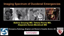
Babina Gosangi MD Tatiana Rocha MD Alejandra DuranMendicuti MD Emergency Radiology Brigham and Womens Hospital Boston MA Goals and Objectives Review the anatomy of the duodenum with an emphasis on its relationships with adjacent structures ID: 909896

duodenum duodenal segment arrow duodenal duodenum arrow segment images year yellow show shows ulcer image orange man contrast superior

Share:

Link:

Embed:

Download Presentation from below link

Download Presentation The PPT/PDF document "Imaging Spectrum of Duodenal Emergencies" is the property of its rightful owner. Permission is granted to download and print the materials on this web site for personal, non-commercial use only, and to display it on your personal computer provided you do not modify the materials and that you retain all copyright notices contained in the materials. By downloading content from our website, you accept the terms of this agreement.


Presentation Transcript

Slide1

Imaging Spectrum of Duodenal Emergencies

Babina Gosangi MD, Tatiana Rocha MD, Alejandra Duran-Mendicuti MDEmergency Radiology, Brigham and Women’s Hospital, Boston, MA

Slide2

Goals and Objectives

Review the anatomy of the duodenum, with an emphasis on its relationships with adjacent structures.Describe the role of various imaging modalities in diagnosing and assessing duodenal emergencies, including upper gastrointestinal series and multidetector CT, including dual-energy CT.

Discuss duodenal emergencies in a systematic manner based on cause and review case examples.

Slide3

SMA

Inferior pancreaticoduodenal artery

Superior

pancreaticoduodenal artery

Gastroduodenal artery

Celiac artery

Segments of the Duodenum and its

Blood Supply

Stomach

1st segment

Superior duodenal flexure

Inferior duodenal flexure

2

nd

segment

4

th

segment

3

rd

segment

Aorta

Superior mesenteric artery (SMA)

Illustrations show the anatomy of the duodenum and its blood supply.

Slide4

Anatomy of the Duodenum

It is the smallest and widest C-shaped portion of the small intestine, measuring 25–40 cm in length.It begins at the duodenal bulb and ends at the ligament of Treitz.

It is divided into four segments. The first segment of the duodenum lies within the peritoneum, but the other three segments are retroperitoneal in location.

Gallbladder

1

st

segment

Common bile duct (CBD)

Portal vein

Inferior vena cava (IVC)

2

nd

segment

L3 vertebra

Right kidney and ureter

Liver

Head of the pancreas

CBD opening

First Segment of the Duodenum

Second Segment of the Duodenum

Extends from the duodenal bulb to the superior duodenal flexure.

Anteriorly: Gallbladder and liver

Posteriorly: Bile duct, portal vein, and IVC

Superiorly: Epiploic foramen

Inferiorly: Pancreatic head

Extends from the superior duodenal flexure to the inferior duodenal flexure.

Pancreatic duct and CBD enter through the ampulla of Vater.

Superiorly: Liver, gallbladder

Medially: Pancreatic head

Posteriorly: Right kidney, ureter, adrenal gland, and L3 vertebra

Slide5

Anatomy of the Duodenum

3

rd

segment

Superior mesenteric vein

SMA

Aorta

IVC

L3 vertebra

4

th

segment

Duodenojejunal flexure

Aorta

Third Segment of the Duodenum

Fourth Segment of the Duodenum

Extends from inferior duodenal flexure, crosses L3 vertebra.

Anteriorly: Superior mesenteric artery and vein

Posteriorly: Aorta, IVC, and L3 vertebra

Passes to the left of the aorta and terminates at the duodenojejunal flexure.

Slide6

Arterial blood supply

The duodenum has a dual blood supply.The first and second segments of the duodenum are supplied by the superior pancreaticoduodenal branches of the gastroduodenal artery (branch of common hepatic artery originating from the celiac axis).The third and fourth segments of the duodenum are supplied by the inferior pancreaticoduodenal branches of the SMA.Venous drainage

Eventually drains into the portal vein through the splenic vein and superior mesenteric vein.

Lymphatic drainageDrains into the pancreaticoduodenal lymph node, which finally drains into the superior mesenteric nodes.

Blood Supply of the Duodenum

Slide7

Role of Imaging in Duodenal Emergencies

Barium Study

Can be used as a first-line imaging modality in some cases

Barium swallow studies can be useful in the identification of strictures, perforations, fistulizations, or obstruction.

CT

Dual-energy CT has been particularly useful in the identification of hemorrhages or perforation of the duodenum.

Conventional CT is useful for the diagnosis of most duodenal pathologic conditions.

MRI and US

Limited roleBarium swallow shows a duodenal stricture (arrow), depicted as a filling defect.

CT image shows the stricture (arrow) as short segment narrowing.

Slide8

Duodenal Emergencies

Peptic ulcer disease: duodenal ulcer perforation and/or hemorrhageInfectious or inflammatory: duodenitis, abscess

Trauma: duodenal transection, duodenal rupture

Vascular: Aortoenteric fistula (AEF), SMA syndrome

Postsurgical: polypectomy syndrome, choledochoduodenal fistula, cholecystectomy abscess extending into the duodenum

Duodenal mass- or tumor-related emergencies: obstruction, hemorrhage, pancreatitis

Miscellaneous: bezoars

Slide9

Peptic Ulcer Disease and Duodenal Ulcers

CT images (

a, b

) in an 81-year-old woman show free air (yellow arrow in

a

) and extravasation of oral contrast material (orange arrow in b) along the second part of the duodenum. Extraluminal contrast material (green arrow in

c and d) is better depicted on the dual-energy CT images (c, d).

Perforated Duodenal Ulcer

Uncomplicated duodenal ulcers can be diagnosed at fluoroscopy as a pocket of barium contrast material filling a crater.

A perforated ulcer can be easily diagnosed by the presence of pneumoperitoneum on plain radiographs, while CT images can show discontinuity in the mucosa.

Hemorrhage can be identified by the presence of high-attenuation material in the duodenum or surrounding the duodenum, with active contrast material extravasation.

a

b

c

d

Slide10

Peptic Ulcer Disease and Duodenal Ulcers

CT images (

a, b

) in a 38-year-old woman show contrast blush on the arterial phase image (yellow arrow in

a

), which increases on the portal venous phase image (orange arrow in b). Iodinated contrast material (green arrow in

c and d) is depicted on the dual-energy CT images (c, d), suggesting hemorrhage in the duodenum, the most likely cause being duodenal ulcer.

Hemorrhage from a Duodenal Ulcer

a

b

c

d

Slide11

Peptic Ulcer Disease and Duodenal Ulcers

Uncomplicated Ulcer at the Duodenal Bulb

(

a, b

) CT images in a 63-year-old man with epigastric pain show stranding and a small amount of free fluid around the duodenal bulb, with thinning and outpouching of the wall of the proximal duodenum (yellow arrow). (

c

) Endoscopic image shows an ulcer (arrow) in the duodenal bulb.

a

bc

Slide12

Inflammatory and Infectious Conditions

Inflammatory conditions can manifest with thickening of the duodenal wall with adjacent stranding.

Infections can manifest with wall thickening and stranding. Collections and/or abscesses can manifest in some cases.

Perforated Duodenal Ulcer Complicated by Abscess

(

a

) CT image in a 58-year-old woman shows a duodenal ulcer complicated by a gas-containing collection (yellow arrow). (

b

) Fluoroscopic image with pigtail placed in the collection shows a fistula and rapid filling of the second segment of the duodenum. (c) IR-guided pigtail was placed to drain the abscess, which gradually resolved.bac

Slide13

Inflammatory and Infectious Conditions

(

a, b

) CT images in a 66-year-old woman show short segment abrupt narrowing of the second segment of the duodenum, with massive dilatation of the stomach proximally (yellow arrow). (c) Dual-energy CT image shows enhancement at the stricture site, with focal narrowing and wall thickening (orange arrow). Endoscopy helped confirm a nonmalignant ulcer (not shown).

Inflammatory Duodenal Stricture

a

b

c

Slide14

Inflammatory and Infectious Conditions

CT images in a 62-year-old man with duodenal diverticulum show extensive stranding along the diverticulum, which suggests duodenal diverticulitis (yellow arrow)

.

Duodenal Diverticulitis

a

b

Slide15

Inflammatory and Infectious Conditions

CT images in a 64-year-old man with acute epigastric pain. There is mild wall thickening and stranding involving the second and third segments of the duodenum, with minimal adjacent fluid (yellow arrow). The findings were consistent with duodenitis, and the patient was administered antacid medication, after which the symptoms were relieved.

Acute Duodenitis

Slide16

The third segment of the duodenum is the most prone to traumatic injury owing to its prevertebral location. Duodenal transection is associated with greater morbidity, and there is discontinuity of the duodenum. Duodenal rupture manifests with discontinuity of the duodenum and extraluminal air.

Trauma

CT images in a 45-year-old man who fell from a height show duodenal wall thickening with foci of extraluminal air surrounding the duodenum (yellow arrows in

a

and

b

) suggestive of rupture. Note the retroperitoneal hemorrhage (orange arrow in

c).Duodenal Rupture

a

b

c

Slide17

Trauma

CT images in a 19-year-old man following a motor vehicle collision show discontinuity in the duodenum (yellow arrows in

a

and

b

), liver laceration (orange arrow in

c

), and hemoperitoneum (green arrow in

d).Duodenal Transection

a

b

c

d

Slide18

Vascular

The third segment of the duodenum is more prone to vascular complications like SMA syndrome and AEF owing to its close proximity to the aorta. AEF manifests with extensive stranding along the aorta, with contrast in the duodenum.

CT images in a 61-year-old woman with left lower limb pain show AEF with communication between the aorta and duodenum. Note the small amount of contrast in the duodenum (yellow arrow in

a

and

c

), with extensive stranding around the aorta (orange arrow in

b)

Aortoenteric Fistula

b

a

c

Slide19

Duodenal Hemorrhage Secondary to Inferior Pancreaticoduodenal Artery Aneurysm Rupture

Vascular

(

a, b

) MR cholangiopancreatographic images in a 65-year-old man show T1-weighted hyperintense periduodenal collection (yellow arrow in

a

and

b). Correlation with the CT image shows a hyperattenuating periduodenal collection, with a 7-mm aneurysm on the CT angiogram (orange arrow in d

). (e, f) Angiograms show an aneurysm of the inferior pancreaticoduodenal artery (green arrow in e and f), which was coiled.

a

b

c

d

e

f

Slide20

Vascular

Enhanced CT images of the abdomen in a 37-year-old man show an aortomesenteric angle of

12° (orange arrow) and aortomesenteric distance of 4 mm (yellow arrow), suggestive of SMA syndrome.

SMA Syndrome

Slide21

Vascular

Duodenal Varices Embolization and Complication

Portal hypertension and hematemesis in a 55-year-old man. IR angiogram (

a

) shows multiple duodenal varices. TIPSS was performed (

b), and the varices were embolized using gelfoam. Follow-up (

c, d) CT images shows a small varix (yellow arrow in

d) communicating with an overlying duodenal ulcer identified by the presence of air foci (orange arrow in d).

abc

d

Slide22

Postsurgical Complications

CT images in a 42-year-old man who presented with pain following the removal of a duodenal polyp by using a hot snare show mucosal enhancement and stranding adjacent to the second and third parts of the duodenum (yellow arrow), consistent with inflammation.

Postpolypectomy Syndrome

The duodenum owing to its close proximity to gall bladder, pancreas, and liver can get involved during surgical procedures of the adjacent organs.

Postsurgical complications such as abscess formation, inflammation with phlegmon, and fistulas can be better visualized at CT.

Slide23

Postsurgical Complications

(a)

CT image in a 58-year-old man who underwent cholecystectomy shows a rim-enhancing abscess in the gall bladder fossa (yellow arrow in

a and

b) extending into the duodenum (orange arrow). (b) Dual-energy CT image confirms the findings.

Postcholecystectomy Abscess Communicating with Duodenum

b

a

Slide24

Emergencies Related to Duodenal Masses/Tumors

Abdominal pain in a 62-year-old man. (

a

) Upper gastrointestinal study shows a long segment with nonopacification of the third segment of the duodenum (yellow arrow). (

b) CT image obtained after the administration of oral contrast material shows an infiltrative mass involving the third segment of the duodenum (orange arrow), which was surgically biopsied. The findings were consistent with adenocarcinoma of the duodenum.

Infiltrative Primary Duodenal Mass causing Obstruction

Primary or metastatic lesions in the duodenum can cause emergent manifestations, such as epigastric pain, obstruction, hemorrhage.

Barium studies may show short- or long-segment filling defects, while CT can show infiltrative mass, obstruction, and/or hemorrhage.

Endoscopy with biopsy is the confirmatory investigation.ab

Slide25

Bleeding Distal Duodenal Metastasis

(

a, b

) CT images in a 62-year-old woman who presented with pain in the abdomen shows contrast material extravasation in distal second segment of the duodenum (yellow arrow in

a

), which increases on delayed images (orange arrow in

b

). The patient was watched, but subsequently she developed pain in the abdomen again.

(

c, d

) Follow-up CT images show small bowel intussusception with multifocal metastases, consistent with non–small cell lung cancer (green arrows).

Emergencies Related to Duodenal Masses/Tumors

a

b

c

d

Slide26

Emergencies Related to Duodenal Masses/Tumors

Primary Duodenal Mass Manifesting with Pancreatitis

CT images in a 77-year-old man woman with epigastric pain shows a masslike appearance at the pancreaticoduodenal groove, duodenum, and head of the pancreas, which inseparable at CT. The patient had elevated serum lipase levels. Endoscopic image shows a distorted appearance of the second segment of the duodenum. The results of a biopsy confirmed adenocarcinoma of the duodenum.

a

b

c

Slide27

Foreign Body/Bezoars

(

a, b

) CT images in a 33-year-old woman with abdominal pain show a low density object in the duodenum and stomach causing massive dilatation (yellow arrow). (

c

) Endoscopic image shows hair. (d

) Postoperative photograph shows a large bezoar.

Bezoar

abc

d

Slide28

Conclusion

The particular location of the duodenum, with both intraperitoneal and retroperitoneal segments, and its relationships with adjacent structures results in its involvement in a multitude of primary and secondary processes.Knowledge of the entities and radiologic findings of emergent duodenal pathologic conditions is important for accurate differential diagnosis and prompt patient management.

Slide29

References

1. Jayaraman MV, Mayo-Smith WW, Movson JS, Dupuy DE, Wallach MT. CT of the duodenum: an overlooked segment gets its due. Radiographics 2001;21(Spec Issue):S147–160.2. Heller MT, Haarer KA, Itri JN, Sun X, Duodenum: MDCT of acute conditions. Clin Radiol 2014;69(1):e48–55.

3. Shaffer HA Jr. Perforation and obstruction of the gastrointestinal tract: Assessment by conventional radiology. Radiol Clin North Am 1992;30(2):405–426.

Slide30

Thank you

bgosangi@bwh.harvard.edu