/
The Molecular Basis of Blood Coagulation Review The Molecular Basis of Blood Coagulation Review

The Molecular Basis of Blood Coagulation Review - PDF document

cecilia
cecilia . @cecilia
Follow
342 views
Uploaded On 2022-08-23

The Molecular Basis of Blood Coagulation Review - PPT Presentation

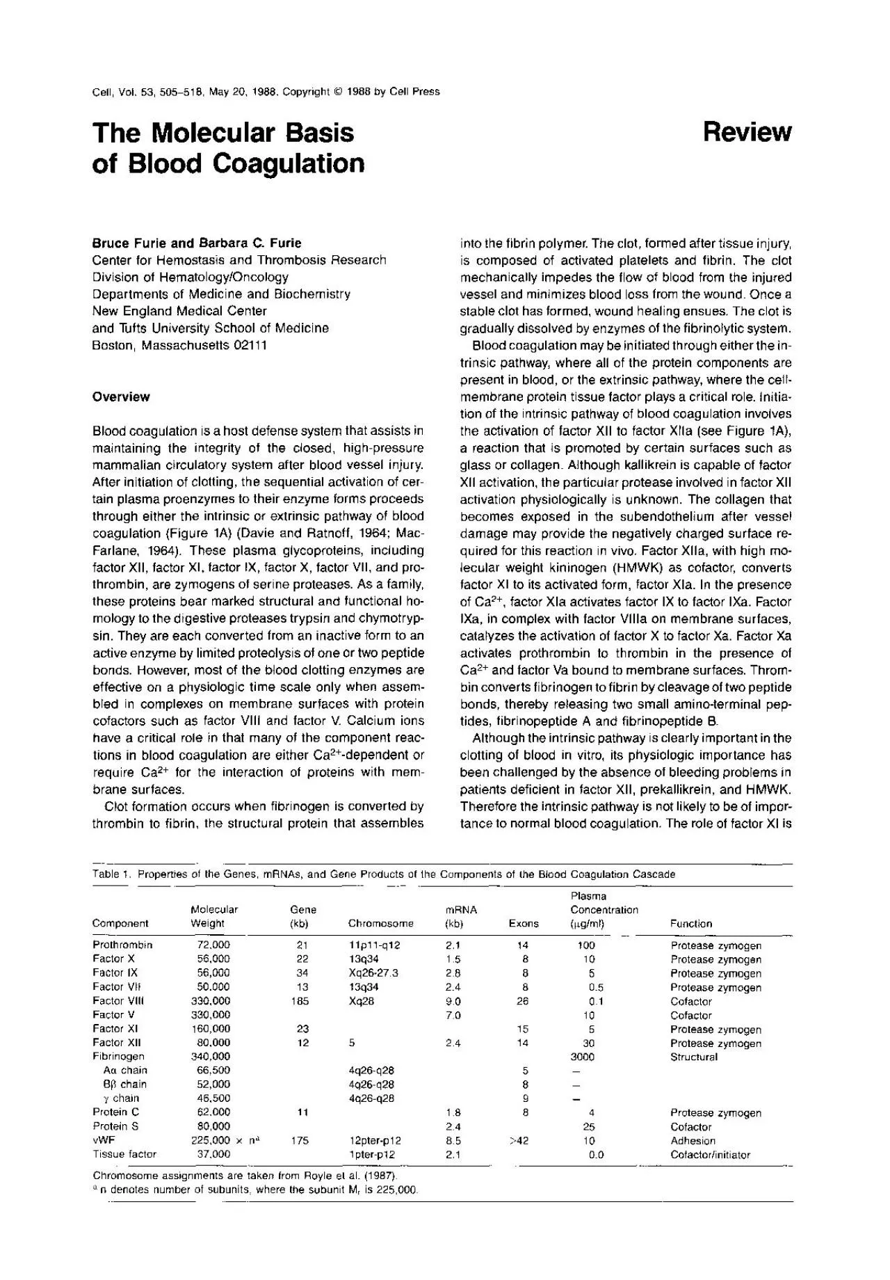
Bruce Furie and Barbara C Furie Center for Hemostasis and Thrombosis Research Division of HematologyOncology Departments of Medicine and Biochemistry New England Medical Center and Tufts University ID: 940689

protein factor proteins blood factor protein blood proteins coagulation 000 chain gene pro zymogen 1987 domains domain egf acid

Share:

Link:

Embed:

Download Presentation from below link

Download Pdf The PPT/PDF document "The Molecular Basis of Blood Coagulation..." is the property of its rightful owner. Permission is granted to download and print the materials on this web site for personal, non-commercial use only, and to display it on your personal computer provided you do not modify the materials and that you retain all copyright notices contained in the materials. By downloading content from our website, you accept the terms of this agreement.


Presentation Transcript

The Molecular Basis of Blood Coagulation Review Bruce Furie and Barbara C. Furie Center for Hemostasis and Thrombosis Research Division of Hematology/Oncology Departments of Medicine and Biochemistry New England Medical Center and Tufts University School of Medicine Boston, Massachusetts 02111 Overview Blood coagulation is a host defense system that assists in maintaining the integrity of the closed, high-pressure mammalian Table 1. PropertIes of the Genes, mRNAs, and Gene Products of the Components of the Blood Coagulation Cascade Plasma Molecular Gene mRNA Concentration Compo

nent Weight W Chromosome WW Exons WW Function Prothrombin 72,000 21 llpll-q12 2.1 14 Protease zymogen Factor X 56,000 22 1.5 8 Protease zymogen Factor IX 56,000 34 Xq26-27.3 2.8 8 Protease zymogen Factor VII 50,000 13 2.4 8 0.5 Protease zymogen Factor VIII 330,000 185 Xq28 9.0 26 0.1 Cofactor Factor a 4 Protease zymogen Protein S 80,000 2.4 25 Cofactor vWF 225,000 x na Ppter-p12 85 �42 10 Adhesion Tissue factor 37,000 1 pter-p12 2.1 Cofactorlinitlator Chromosome assignments are taken from Royle et al. (1987). a denotes number of subunits, where the subunlt M, is 225,000 Cd

l 506 Intrinsic Pathway Figure 1. The Protems of Blood Coagulation (A) The blood coagulation cascade. Glycoprotein components of the intrinsic pathway include factors XII, XI, VIII, X, and V, prothrombin, and fibrinogen. Glycoprotein components of the extrinsic pathway, initiated by the action of tissue factor located on cell surfaces, include factors VII, X, and V, prothrombin and fibrinogen. The cascade reactions generation of anticoagulants such as activated protein C are processes that prevent clot formation in normal blood vessels. After tissue injury, thrombogenic subendothel

ial components of the blood vessel are exposed. Receptors for blood clotting proteins may be expressed on the en- dothelial cell membrane, allowing for the sequential Structural Features of the Blood Clotting Proteins and Their Encoding Genes The plasma proteins involved in hemostasis can be sepa- rated into three functional groups with interrelated but Figure 2. Stuctural Domains of the Proteins involved in Hemostasis and of Related Proteins Domains, identified in the key, are in the text. Sites of proteo- lytic cleavage associated with synthesis of the mature protein are indi-

cated by thin arrows. Sites of proteolytic cleavage associated with zymogen activation are indicated by the thick arrows. Shown, top to bottom, are schematic structures for prothrombin (Degen et al., Some of Common Domain Structures The structure and organization of the genes coding for the blood coagulation proteins emphasize that the evolution of new protein function occurs via gene duplication, gene modification, and exon shuffling (Gilbert, 1978; Patthy, 1985). Each of the exons may be considered a module coding for a homologous domain in each protein (Figures 2 3). The three

-dimensional structures of the polypep- tide backbones of these homologous domains are likely to be nearly Factor VII Factor IX Factor X Protein C Prothrombin Factor XI Factor XII 0 CATALYTIC El REPEAT ii FIBRONECTIN TYPE I FIBRONECTIN TYPE ,I Figure 3. Exon-lntron Structures of the Genes Encoding the Blood Coagulation Serine Proteases Exons are shown schematrcally (see key) and are drawn to scale. Introns, indicated by lines connecting the exons. are not drawn to scale. The exons I, II, on a and a and gene a gene a gene cleavage of two peptide bonds and the release of an acti-

vation peptide. Twelve y-carboxyglutamic acid residues are located near the amino terminus of the mature zymo- gen. A single f3-hydroxyaspartic acid at position 64 in the EGF domain has uncertain function. Factor X (M, 56,000) is synthesized as a single chain (Fung et al., 1985; Leytus et al., 1984) but is converted to a two-chain zymogen that circulates in the plasma. The light chain of human factor X includes eleven y-carboxyglutamic acid residues and the EGF domain, containing a single f3-hydroxyaspartic acid. The activation peptide is cleaved from the heavy chain during activat

ion of factor X by the factor Protein Cofactors The majority of the serine proteases of blood coagulation require protein cofactors for efficient proteolytic activity. Tissue factor is a transmembrane protein (M, 37,000) that activates blood coagulation through the extrinsic pathway by forming a complex with factor Vlla that activates factor X. This protein is not present in plasma, but rather is a component of many cell surfaces with the exception of resting endothelial cells and monocytes. The amino acid to regions of other proteins (Dahlback et al., 1986). Protein S is synthes

ized as a single chain containing a signal pep- tide, a propeptide, a Gla domain, four EGF domains con- taining 8-hydroxyaspartic acid and 8-hydroxyasparagine, and additional large domain likely responsible for rec- ognition of activated protein C (Lundwall et al., 1986; Hoskins et al., 1987). Protein S exists in two forms in the blood: as a species bound to C4b-binding binding protein and as a free form. The gene encoding facror V//l is one of the largest known, spanning 186 kb on the chromosome. The coding DNA is divided into twenty-six exons, including 2%-3% of the plasma pro-

tein. It is composed of two pairs of each chain: the Aa chain, the B8 chain, and the y chain. Removal of fibrino- peptide A from the Aa chain and removal of fibrinopeptide B from the B8 chain leads to the generation of fibrin mono- mer. Fibrin is a prototypic example of protein self-assembly: it rapidly polymerizes to form long structural strands that represent the fibrin clot. The clot Factor IX Prothrombin Factor x Protein C Factor VII Protein S Bone Cta protein Ser Leu Vat His Ser Leu Gty Ctu Thr Pro Ata Pro Leu Asp Trp Lys Pro Cty Pro His Vat Leu Pro Vat Leu Glu Gty Ala Gtu Se

r Figure 5. Sequences of the Propeptide Domains of Vitamin K-Dependent Blood Coagulation Proteins The size of the propeptide has been established for factor IX (Diuguid et al., 1986; Bentley et al., 1988) and protein C (Long et al., 1984; Foster et Basis of Blood Coagulation 513 EGF precursor in certain vitamin K-dependent proteins (Stenflo et al., 1987) as well as in proteins outside of this family, including the complement proteins Clr and Cls, thrombomodulin, uromodulin, and the low density lipopro- tein receptor (Stenflo et al., 1987, Przysiecki et al., 1987). A consensus s

equence encompassing the P-hy- droxylated Asp and Asn residues within a number of EGF domains has been noted by Stenflo et al. (1987): Cys-X-AspIAsn-X-X-X-X-PhelTyr-X-Cys-X-Cys. EGF domains that lack the consensus sequence do not contain this posttranslational modification. Multimerization of von Willebrand Factor vWF is initially synthesized as a large Cleavage of Prosequences The propeptides in the vitamin K-dependent proteins and vWF are critical to posttranslational processing. After pro- cessing, these peptides are cleaved and the mature pro- teins circulate in the plasma.

Two contiguous basic amino acids appear to the amino-terminal side of the site of propeptidase Molecular Genetics of Hemophilia Although a wide diversity of bleeding disorders as- sociated with almost all phases of clot formation and its regulation are known, the structural basis for the defect has been identified in only a few instances. Genetic defects in the blood coagulation proteins are associated with a bleeding disorder known as hemophilia. Defects in factor VIII (hemophilia A) account for about 85% of this heredita.ry disorder, while defects in factor IX (hemophilia 9) are

responsible for 100/o-12%. Other defects in coagu- Cell 518 K. (1985). Nucleotide sequence of the gene for human Factor IX. Bio- chemistry 24, 3736-3750. Youssoufian, H.. Kazazian, H. H.. Jr., Phillrps, D. G., Aronis, S., Tsiftis, G.. Brown, V. A., and Antonarakis, S. (1986). Recurrent mutations In haemophilia A gwe evidence for CpG mutatron hotspots. Nature 324, 380-382. Youssoufian. H., Antonarakis. S. E., Aronis, S.. Tsiftis. G., Phillips, D. G., and Kazazian. H. H., Jr. (1987). Characterization of five partial deletions of the factor VIII gene. Proc. Natl. Acad. Sci. USA 84, 3