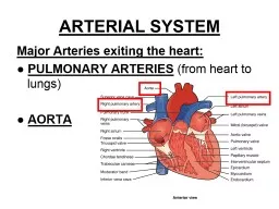
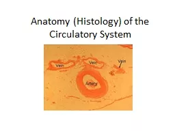
/
 Vascular shunt Anatomy of the arteries veins and capillaries.  Vascular shunt Anatomy of the arteries veins and capillaries.

Vascular shunt Anatomy of the arteries veins and capillaries. - PowerPoint Presentation

celsa-spraggs
celsa-spraggs . @celsa-spraggs
Follow
342 views
Uploaded On 2020-04-04

Vascular shunt Anatomy of the arteries veins and capillaries. - PPT Presentation

Anatomy and Physiology On the white boards outline the process of fainting after an athlete performs exercise and then stops abruptly clue vascular return Cause During exercise high Q what is Q ID: 775365

blood exercise arterioles capillary blood exercise arterioles capillary muscles capillaries organs arteries flow vascular pressure working sphincters amp structure

Share:

Link:

Embed:

Download Presentation from below link

Download Presentation The PPT/PDF document " Vascular shunt Anatomy of the arteries ..." is the property of its rightful owner. Permission is granted to download and print the materials on this web site for personal, non-commercial use only, and to display it on your personal computer provided you do not modify the materials and that you retain all copyright notices contained in the materials. By downloading content from our website, you accept the terms of this agreement.


Presentation Transcript

Slide1

Vascular shuntAnatomy of the arteries veins and capillaries.

Anatomy and Physiology

Slide2

On the white boards outline the process of fainting after an athlete performs exercise and then stops abruptly. (clue, vascular return)

Cause During exercise – high Q (what is Q)After and stop – drop in muscle pump and less VRSO – less SV, Q, BP drops and less flow O2 to brain = dizzy

Solutions

Put head below heart to aid blood flow there

Lie down - restores BP and aids VR.

Active cool down will aid reduction in VR

Slide3

Venule Structure & Function

Venule Structure

Venules are minute vessels that drain blood from capillaries and into veins. Many venules unite to form a vein.

Venule Function

Drains blood from capillaries into veins, for return to the heart

Slide4

Artery & Vein Composition

1 - an outer fibrous layer — tunica externa2 - a thick middle layer — tunica media3 - a thin lining of cells to the inside — tunica interna.

The tunica media is comprised of smooth muscle and elastic tissue, which enables the arteries and arterioles to alter their diameter.

1

2

3

Slide5

Artery Structure

The artery wall has a thick muscular / elastic layer to expand as the blood surges through, and recoil after.

Artery Function

The blood is under HIGH PRESSURE and HIGH VELOCITY as it has been ejected from the heart.

Slide6

Arteriole Structure & Function

Arterioles are tiny branches of arteries that lead to capillaries.

Slide7

Capillary Structure & Function

Capillaries are tiny (extremely narrow) blood vessels – 1 cell thick Capillary beds surround organs. These capillaries are supplied with blood by arterioles and drained by venules. Capillary walls are only one cell thick which permits exchanges of material between the contents of the capillary and the surrounding tissue.

Supply gases & nutrients – remove wasteDiffusion of oxygen, carbon dioxide, water, salts, between the blood and the organs BP and BLOOD FLOW here are much less, as the total area of capillaries is immense.

Slide8

Vascular shunting.

What percentage of blood is used in the muscles while we are at rest?

20%

Have a look at page 85 to discover how this changes when we take part in high intensity exercise.

Slide9

Chart on page 85

Don’t panic the blood supply to the brain is maintained.

Slide10

Slide11

So how does this all transfer into an exam question?

See page 120 and hand out.Organs (during exercise)There is vasoconstricts in the arterioles and pre-capillary sphincter. These both decrease the distribution of blood flow from the essential organs.

Muscles (during exercise)

The arterioles and pre capillary sphincters vasodilate.

Both increase the blood flow towards the capillaries in the working muscles.

Slide12

Exam Q Explain the vascular shunt mechanism during exercise. 4

Vasodilation

of

arteries/arterioles/blood vessels/leading to working muscles/vascular shunt

Opening/vasodilation of pre capillary sphincters leading to working

muscles

Vasoconstriction of arteries/arterioles/blood vessels leading to non-essential organs

Closing of pre capillary sphincters leading to non-essential

organs

Slide13

Exam Question

During exercise the body has to respond to an increased demand for oxygen by the working muscles.

Figure

2 shows the distribution of cardiac output during exercise.

Describe

the mechanism that allows for the redistribution of blood flow during exercise and explain how it is controlled

.

(10 marks)

Slide14

Reading the flipping question.

Break it down into the command words and the subject qualifiers.

What is it asking for?

Section 1 we can answer.

Section 2 Look at page 86 and bullet point on the white boards the answer. See where you will get ten marks in total.

Slide15

Mark Scheme

Distribution

of blood flow during exercise

(

mechanism

) (

subsub

max 4)

vascular

shunt mechanism


(

redistributes blood during exercise so that) areas with the greatest need receive more blood/areas with low demand receive less blood through vasodilation of arterioles/blood vessels feeding working muscles

and vasodilation/opening of

precapillary

sphincters feeding working muscles

through vasoconstriction of arterioles feeding others organs (e.g. liver/kidney/intestines)

and vasoconstriction/closing of

precapillary

sphincters feeding these organs

Slide16

vascular shunt mechanism

)

(control explanation)

(

subsubmax

4)

controlled by the vasomotor control

centre

CCC;

located in the medulla (oblongata) of the brain

VCC responds to changes in blood pressure/muscle/blood chemistry

chemoreceptors detect changes in lactic acid/carbon dioxide/oxygen/pH/ content of blood

chemoreceptors located in muscles/aorta/carotid arteries

baroreceptors detect changes in blood pressure

baroreceptors located in aorta/carotid arteries

VCC uses sympathetic nervous system which acts on the middle layer of smooth muscle in an arteriole/the ring of smooth muscle at the opening of a capillary (

precapillary

sphincter)/control diameter of arterioles/

precapillary

sphincters

Slide17

Using the white boards draw the transfer of blood highlighting the affiliation to Hb. Give values where appropriate.

Next person add in the affiliation to monoxide (smoke).

Slide18

Application of understanding.Can you check your answers against someone else in the group. See if you have the same answer. (speed dating).

Can you explain why the blood pressure in the endurance training will plateau at around 140-160?

Can you account for the increase in systolic

Bp

in line with exercise intensity?

Why during exercise does the diastolic pressure stay

roughy

the same?

Can you explain why localised muscular diastolic pressure may fall?