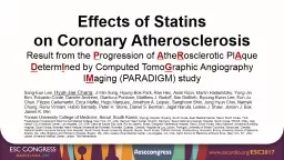
Result from the P rogression of A the R osclerotic Pl A que D eterm I ned by Computed Tomo G raphic Angiography I M aging PARADIGM study SangEun Lee HyukJae Chang ID: 774858
Download Presentation The PPT/PDF document " Effects of Statins on Coronary Atherosc..." is the property of its rightful owner. Permission is granted to download and print the materials on this web site for personal, non-commercial use only, and to display it on your personal computer provided you do not modify the materials and that you retain all copyright notices contained in the materials. By downloading content from our website, you accept the terms of this agreement.
Slide1
Effects of Statinson Coronary AtherosclerosisResult from the Progression of AtheRosclerotic PlAque DetermIned by Computed TomoGraphic Angiography IMaging (PARADIGM) study
Sang-Eun Lee,
Hyuk-Jae Chang
, Ji Min Sung, Hyung-Bok Park, Ran
Heo
,
Asim
Rizvi, Martin
Hadamitzky
, Yong-
Jin
Kim,
Edoardo
Conte, Daniele
Andreini
, Gianluca
Pontone
, Matthew J.
Budoff
,
Ilan
Gottlieb,
Byoung
Kwon Lee, Eun Ju Chun, Filippo
Cademartiri
, Erica Maffei, Hugo Marques, Jonathon A. Leipsic,
Sanghoon
Shin, Jung Hyun Choi,
Namsik
Chung,
Renu
Virmani
, Habib
Samady
, Peter H. Stone, Daniel S. Berman,
Jagat
Narula
,
Leslee
J. Shaw, Jeroen J.
Bax
, James K.
Min
Yonsei University College of Medicine, Seoul, South Korea;
Myongji
Hospital,
Goyang
, South Korea;
Asan
Medical Center, Seoul, South Korea; York-Presbyterian Hospital and Weill Cornell Medical College, New York, NY, USA; German Heart Center, Munich, Germany; Seoul National University Hospital, South Korea; IRCCS, Milan, Italy; Harbor UCLA Medical Center, Los Angeles, CA, USA; Casa de
Saude
São Jose, Rio de Janeiro, Brazil; Gangnam Severance Hospital, Seoul, South Korea; Seoul National University
Bundang
Hospital, South Korea; Montréal Heart Institute/
Université
de Montréal, Montréal, Quebec, Canada; Hospital da Luz, Lisbon, Portugal; University of British Columbia, Vancouver, Canada; National Health Insurance Service
Ilsan
Hospital, South Korea; Busan University Hospital, Busan, South Korea;
CVPath
Institute, Gaithersburg, Maryland; Emory University School of Medicine, Atlanta, GA, USA; Brigham and Women's Hospital, Boston, MA, USA; Cedars-Sinai Medical Center, Los Angeles, CA, USA; Icahn School of Medicine at Mount Sinai, New York, NY, USA; Leiden University Medical Center, Leiden, The Netherlands
Slide2Statins have been effective at reducing risk of major adverse cardiac events.
Source:
Sacks et al. NEJM 1996;335:1001-9.
In previous studies, statin decreased the lipid burden and increased the fibrous components.
But limited to small number of patients, short durations of follow-up, and qualitative metrics.
Source:
Hattori et al. J Am Coll
Cardiol Img 2012;5:169 –77
Background
Slide3Background
Uncertainty exists as to the precise effects of statins on changes in coronary atherosclerotic plaque characteristics.
Coronary CT angiography (CCTA) is a non-invasive test with high performance that enables both quantitative analysis of plaque burden and plaque characterization.
Assessing impact of statins on clinical outcomes
Assessing impact of statins on atherosclerosis
Target patient group of previous studies evaluating impact of statins
Severity of coronary heart disease
Slide4To determine the long-term effects of statins on coronary atherosclerosis.
Purpose
Slide5Study Design & Population
Dynamic multinational observational prospective registry Study Criteria
Inclusion criteria
Consecutive patients with suspected or known coronary artery disease undergoing serial CCTAs at an inter-scan interval of
≥
2 years
Exclusion criteria
Prior coronary artery disease before baseline CCTA (CCTA-1)
No information on statin at both baseline (CCTA-1) and follow-up CCTA (CCTA-2)
Patients who discontinued statin use after the CCTA-1
Patients with non-interpretable CCTA
Slide6CONSORT diagram
13 sites from 7 countries enrolled between 2003 and 2015
Slide7CCTA Analysis
Core laboratoryIntegrative Cardiac Imaging Research Center, Severance Hospital, Seoul, KoreaLevel III experienced readersQAngioCT Research Edition v2·1·9·1 (Medis Medical Imaging Systems, the Netherlands)
Measured parameters
Plaque volume (PV, mm
3
): sub-classified by pre-defined Hounsfield unit cutoff values
% diameter stenosis (%DS)
High-risk plaque (HRP) features: positive remodeling, low-attenuation plaque, spotty calcification
* Segment matching between baseline & follow-up CT – using branch point
Slide8Primary EndpointThe annualized per-lesion change in plaque volumes between CTA-1 and CTA-2 for statin-naïve versus statin-taking patientsSecondary EndpointAnnualized change in PV by compositionsDevelopment of high-risk plaque and its constituent featuresDevelopment of obstructive coronary lesions (≥50% diameter stenosis)
Study Endpoints
Slide9Patient Characteristics
Total
(n=1,255)
Statin-naïve patients
(n=474)
Statin-taking patients
(n=781)
P
P
Age,
years
60.4±9.2
59.2±9.6
61.1±8.8
0∙0002
<0.001
Male sex
712 (57%)
260 (55%)
452 (58%)
0∙2948
0.295
CTA interval, years
3.8±1.6
3.7±1.6
3.9±1.6
0∙0155
0.016
Hypertension
654 (52%)
217 (46%)
437 (56%)
0∙0004
<0.001
Diabetes mellitus
261 (21%)
70 (15%)
191 (25%)
<∙0001
<.001
Family history of CAD
337 (27%)
117 (25%)
220 (28%)
0∙1768
0.177
Smoking history
467 (37%)
178 (38%)
289 (37%)
0∙8280
0.828
Cardiac
symptom
1,062 (97%)
400 (97%)
662 (97%)
0∙5396
0.50
Baseline Lipid Profile
Total
cholesterol,
mg/
dL
190.4±40.2
186.6±34.5
192.7±43.1
0.008
LDL, mg/
dL
116.0±34.9
114.1±28.7
117.1±38.2
0.123
HDL, mg/
dL
50.9±14.1
51.3±14.2
50.7±14.0
0.490
Slide10CTA Findings According to Statin Therapy
Lesions in statin-naïve patients
(n=1,079)
P
value between baseline
vs. FU
Lesions in statin-taking patients
(n=2,496)
P
value between baseline
vs. FU
P
value between groups
Baseline
FU
Baseline
FU
Baseline
FU
Stenosis severity
%DS ≥ 50%
12 (1
%
)
23 (2
%
)
0.028
40 (2
%
)
76 (3
%
)
<.001
0.261
0.127
Stenosis severity, %
11.6±12.7
18.2±12.4
<.001
14.4±13.8
20.1±13.2
<.001
<.001
<.001
Annual change in %DS, %/year
1.9±3.8
1.5±3.8
0.009
High-risk plaque characteristics
High-risk plaque
108 (10%)
170 (16%)
<.001
342 (14%)
428 (17%)
<.001
0.002
0.306
Positive remodeling
514 (48
%
)
799 (74
%
)
<.001
1,398 (56
%
)
1,901 (76
%
)
<.001
<.000
0.178
Low-attenuation plaque
91 (8
%
)
111 (10
%
)
0.027
212 (8
%
)
233 (9
%
)
0.096
0.953
0.375
Spotty calcification
73 (7
%
)
113 (10
%
)
<.001
254 (10
%
)
332 (13
%
)
<.001
0.001
0.019
Slide11Plaque Volume at Baseline
Lesions in statin-naïve patients
(n=1,079)
Lesions in statin-taking patients
(n=2,496)
P
value
Total PV, mm
3
27.32±57.82
37.24±72.32
<.001
Calcified PV, mm
3
7.68±22.49
14.00±39.08
<.001
Fibrous
PV
, mm
3
12.69±28.21
16.20±31.30
0.001
Fibrous fatty
PV
, mm
3
6.20±17.39
6.28±16.39
0.898
Necrotic core
PV
, mm
3
0.75±3.39
0.77±3.39
0.950
Slide12Annualized Change in Plaque Volume
Lesions in statin-naïve patients
(n=1,079)
Lesions in statin-taking patients
(n=2,496)
P
value
Total PV, mm
3
/year
5.35±10.06
4.61±9.67
0.039
Calcified PV, mm
3
/year
2.35±4.72
3.10±6.55
<0.001
Fibrous PV, mm
3
/year
2.24±5.80
1.42±5.79
<0.001
Fibrous fatty PV, mm
3
/year
0.72±5.50
0.10±4.01
<0.001
Necrotic core PV, mm
3
/year
0.04±1.14
-0.0003±0.88
0.271
Slide13Effects of Statin on Atherosclerosis
Slide14Conclusions
Statins are associated with
Slower progression of overall coronary atherosclerosis volume
Increased plaque calcification
Reduction of high-risk plaques
Statins are not associated with lesions progressing to high-grade stenoses.
Slide15Thank You