/
Enzymes Clinical significance & Methods Enzymes Clinical significance & Methods

Enzymes Clinical significance & Methods - PowerPoint Presentation

christina
christina . @christina
Follow
343 views
Uploaded On 2022-06-15

Enzymes Clinical significance & Methods - PPT Presentation

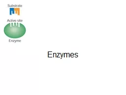
Measurements of enzymes in serum within a tissue enzymes as markers of disease Injury to tissue releases cellular substances that can be used as plasma markers of tissue damage enzyme release is highly specific for cell death in some settings ID: 918494

serum activity acp ast activity serum ast acp phosphatase acid alp disease bone enzymes methods liver increased isoenzymes clinical

Share:

Link:

Embed:

Download Presentation from below link

Download Presentation The PPT/PDF document "Enzymes Clinical significance & Meth..." is the property of its rightful owner. Permission is granted to download and print the materials on this web site for personal, non-commercial use only, and to display it on your personal computer provided you do not modify the materials and that you retain all copyright notices contained in the materials. By downloading content from our website, you accept the terms of this agreement.


Presentation Transcript

Slide1

Enzymes

Clinical significance & Methods

Slide2

Measurements of enzymes

in serum

within a tissue

enzymes as markers of disease,

Injury to tissue releases cellular substances that can be used as plasma markers of tissue damage.

enzyme release is highly specific for cell death in some settings.

Slide3

Some enzymes are predominantly found in the specialized tissue

while others, more widely distributed, have tissue specific

isoenzymes

or isoforms

The timing of the enzyme's diagnostic window

early indicators

late indicators

Several enzymes have diagnostic utility

Slide4

Overlap occurs for some enzymes

may be used

for investigating disease

in several organs.

Slide5

Enzymes of clinical

interest

MUSCLE ENZYMES

More commonly measured

CK, LD

CK

; adenosine

triphosphate

:

creatine

N-

phosphotransferase

Slide6

Slide7

Inhibitors

excess Mg

2+

Many metal ions, such as Mn

2+

, Ca

2+

, Zn

2+

,Cu

2+

sulfhydryl

-binding reagents

Iodoacetate

Slide8

CK is a

dimer

(B and M)

the products of loci on chromosomes 14 and 19, respectively.

BB (or CK-1), MB (or CK-2), and MM (or CK-3).

numbered on the basis of their

electrophoretic

mobility, with the most anodal form receiving the lowest number.

The distribution of these

isoenzymes

in the various tissues

Slide9

Slide10

Serum CK activity is subject to a number of physiological variations.

Sex, age, muscle mass, physical activity, and race all interact to affect serum activities

Slide11

Clinical Significance

Serum CK activity is greatly elevated in all types of muscular dystrophy.

may be increased long before the disease is clinically apparent.

Serum CK activity characteristically falls as patients get older and as the mass of functioning muscle diminishes with the progression of the disease.

Slide12

Long time Physical inactivity reduce serum CK

Skeletal muscle that is diseased or damaged

(fetal reversion)

Renal failure , increase CK

Serum CK activity demonstrates an inverse relationship with thyroid activity.

Following a

myocardial

infarction

MB

isoenzyme

Slide13

Slide14

The assay of CK activity

Coupled enzyme methods

NADP+ to NADPH, monitored

spectrophotometrically

.

Slide15

CK activity in serum is relatively unstable and is rapidly lost during storage.

Average stabilities are less than 8 hours at room temperature, 48 hours at 4°C, and 1 month at -20°C.

Methods for the Separation & Quantification of CK

Isoenzymes

Electrophoresis & various immunological methods.

Slide16

Enzymes of clinical interest

Widely

used

enzymes

Aspartate aminotransferases (AST)

Alanine aminotransferases (ALT)

ALP

Slide17

The aminotransferases

The aminotransferases

catalyze the

interconversion

of amino acids to 2-oxo-acids

L-aspartate:2-oxoglutarate

aminotransferase;AST

L-alanine:2-oxoglutarate aminotransferase; ALT

Slide18

Slide19

Slide20

Pyridoxal-5'-phosphate ↔ pyridoxamine-5' –phosphate

Both the coenzyme-deficient

apoenzymes

and the

holoenzymes

may be present in serum.

All factors affecting the rate of reaction must be optimized and controlled

Slide21

Transaminases

are widely distributed throughout the body.

Slide22

Clinical Significance

Liver disease is the most important cause of increased

transaminase

activity in serum.

In most types of liver disease, ALT activity is higher than that of AST;

exceptions

alcoholic hepatitis, hepatic cirrhosis, and liver

neoplasia

.

elevated even before the clinical signs and symptoms of disease

Slide23

Peak values

of

transaminase

activity occur between the 7th and 12th days

Medications

nonsteroidal

antiinflammatory

drugs, antibiotics.

ALT is the more liver-specific enzyme.

elevations of ALT activity persist longer than do those of AST activity.

Slide24

After AMI, increased AST activity appears in serum

AST activity also is increased in some types of muscle diseases

Also serum CK

Mitochondrial AST (m-AST) activity

extensive liver cell degeneration and necrosis.

the ratio between m-AST and total AST activities

typical of alcoholic hepatitis

Slide25

The increased AST activity might reflect decreased clearance

Macro-AST

Slide26

Measurement of

Transaminase

Activity

coupling the

transaminase

reactions to specific

dehydrogenase

reactions.

Pyruvate

formed in the ALT reaction is reduced to lactate by LD.

Slide27

The change in absorbance per minute

(

Δ

Almin

) is proportional to the

micromoles of NADH oxidized and in turn to micromoles of substrate transformed per minute.

AST activity in serum is stable for up to 48 hours at 4°C.

ALT stability is better maintained at -70°C.

Slide28

Reference Intervals

AST

; 31 U/L for women and 35 U/L for men

Methods for the Separation & Quantification of AST

Isoenzymes

electrophoresis, selective inhibition, and immunoassays.

anionic (

cytoplasmic

AST) and a cationic band (m-AST)

Slide29

m-AST (healthy individuals )

About 5% to I0% of the activity of total AST in serum

3.0 U/L.

Slide30

ALKALINE PHOSPHATASE

ALP

Orthophosphoric

monoester

phospho

hydrolase

alkaline optimum

hydrolysis of a large variety of naturally occurring and synthetic substrates

ALP activity is present in most organs of the body

especially associated with membranes and cell surfaces

In the mucosa of the small intestine and proximal convoluted tubules of the kidney, in bone (osteoblasts), liver, & placenta

Slide31

Metabolic function

Lipid transport in the intestine

Calcification process in bone

ALP exists in multiple forms

Slide32

Slide33

activators of the enzyme,

divalent ions, Zn

2+

Inhibitors

Phosphate, cyanide ions,…

The ALP activity in the sera of healthy adults

Liver

Skeleton

Slide34

Clinical Significance

Common causes of elevation

Liver

Hepatobiliary

disease

bone

Bone disease associated with increased

osteoblastic

activity

Carcinoplacental

isoenzymes

derepression

of the placental ALP gene

non placental

isoenzymes

Modified forms of ALP

Slide35

Determination of Alkaline

Phosphatase

Activity

The rate of formation of 4-NP at 405 nm is monitored

Slide36

Determination of Alkaline

Phosphatase

Activity

Serum or

heparinized

plasma, free of

hemolysis

, should be used.

Freshly collected serum or up to 4 hours RT

*

Frozen specimens; ALP activity increases

kept at room temperature for 18 to 24 before assay

*Room temperature

Slide37

Reference Intervals

ALP activities in serum vary with age.

Slide38

Methods for the Separation and Quantification of

Alkaline

Phosphatase

Isoenzymes

Assays for ALP

isoenzymes

are needed when:

the source of an elevated ALP in serum is not obvious

to monitor the disease activity and the effect of appropriate therapies

Electrophoretic

mobility

Stability to

denaturation

by heat or chemicals

Response to the presence of selected Inhibitors

Slide39

anodal mobility,

The liver, most rapidly

Bone ALP, slightly lower

Intestinal ALP

Placental

isoenzyme

Discrete band overlying the diffuse bone fraction

Improvement of

electrophoretic

separation

treated briefly with neuraminidase

Electrophoresis in the presence of wheat germ

lectin

Slide40

Placental ALP is heat stable

65°C for 30 min

Other evidences

eg

;measurement of GGT

Immunological methods

Intestinal or placental ALPs.

Slide41

Acid Phosphatase (ACP)

Include all phosphatases

that hydrolyze

phosphate esters with an optimum pH of less

than 7.0.

Produced

by

Primarily, prostate

gland,

also

found

in

erythrocytes, platelets

, leukocytes, bone marrow, bone, liver, spleen

, kidney

, and intestine.

Slide42

Acid Phosphatase (ACP)

ACP is present

in

Lysosomes,

Extra

lysosomal

The

lysosomal

and

prostatic

enzymes

strongly

inhibited by

D-tartrate

ions

,

the

erythrocyte and

bone

isoenzymes

are not inhibited.

Slide43

Acid Phosphatase (ACP)

Normal serum ACP

The

majority of the normally low

ACP activity

of (

unhemolyzed

) serum is of

a

Tartrate-resistant type

(TR-ACP)

Probably originates

mainly in osteoclasts

.

Increased

Physiologically in

growing children

Pathologically in conditions of increased

osteolysis

and bone remodeling.

High concentrations of TR-ACP

in

serum

Reflect increased

osteoclastic

activity

, whether appropriate as in normal

bone growth

, or

damaging

Slide44

The only

nonbone

condition

Gaucher's

disease

of

spleen,

a

lysosomal

storage

disorder,

elevated activities of TR-ACP

are found in

serum,

abnormal macrophages

in spleen and other tissues,

overexpress ACP

Slide45

Acid Phosphatase (ACP)

ACP-determining genes

At

least four

have

been

identified and

mapped

.

ACPs are labile

more

than 30% of

the ACP

activity may be lost in 3 hours at room temperature.

Acidification of the

serum

specimen

to a pH below 6.5

aids in

stabilizing the enzyme activity.

Slide46

Acid Phosphatase

Prostatic acid phosphatase (PAP

),

an

optimum

pH of 5

to 6,

very

labile at a pH of greater than

7.0

very labile at

temperature greater

than 37°C

.

Distinguished from other

acid phosphatases

by

Using tartrate

,

Strongly inhibits

the prostatic form.

Select substrates

that are more specific for

PAP

thymolphthalein

monophosphate

and

β

-

naphthol

phosphate

.

Hydrolyzed by

PAP much more quickly

Slide47

Acid Phosphatase

The clinical use of PAP

as

a screening tool for

prostate cancer.

to

help stage prostate cancer

,

to correlate with

the prognosis of the disease

,

to monitor therapy.

Elevated serum PAP may be seen in

malignant conditions,

osteogenic

sarcoma, multiple myeloma,

and bone

metastases of other cancers.

in some

benign

conditions,

Osteoporosis, benign prostatic hyperplasia

and hyperparathyroidism

.

Slide48

Acid Phosphatase

The clinical use of

PAP

has

been replaced by

PSA

PAP

is not

as sensitive as PSA for screening or for detection of

early cancer.

restricted to

confirmation of

metastatic prostate cancer and staging of

prostate cancer.

Currently the method of choice for PAP

Measurement of enzymatic

activity.

Slide49

Acid Phosphatase

Measurement of ACP

Methods

Continuous-monitoring

methods of ACP activity

Immunological

methods

Principle:

Thymolphthalein

monophosphate is hydrolyzed by prostatic ACP at pH 5.4

and

37°C.

The reaction stopped after 30 min by addition of NaOH-Na

2

CO

3

solution

This develops the alkaline color of the liberated

Thymolphthalein

Measured at 595 nm

Slide50

Acid Phosphatase

Specimens:

Serum

should be immediately separated from

erythrocytes and

stabilized by the addition of

acetic

acid

to

lower the pH to

5.4

Under these conditions,

ACP activity

is maintained at room temperature for several hours,

for up to a week if the serum is refrigerated, and for 4

months if

stored at

-20°C

.

Slide51

Acid Phosphatase

Specimens

:

Hemolyzed

serum specimens are contaminated with considerable amounts of the erythrocyte tartrate-resistant

isoenzyme

and should be rejected

.

Chylous

sera should be avoided

Interference with measurement due to turbidity

Slide52

Acid Phosphatase