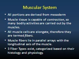
/
Introduction The core musculature provides stability and the ability to produce force Introduction The core musculature provides stability and the ability to produce force

Introduction The core musculature provides stability and the ability to produce force - PowerPoint Presentation

christina
christina . @christina
Follow
66 views
Uploaded On 2023-08-30

Introduction The core musculature provides stability and the ability to produce force - PPT Presentation

Exercising the core muscles can reduce lower back pain 2  Core stability exercises recruit the anterior posterior medial and lateral muscles of the core 7 Core exercises have a broad appeal from athletic professionals to the everyday person and has many fields of application including re ID: 1014758

left muscle movement activation muscle left activation movement oblique core exercises muscles external rotational journal weight internal anti research

Share:

Link:

Embed:

Download Presentation from below link

Download Presentation The PPT/PDF document "Introduction The core musculature provid..." is the property of its rightful owner. Permission is granted to download and print the materials on this web site for personal, non-commercial use only, and to display it on your personal computer provided you do not modify the materials and that you retain all copyright notices contained in the materials. By downloading content from our website, you accept the terms of this agreement.


Presentation Transcript

1. IntroductionThe core musculature provides stability and the ability to produce force throughout the pelvis and the trunk (1). Exercising the core muscles can reduce lower back pain (2). Core stability exercises recruit the anterior, posterior, medial, and lateral muscles of the core (7).Core exercises have a broad appeal from athletic professionals to the everyday person and has many fields of application including recreational and therapeutic (1,3).Prior research on torso muscle effectiveness has focused on the frontal and sagittal planes but there is little research in the transverse plane (8,9).Our study aimed to determine the differences in muscle activation of rotational and anti-rotational exercises for the rectus abdominis, internal oblique, external oblique, and erector spinae muscles. This is a methodological study with four researchers as subjectsWe hypothesized that anti-rotational exercises would elicit greater muscle activation and recruitment of the superficial trunk muscles than rotational exercises. Anti-Rotational and Rotational Abdominal Exercises and the Concurrent Muscle Activation: A Methodology StudyJ.T. Stephens, E.M. Bacon, C.B. Evans, S.L. Locke, R.S McCullochDepartment of Human Physiology: Gonzaga University, Spokane, WA 99258 Contact: jstephens3@zagmail.gonzaga.eduAbstractStrengthening the core musculature is essential because of the beneficial effects that highly trained trunk muscles have on strength, sports performance, and functional activities. With vast amounts of knowledge on frontal and sagittal plane stabilization core exercises from previous research, it is of increasing importance to determine if stabilization exercises in the transverse plane have similar muscle activations. PURPOSE: This study aims to use quantitative measurements of core muscle activation when completing anti-rotational and rotational exercises to determine the more effective method for strengthening the core. METHODS: Surface electromyography (EMG) electrodes were placed on the rectus abdominis, internal oblique, external oblique, and erector spinae. Participants performed maximum voluntary isometric contraction (MVIC) tests then completed 12 trials of an anti-rotation and rotational variation of the Pallof press. On both right and left sides, and at loadings of 7.5%, 10%, and 12.5% of fat free mass, subjects rotated 90° away from a pulley system and then returned, as well resisted rotation for 10 seconds. Results were analyzed with a two-way analysis of variance and a Bonferroni post hoc test. RESULTS: The weight x movement interaction was significantly different for the left internal oblique (p = 0.039) and left external oblique (p = .047), but not for the right internal oblique (p = 0.068) and right external oblique (p = 0.699). There was no significant main effect of weight variation for the 8 muscles recorded. There was a significant main effect of type of movement on activation of the participants’ right internal oblique (F(3,9) = 119.4, p < .05), left internal oblique (F(3,9) = 10.5, p < .05), right external oblique (F(3,9) = 5.1, p < .05) and right erector spinae (F(3,9) = 40.8, p < .05). CONCLUSION: In conclusion, rotational abdominal exercises elicit greater muscle activation of the deep core musculature in comparison to anti-rotational exercises. Training rotational abdominal exercises could be used to benefit athletes, preventing injury, and rehabilitating back injuries.AcknowledgementsThank you to Gonzaga University for sponsoring this study and our advisor Dr. McCulloch who supported and encouraged us throughout this journey. DiscussionThe obliques had consistent greater muscles activation than the erector spinae and rectus abdominus muscles.This was expected as the obliques act in the same plane as the tested movement – transverse planeMuscle activation was greater in the rotational movements compared to anti-rotational movements; however, no significance was found in pairwise comparisons.Likely due to the need of overcoming the force of gravity rather than equaling itThe significant interaction between weight of external load and type of movement for the left internal oblique and left external oblique showed that as the weight of the external load increased so did the degree of muscle activation.At each weight, the left internal oblique elicited a greater percentage of muscle activation for movement to the left and static hold on the left. In contrast, the left external oblique had greater muscle activation for movement to the right and static hold on the right.  Muscles that supported movement on the subject’s dominant side were less activated than muscles that supported movement on the subject’s non-dominant side All participants were right-handed, therefore, muscles on the subject’s left side or muscles that supported movement to the left were more active than the opposing right side/right-moving muscles The advantage of working in the traverse plane as opposed to the sagittal plane is decreased reliance on hip flexors to perform the movement, less lumbar spinal flexion, and ability to engage deeper abdominal muscles.Limitations include only 4 subjects being included in this study due to restrictions from the COVID-19 pandemic, the accuracy in the placement of surface EMG electrodes, and the potential that weight loads were too heavy, and participants had compensations in their form.Future research including equal numbers of left and right-handed individuals, changing weight, speed, and/or keeping the same weight but manipulating the speeds the trials are done at. The  findings of this  methodology  suggest  reliable carry over to a full scale study and  merit a deeper investigation into the core musculature acting int the transverse plane.ReferencesSaeterbakken A, Fimland M. Muscle activity of the core during bilateral, unilateral, seated and standing resistance exercise. Eur J Appl Physiol. 2012; 112(5):1671-8. Kibler WB, Press J, Sciascia A. The Role of Core Stability in Athletic Function. Sports Med. 2006; 36(3):189-98. Horsak B, Wunsch R, Bernhart P, Gorgas A, Bichler R, Lampel K. Trunk muscle activation levels during eight stabilization exercises used in the functional kinetics concept: A controlled laboratory study. Journal of back and musculoskeletal rehabilitation. 2017; 30(3):497-508. Hibbs AE, Thompson KG, French D, Wrigley A, Spears I. Optimizing Performance by Improving Core Stability and Core Strength. Sports Med. 2008; 38(12):995-1008. B. F. Walker, “The prevalence of low back pain: a systematic review of the literature from 1966 to 1998,” Journal of Spinal Disorders, vol.13, no.3, pp.205–217,2000. Beim G, Giraldo A, Pincivero D. Abdominal Strengthening Exercises A Comparative EMG Study. Journal of Sport Rehabilitation. 1997; Oliver G, Stone A, Plummer H. Electromyographic Examination of Selected Muscle Activation During Isometric Core Exercises. Clinical Journal of Sport Medicine. 2010; 20(6):452-7. Sternlicht, E., & Rugg, S. (2003). Electromyographic analysis of abdominal muscle activity using portable abdominal exercise devices and a traditional crunch. Journal of Strength and Conditioning Research, 17(3), 463-468. doi:10.1519/00124278-200308000-00006Gottschall J, Mills J, Hastings B. Integration Core Exercises Elicit Greater Muscle Activation Than Isolation Exercises. Journal of Strength and Conditioning Research. 2013; 27(3):590-6.Andre M, Fry A, Heyrman M, et al. A Reliable Method for Assessing Rotational Power. Journal of Strength and Conditioning Research. 2012; 26(3):720-4. Zemková E, Jeleň M, Zapletalová L, Hamar D. Muscle Power during Standing and Seated Trunk Rotations with Different Weights. Sport Mont. 2017; 15(3):17-23.Zemková E, Cepková A, Uvaček M, Šooš L. A Novel Method for Assessing Muscle Power During the Standing Cable Wood Chop Exercise. Journal of Strength and Conditioning Research. 2017; 31(8):2246-54.Lukaski HC, Johnson PE, Bolonchuk WW, Lykken GI. Assessment of fat-free mass using bioelectrical impedance measurements of the human body. The American journal of clinical nutrition. 1985; 41(4):810-7. Durnin, J. V. G. A., Womersley J. Body fat assessed from total body density and its estimation from skinfold thickness: measurements on 481 men and women aged from 16 to 72 Years. Br J Nutr. 1974; 32(1):77-97.HAMLYN N, BEHM D, YOUNG W. TRUNK MUSCLE ACTIVATION DURING DYNAMIC WEIGHT-TRAINING EXERCISES AND ISOMETRIC INSTABILITY ACTIVITIES. Journal of Strength and Conditioning Research. 2007; 21(4):1108-12. Ekstrom RA, Donatelli RA, Carp KC. Electromyographic Analysis of Core Trunk, Hip, and Thigh Muscles During 9 Rehabilitation Exercises. The Journal of orthopaedic and sports physical therapy. 2007; 37(12):754-62.Morini S, Ciccarelli A, Cerulli C, et al. Functional Anatomy of Trunk Flexion-Extension in Isokinetic Exercise: Muscle Activity in Standing and Seated Positions. Journal of Sports Medicine and Physical Fitness. 2008;48:17-23Bartuzi P, Tokarski T, Roman-Liu D. The effect of the fatty tissue on EMG signal in young women. Acta of bioengineering and biomechanics. 2010; 12(2):87.McGill S. Core Training: Evidence Translating to Better Performance and Injury Prevention. Strength and Conditioning Journal. 2010; 32(3):33-46.ResultsThere was a movement effect on the participants’ left internal oblique muscle activation.  An effect of type of movement was detected in the participants'’  right internal oblique muscle activation. An effect of type of movement was detected in the participants''  right external oblique muscle activation. An effect of type of movement was detected in the participants''  right  erector spinae muscle activation.MethodsLeft – Fig. 1 equipment and apparatus to perform movements Right – Fig. 2 Electrode placement (front) EMG (electromyography) Wireless electrodes were placed on four muscle groups: left and right rectus abdominis  (RA), left and right internal oblique (IO), left and right external oblique (EO), and left and right erector spinae (ES) for a total of 8 electrodes (Fig. 2) The left and right IO and EO electromyography (EMG) electrodes were placed first, and then the IO and EO maximum voluntary isometric contractions (MVIC) were performed. Then the RA electrodes were placed followed by the RA MVIC. Finally, the ES electrodes were placed and then the ES MVIC was performed. The MVIC’s were used to compare to the normalized testing dataTesting A pulley system as deigned and created for this study (Fig. 1)Each subject completed an anti-rotation (static) test and rotation (dynamic) test at three weights and in 2 directions for a total of 12 tests. Direction – left or right Anti-rotation – subject seated and extended their arms straight forward with the handle at shoulder higher and maintained this position for 10 seconds. Rotation – subject seated and rotated 90° away from their center and then returned to center. Three rotations were completed within 10 seconds at 32 BPM.Weight – 7.5%, 10%, and 12.5% of body weight This number was determined through a body fat analysis Data and Statistical Analysis The root mean square (RMS) of each muscle was normalized to the RMS from the MVIC trials .A repeated measures ANOVA was conducted to determine the differences in muscles activation for each weight loading and movement type A Bonferroni test was used to identify pairwise comparison in muscle action for each weight load and movement type Alpha level was set at p < .05Mean Muscle Activation of the External Obliques for Each Movement (n=4): Muscle activation of each subject was measured and normalized to the respective maximal voluntary isometric contraction. The two muscles (EOL, EOR) were compared across each movement variation (MR, ML, SR, SL). The loading used was 12.5% of fat free mass for each subject. The error bars represent the standard error of the group. Abbreviations: EOL – External Oblique Left, EOR – External Oblique Right, MR – Movement to the Right, ML – Movement to the Left, SR – Static hold Right, SL – Static hold Left.Mean Muscle Activation of the External Obliques for Each Movement (n=4): Muscle activation of each subject was measured and normalized to the respective maximal voluntary isometric contraction. The two muscles (EOL, EOR) were compared across each movement variation (MR, ML, SR, SL). The loading used was 12.5% of fat free mass for each subject. The error bars represent the standard error of the group. Abbreviations: EOL – External Oblique Left, EOR – External Oblique Right, MR – Movement to the Right, ML – Movement to the Left, SR – Static hold Right, SL – Static hold Left.