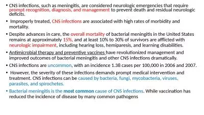
Xiaowei Su PGY3 amp Dr Geoffrey Murdoch Adapted from slides by jen nichols and robyn massa Bacterial Meningitis Meningitis is an inflammatory disease of the leptomeninges Abnormal number of white blood cells in the CSF ID: 919770
Download Presentation The PPT/PDF document "CNS Infections Neuropathology Conference..." is the property of its rightful owner. Permission is granted to download and print the materials on this web site for personal, non-commercial use only, and to display it on your personal computer provided you do not modify the materials and that you retain all copyright notices contained in the materials. By downloading content from our website, you accept the terms of this agreement.
Slide1
CNS Infections
Neuropathology Conference (11/5/2018)
Xiaowei Su, PGY-3 & Dr. Geoffrey Murdoch
Adapted from slides by
jen
nichols
and
robyn
massa
Slide2Bacterial Meningitis
Meningitis is an inflammatory disease of the leptomeninges
Abnormal number of white blood cells in the CSF
Infection of the arachnoid mater and the CSF in both the subarachnoid space and the cerebral ventricles
Slide3History of Meningitis
Thomas Willis described patients with "inflammation of the meninges with a continual fever“. He also described an early epidemic of meningitis in 1661
Heinrich Quincke utilized his new technique of lumbar puncture in 1891 to analyze CSF
William
Mestrezat
and H. Houston Merritt compiled large series of CSF profiles, identifying three major organisms (Streptococcus pneumoniae, Neisseria meningitidis and Haemophilus influenza) in the late 19th century
Vladimir
Kernig
and Josef
Brudzinski
described their eponymous signs in 1882 and 1909
Antibiotic therapy began in the 20th century with the use of sulfonamides by Francois
Schwentker
and penicillin by Chester Keefer
Vaccination against meningitis debuted in the early 20th century
Slide4Epidemiology
Approximately 1.2 million cases per year worldwide, responsible for 135,000 deaths each year
Community Acquired Bacterial Meningitis
Strep pneumoniae, Neisseria meningitidis, Listeria monocytogenes >> Haemophilus influenzae (due to vaccines)
Healthcare associated bacterial meningitis
Usually staphylococci and aerobic gram-negative bacilli
If due to neurosurgery, can vary with whether or not antimicrobial prophylaxis was given to prevent surgical site infection
Can also occur following ventricular drains or cranial trauma, and other predisposing conditions include acute sinusitis and mastoid infections
Slide5Pathogenesis & Pathophysiology
Bacteria that cause meningitis are able to colonize the host mucosal epithelium, invade, survive within the bloodstream, then cross the BBB, multiplying within the CSF
Much of the damage results from cytokine release within the CSF as the host mounts an inflammatory response
Inflammatory response is initiated both in the bloodstream and the CSF, damaging the endothelium of the BBB (tight junctions)
Slide6Clinical Features
Patients with bacterial meningitis usually present soon after symptom onset
Classic triad
Fever, nuchal rigidity, change in mental status More common in Pneumococcal meningitis than meningococcal meningitis (58% vs 27%)
99 to 100% sensitive if patient has 0 of these 3
95% of adults presented with at least 2 of the following 4 symptoms: headache, fever, nuchal rigidity, change in mental status
Other resulting symptoms: seizures, focal neurologic deficits (CN palsies), papilledema, ischemic stroke (pneumococcal meningitis), hearing loss (late), skin manifestations (N.
meningitidis
)
Slide7Clinical Signs
Brudzinski’s Sign Kernig’s Sign
Slide8Diagnostic Workup
Laboratory Workup
CBC: leukocytosis (majority PMNs)
50-90% of patients have positive blood culturesRoutine blood work is usually otherwise unrevealing
Lumbar Puncture
Crucial for establishing the diagnosis, identifying causative organism, performing susceptibility testing
CT scan should be performed before LP if there is concern for increased ICP
Immunocompromised state, history of CNS disease (mass lesion, stroke, focal infection), new onset seizure within 1 week of presentation, papilledema, abnormal level of consciousness (GCS <11), focal neurologic deficit
Slide9CSF Findings
Slide10MRI Findings
T1 plus contrast:
Meningeal enhancement; especially characteristic when thicker, longer, more intensely enhancing, and if there is nodular enhancement
The degree of abnormal enhancement correlates with the degree of inflammatory cellular infiltration of the meninges
T2 FLAIR:
Leptomeningeal hyperintensities; not specific for infection vs inflammation
CSF
hyperintensities
, usually present in the lateral ventricles and cortical sulci
DWI:
Abscesses (Strep
pneumo
), especially in the subarachnoid or
intraventricular
regions
Resultant ischemic infarction or vasculitis
Slide11MRI Findings
Slide12Neuropathology
Streptococcal meningitis
H&E
(hematoxylin stains nucleic acids blue
, eosin stains amino acids
red
)
Gram Stain
(crystal
violet
stains thick peptidoglycan cell walls)
Slide13Empiric treatment
Vancomycin 25-30mg/kg load with 15mg/kg maintenance
(surgicalcriticalcare.net/Resources/
vancomycin.php)
Ceftriaxone 2gm q12H (Aztreonam 2gm q6H for penicillin allergy)
Ampicillin 2gm q4H if >50-yrs (add Bactrim 15-20mg/kg/day divided q6-8H to aztreonam for allergy)
Cefepime 2gm q8H is recommended for penetrating trauma, post-NSGY, or shunt;
alternative is ciprofloxacin 400mg q8H or aztreonam 2gm q6H
Acyclovir 10mg/kg q8H (ideal or adjusted body weight) if suspecting viral encephalitis
Dexamethasone for all adults with suspected bacterial meningitis not in septic shock
(0.15 mg/kg q6H x4-days, first dose within 20-min prior to first antimicrobial dose)
Dexamethasone decreases mortality, neurologic complications, hearing loss for pneumococcal meningitis (most common organism >18-yrs)
Begin antimicrobials after blood cultures, do not delay for LP
Slide14Viral encephalitis
Clinical course is more slowly progressive, altered level of awareness, personality changes, headache may worsen over days to >1-week
HSV & VZV encephalitis treated with acyclovir
CMV in immunocompromised patients treated with ganciclovir
EBV responds with supportive therapy alone
Arboviruses (West Nile, St. Louis, Japanese), lymphocytic choriomeningitis virus (LCMV), enteroviruses (acute flaccid myelitis) have no targeted treatments
Radiopaedia.org
Slide15Neuropathology
Enteroviral meningitis
H&E
(hematoxylin stains nucleic acids blue
, eosin stains amino acids
red
)
CMV radiculitis
H&E
(hematoxylin stains nucleic acids
blue
, eosin stains amino acids
red
)
Slide16Fungal Meningitis - Cryptococcal
Encapsulated saprophytic yeast, transmitted by inhalation
C. neoformans - most common, 82% of disease worldwide
C. gattii - immunocompetent individuals in tropical & subtropical regions (sporadic cases in North America)
Mostly affects those with impaired cell-mediated immunity
HIV - 95% in middle to low income countries, 80% in high income countries
Immunosupressant medications
Immunocompetent hosts - autoimmune disease, malignancy, occult immune deficiencies
Slide17Clinical Features
Subacute headache
Confusion
Increased ICP
CN palsies, seizures
Meningismus
<20% of patients
Cryptococcomas (granulomas)
hydrocephalus
Ocular (papilledema, uveitis, chorioretinitis, optic nerve dysfunction)
Pulmonary, cutaneous, and bloodstream infections also occur
Slide18Diagnosis
Lumbar puncture
Elevated opening pressure (can fluctuate)
Lymphocytic pleocytosis, low glucose, elevated protein (can be normal, especially in HIV)
India ink staining under light microscopy (low sensitivity)
CSF cryptococcal antigen >93% sensitive and >93% specific, urine antigen less specific
Fungal culture on Sabouraud media, grows after 36 hours
Radiology
Cryptococcomas
and pseudocysts in midbrain or basal ganglia
Low sensitivity for symptomatic hydrocephalus and dilated perivascular spaces
Slide19Imaging
Slide20Neuropathology
Slide21Neuropathology
Slide22Neuropathology
Slide23Neuropathology
Cryptococcus meningitis
H&E
(hematoxylin stains nucleic acids blue
, eosin stains amino acids
red
)
PAS
(period acid oxidizes polysaccharides, creating aldehydes that stain
purple
with Schiff reagent)
Mucicarmine
(stains mucopolysaccharide capsules
red
)
GMS
(methanamine oxidizes mucopolysaccharides, creating aldehydes that reduce silver reagent
black
)
Slide24Imaging
Slide25IRIS
Host immune recovery triggers inflammatory response to antigens
Paradoxical unmasking after initial response to antifungals
Occurs after starting ART or pausing anti-rejection medications
May occur in immunocompetent hosts as their immune system recovers from high fungal burden
Risk factors: severe disease, slow fungal elimination
Benefit of steroids is unclear
Mortality up to 36%
Slide26Neuropathology
Immune reconstitution inflammatory syndrome
H&E
(hematoxylin stains nucleic acids blue
, eosin stains amino acids
red
)
CD3
(anti-CD3 binds T-cells, which turns
brown
with biotin-based detection)
IBA1
(anti-IBA1 binds microglia, which turns
brown
with biotin-based detection)
Slide27Treatment
Slide28Treatment
Amphotericin B
Side effects: nephrotoxicity, hypokalemia, hypomagnesemia
Liposomal formulations are less nephrotoxic
Greatest early fungicidal activity
Flucytosine
Side effects: bone marrow suppression
Reduction of raised ICP
Serial LPs, CSF drainage catheter, VP shunt (acetazolamide may cause harm)
Changes in exam are the earliest indication of worsening ICP
Management of immune reconstitution inflammatory syndrome (IRIS)
ART for HIV patients: start 4-10 weeks after initiating antifungal treatment
Slide29Other Fungal Meningitis
Aspergillus
H&E
(hematoxylin stains nucleic acids blue
, eosin stains amino acids
red
)
GMS
(methanamine oxidizes mucopolysaccharides, creating aldehydes that reduce silver reagent
black
)
Tuberculosis
H&E
(hematoxylin stains nucleic acids
blue
, eosin stains amino acids
red
)
FITE
(carbol fuchsin stains cell walls
red
, other cells destained by acid-alcohol and counterstain
blue
)
Slide30Questions
“The brain is a wonderful organ. It starts working the moment you get up in the morning and does not
stop until you get into the office.”
- Robert Frost