/
Lecture 8 :Protein synthesis Lecture 8 :Protein synthesis

Lecture 8 :Protein synthesis - PowerPoint Presentation

dandy
dandy . @dandy
Follow
66 views
Uploaded On 2023-09-02

Lecture 8 :Protein synthesis - PPT Presentation

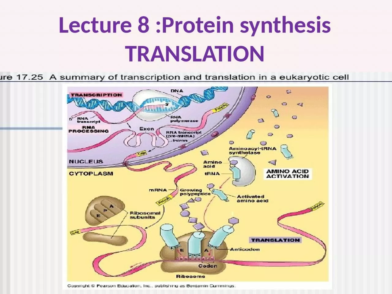
TRANSLATION Initiation Elongation and Termination of Protein Synthesis in Eukaryotes Initiation Initiation of protein synthesis differs significantly between prokaryotes and eukaryotes Eukaryotic mRNA has no ribosomebinding site RBS Instead recognition and binding to the ribosome ID: 1015369

binding complex protein trna complex binding trna protein gtp mrna ribosome codon subunit structure cap eukaryotic elongation site initiation

Share:

Link:

Embed:

Download Presentation from below link

Download Presentation The PPT/PDF document "Lecture 8 :Protein synthesis" is the property of its rightful owner. Permission is granted to download and print the materials on this web site for personal, non-commercial use only, and to display it on your personal computer provided you do not modify the materials and that you retain all copyright notices contained in the materials. By downloading content from our website, you accept the terms of this agreement.


Presentation Transcript

1. Lecture 8 :Protein synthesis TRANSLATION

2. Initiation, Elongation, and Termination of Protein Synthesis in EukaryotesInitiation: Initiation of protein synthesis differs significantly between prokaryotes and eukaryotes. Eukaryotic mRNA has no ribosome-binding site (RBS). Instead recognition and binding to the ribosome is by The cap structure at the 5- end, which is added to eukaryotic mRNA before it leaves the nucleus.Two different complexes assemble will form in initiation stage: The first is the 43S pre-initiation complex. This is an assembly of the small 40S subunit of the ribosome attached to eIF1, eIF1A, eIF3, and eIF5. This binds the charged initiator tRNA, Met-tRNA, plus eIF2. The second complex, the cap-binding complex, contains cap-binding protein (eIF4E), eIF4G, eIF4A, eIF4B, and poly(A)-binding protein (PABP).

3. Assembly of the Eukaryotic Initiation Complex (A) The cap-binding complex includes poly(A)-binding protein (PABP), eIF4A, eIF4B, eIF4E, and eIF4G, this complex is in an unphosphorylated state when unbound to mRNA. ATP transfers phosphates to the complex to make it competent for binding the mRNA. (B) The 43S initiation complex forms bringing the small ribosomal subunit together with the tRNA met. This complex uses GTP to attach the tRNA to the 40S subunit via eIF2. In addition, initiation factors eIF1, eIF1A, eIF3, eIF5, and eIF2B guide and make the complex competent to bind to the 5 -UTR of mRNA. (C) The mRNA is recognized by the cap-binding complex via the connections between eIF4E and PABP which bind the 5- and 3- ends of the mRNA, respectively. These two connections cause the rest of the mRNA to loop out. When this is established, then the 43S preinitiation complex can attach and start scanning for the first AUG. After pausing at the first AUG, then the 60S subunit of the ribosome can bind and initiate translation.

4. In general the initiation requires: 1- mRNA (processed in Eukaryotic cell) 2- tRNA which will come charged (bind to A.A) and the first A.A is methionin because the first codon is AUG . the AUG first codon in mRNA will face the triplet codon in anti codon arm in tRNA .3- small 40S ribosomal subunits thus the mRNA will bind.

5. poly(A)-binding protein (PABP).This complex uses GTP to attach the tRNA to the 40S subunit via eIF2the 43S pre-initiation complex. This is an assembly of the small 40S subunit of the ribosome attached to eIF1, eIF1A, eIF3, and eIF5. This binds the charged Met-tRNA, plus eIF2B. The cap-binding complex, contains cap-binding protein (eIF4E), eIF4G, eIF4A, eIF4B, and poly(A)-binding protein (PABP).

6. During eukaryotic initiation, cap-binding complex first attaches to the mRNA via its cap. Next, the poly(A) tail is bound by PABP so that the mRNA forms a ring. This structure can now bind the 43S assembly. In order to align the Met-tRNA, Methionin with the correct AUG codon, the two structures work together to scan each codon from the 5- end. This scanning process uses energy from ATP (Figure). Normally, the first AUG is used as the start, Once a suitable AUG has been located, eIF5 joins the complex, which in turn allows the 60S subunit to join and the cap-binding protein, eIF2, eIF1, eIF3, and maybe eIF5 to depart. eIF5 uses energy from GTP to accomplish this remodeling of the ribosome.In prokaryotes, the first amino acid added (methionine) has a formyl group on its amino group (i.e., it is N-formyl-methionine), but in eukaryotes unmodified methionine is used.

7. Beginning Eukaryotic Translation Elongation: Once the eukaryotic 40S subunit complex finds the first AUG, then the remaining 60S subunit and associated factors combine to form the final 80S ribosome.Once a suitable AUG has been located, eIF5 joins the complex, which in turn allows the 60S subunit to join and the cap-binding protein, eIF2, eIF1, eIF3, and maybe eIF5 to depart. eIF5 uses energy from GTP to accomplish this remodeling of the ribosome.

8. ElongationThe next stage is elongation. Of all the stages of translation, elongation in bacteria and eukaryotes is the most similar. As in bacteria, elongation factors work to decode the mRNA and bind the tRNA into the A-site of the ribosome. Rather than EF-Tu and EF-Ts, eukaryotes use eEF1A to deliver the tRNA using GTP hydrolysis for energy and eEF1B to replace the depleted GDP with fresh GTP. The only difference is that eukaryotic elongation factors include more subunits. The remaining steps are the same. The peptidyl transferase activity of the 28S rRNA of the large subunit links the incoming amino acid to the polypeptide chain. Then elongation factor eEF2 (direct counterpart to bacterial EF-G) uses GTP to drive the conformational changes in the ribosome and ratchet the tRNAs from the P- and A-sites into the E- and P-sites. Elongation continues until a stop codon enters the A-site.

9. peptidyl transferase Then elongation factor eEF2 (direct counterpart to bacterial EF-G) uses GTP to drive the conformational changes in the ribosome and ratchet the tRNAs from the P- and A-sites into the E- and P-sites. Elongation continues until a stop codon enters the A-site.eEF1A to deliver the tRNA using GTP hydrolysis for energy and eEF1B to replace the depleted GDP with fresh GTP.

10.

11.

12. The A.A in the p site will bind with the A.A in A site via peptide bond with the aid of peptidyl transferase .tRNA in p site now is empty ,convert to uncharged and leave the ribosome from Exit site .then the tRNA translocat from A to P site with 2 A.A . Now the A site empty and the complex will move for another triplet codon

13.

14. Eukaryotic termination Eukaryotic termination differs from prokaryotic termination in two ways. First, rather than having two different release factors (RF1 and RF2) to recognize different stop codons (UAA, UAG and UGA), eukaryotes have a single release factor (eRF1) that recognizes all three stop codons. when the ribosome reached stop codon (UAG, UAA,UGA) and there is no anticodon in tRNA. eRF1 binds the stop codon, but this does not affect peptide bond formation. Instead, eRF3 carrying a GTP molecule binds to eRF1. GTP hydrolysis then rearranges the releasing factor and the final amino acid attaches to the polypeptide. Therefore, eukaryotes require GTP for polypeptide completion, whereas in bacteria, RF1 or RF2 is sufficient.

15. eIF3 triggers the release of the 60S subunit, then eIF1 releases the final tRNA. An additional factor, eIF3j, then removes the mRNA. The components are then recycled.

16. when the ribosome reached stop codon eRF1 binds the stop codon, eRF3 carrying a GTP molecule binds to eRF1. GTP hydrolysis then rearranges the releasing factors

17.

18. Basic structure of protein: the structural unit of protein is amino acid (A.A).The main structure of A.A is 1- central C atom called α- carbon. .2-Amino group gives the positive charge 3- carboxylic acid group gives the negative charge 4- R side chain which differ from one amino acid to another, the simplest R side group is H only to form glycin

19. The bond which bind the two adjacent A.A is called peptide bond with releasing of water molecules .

20. Usually the protein chain start with amino group (NH3) and end with Carboxyl group (COOH).binding 2-10 A.A give a rise to oligopeptide while binding more than that will form the polypeptide chain . This linear chain represent 1- the primary structure to the protein .

21. According to structure and configuration ,proteins can be divided to 4 types

22. 2- the secondary structure formed due to folding of the primary structure via hydrogen bonds . Two types are well studied as secondary structure 1- α helix 2- β pleated sheet

23. 3- Tertiary structure is more complicated and we could find α helix and β pleated sheet in the same structure ,other types of bonds exist here like disulfide bond between cystine A.A ,ionic bond ,hydrophobic bond

24.

25. 4- Quaternary structure :more complicated result from folding the other structures and from more than subunits