/
 Cardiovascular System - Ch 11  Cardiovascular System - Ch 11

Cardiovascular System - Ch 11 - PowerPoint Presentation

danika-pritchard
danika-pritchard . @danika-pritchard
Follow
345 views
Uploaded On 2020-04-03

Cardiovascular System - Ch 11 - PPT Presentation

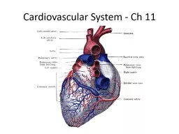
Heart Facts heart pumps 7000 liters of blood daily heart arteries arterioles capillaries site of gas waste nutrient exchange venules veins heart 14 cm long x 9 cm wide ID: 774933

blood heart pressure left blood heart pressure left pulmonary valve atrium figure artery ventricle veins valves semilunar arteries capillary

Share:

Link:

Embed:

Download Presentation from below link

Download Presentation The PPT/PDF document " Cardiovascular System - Ch 11" is the property of its rightful owner. Permission is granted to download and print the materials on this web site for personal, non-commercial use only, and to display it on your personal computer provided you do not modify the materials and that you retain all copyright notices contained in the materials. By downloading content from our website, you accept the terms of this agreement.


Presentation Transcript

Slide1

Cardiovascular System - Ch 11

Slide2

Heart Facts

heart pumps 7,000 liters of blood daily

heart – arteries – arterioles – capillaries (site of gas, waste, nutrient exchange) –

venules

– veins – heart

14 cm long x 9 cm wide

located in the

mediastinum

(btw lungs)

apex: pointed end by 5

th

intercostal

space

weighing 230-340 g. = ½ - ¾ lb

fetal heart begins beating at 3

rd

week

Slide3

Four and Five Weeks Pregnant

Slide4

Figure 11.1a

Midsternal line

2nd rib

Diaphragm

(a)

Sternum

Point of

maximalintensity(PMI)

Slide5

Coverings and Heart Wall

Pericardium

: double layered sac

1.

parietal

: outside of sac and inside lining of fibrous

pericardium (dense

c.t

.)

2.

visceral

: innermost layer of sac covering the heart,

also called

epicardium

(“upon” the heart)

pericardial cavity contains

serous fluid

to reduce friction btw. membranes

Heart Wall

epicardium

: same as visceral pericardium, fat stored

myocardium

: heart muscle striated, involuntary

endocardium

: lines inside of heart; continuous w/ linings of

b.v

.

Slide6

Figure 11.1c

Superior

vena cava

Pulmonarytrunk

Diaphragm

(c)

Aorta

Left lung

Pericardium(cut)

Apex ofheart

Parietal

pleura (cut)

Slide7

Figure 11.2

Pericardium

Myocardium

Pulmonary

trunk

Fibrous pericardium

Parietal layer of serous

pericardium

Pericardial cavity

Epicardium

(visceral layer

of serous

pericardium)

Myocardium

Endocardium

Heart chamber

Heart

wall

Slide8

The Heart: Chambers

Right and left side act as separate pumps

Four chambers

Atria (upper chambers)

Receiving chambers

Right atrium

Left atrium

*

Interatrial

septum

: wall separating them: At one time was a hole for blood to mix in fetus (

foramen

ovale

and is now

fossa

ovale

)

Ventricles (lower chambers)

Discharging chambers

Right ventricle

Left ventricle

*

Interventricular

Septum

: separates the ventricles

Slide9

Figure 11.3b

Superior vena cava

Right atrium

Right pulmonary

veins

Fossa ovalis

Right atrioventricularvalve (tricuspid valve)

Right ventricle

Chordae tendineae

Inferior vena cava

(b) Frontal section showing interior chambers and valves.

Aorta

Left pulmonary artery

Left atrium

Left pulmonary veins

Pulmonary semilunar valve

Left atrioventricular valve(bicuspid valve)

Aortic semilunar valve

Left ventricle

Interventricular septum

Myocardium

Visceral pericardium

Right pulmonary artery

Slide10

Figure 11.5

Right

ventricle

Muscularinterventricularseptum

Leftventricle

Slide11

The Heart’s Role in Blood Circulation

Systemic circulation (body)

Blood flows from the left side of the heart through the body tissues and back to the right side of the heart

Pulmonary circulation (lungs)

Blood flows from the right side of the heart to the lungs and back to the left side of the heart

Slide12

Figure 11.4

Capillary beds

of lungs wheregas exchangeoccurs

Pulmonaryveins

Aorta and branches

Leftatrium

Leftventricle

Heart

Systemic Circuit

Rightatrium

Rightventricle

Venaecavae

Pulmonaryarteries

Pulmonary Circuit

Capillary

beds of allbody tissueswhere gasexchangeoccurs

KEY:

Oxygen-rich,CO2-poor blood

Oxygen-poor,CO2-rich blood

Slide13

The Heart: Valves

Allow blood to flow in only one direction to prevent backflow

Four valves

Atrioventricular

(AV) valves—between atria and ventricles

Tricuspid valve (right side of heart)

Bicuspid (mitral) valve (left side of heart)

Semilunar

valves—between ventricle and artery

Pulmonary

semilunar

valve (right side: to the lungs)

Aortic

semilunar

valve (left side: to the body)

Slide14

Figure 11.3b

Superior vena cava

Right atrium

Right pulmonary

veins

Fossa ovalis

Right atrioventricularvalve (tricuspid valve)

Right ventricle

Chordae tendineae

Inferior vena cava

(b) Frontal section showing interior chambers and valves.

Aorta

Left pulmonary artery

Left atrium

Left pulmonary veins

Pulmonary semilunar valve

Left atrioventricular valve(bicuspid valve)

Aortic semilunar valve

Left ventricle

Interventricular septum

Myocardium

Visceral pericardium

Right pulmonary artery

Slide15

The Heart: Valves

AV valves

Anchored in place by chordae tendineae (“heart strings”)

Open during heart relaxation and closed during ventricular contraction

Semilunar valves

Closed during heart relaxation but open during ventricular contraction

Notice these valves operate opposite of one another to force a one-way path of blood through the heart

Slide16

Figure 11.3a

Left common carotid artery

Left subclavian artery

Aortic arch

Ligamentum arteriosum

Left pulmonary artery

Left pulmonary veins

Left atrium

Auricle of left atrium

Circumflex artery

Left coronary artery in

coronary sulcus (left

atrioventricular groove)

Left ventricle

Great cardiac vein

Anterior interventricular

artery (in anterior interventricular sulcus)

Apex

Brachiocephalic trunk

Superior vena cava

Right pulmonary artery

Ascending aorta

Pulmonary trunk

Right pulmonary veins

Right atrium

Right coronary arteryin coronary sulcus (right atrioventricular groove)

Anterior cardiac vein

Right ventricle

Marginal artery

Small cardiac vein

Inferior vena cava

(a)

Slide17

Cardiac Circulation

Blood in the heart chambers

does not

nourish the myocardium

The heart has its own nourishing circulatory system consisting of

Coronary arteries

—branch from the aorta to supply the heart muscle with oxygenated blood

Cardiac veins

—drain the myocardium of blood

Coronary sinus

—a large vein on the posterior of the heart, receives blood from cardiac veins

Blood empties into the right atrium via the coronary sinus

Slide18

Coronary Arteries

Slide19

Blood Flow Through the Heart

Superior and inferior venae cavae dump blood into the right atrium

From right atrium, through the tricuspid valve, blood travels to the right ventricle

From the right ventricle, blood leaves the heart as it passes through the pulmonary semilunar valve into the pulmonary trunk

Pulmonary trunk splits into right and left pulmonary arteries that carry blood to the lungs

Slide20

Blood Flow Through the Heart

Oxygen is picked up and carbon dioxide is dropped off by blood in the lungs

Oxygen-rich blood returns to the heart through the four pulmonary veins

Blood enters the left atrium and travels through the bicuspid valve into the left ventricle

From the left ventricle, blood leaves the heart via the aortic semilunar valve and aorta

Slide21

The Heart: Valves

AV valves

Anchored in place by

chordae

tendineae

(“heart strings”)

Open during heart relaxation and closed during ventricular contraction

Semilunar valves

Closed during heart relaxation but open during ventricular contraction

Notice these valves operate opposite of one another to force a one-way path of blood through the heart

Slide22

Figure 11.3b

Superior vena cava

Right atrium

Right pulmonary

veins

Fossa ovalis

Right atrioventricularvalve (tricuspid valve)

Right ventricle

Chordae tendineae

Inferior vena cava

(b) Frontal section showing interior chambers and valves.

Aorta

Left pulmonary artery

Left atrium

Left pulmonary veins

Pulmonary semilunar valve

Left atrioventricular valve(bicuspid valve)

Aortic semilunar valve

Left ventricle

Interventricular septum

Myocardium

Visceral pericardium

Right pulmonary artery

Slide23

Heart Bypass Surgery

http://www.youtube.com/watch?v=3Nf6Q2skGOM&feature=endscreen&NR=1

Angioplasty and Stent Insertion

Slide24

The Heart: Conduction System

Special tissue sets the pace

Sinoatrial node = SA node (“pacemaker”), is in the right atrium (70-72 times per minute)

Atrioventricular node = AV node, is at the junction of the atria and ventricles (atria contract)

Atrioventricular bundle = AV bundle (bundle of His), is in the interventricular septum

Bundle branches are in the interventricular septum

Purkinje fibers spread within the ventricle wall muscles (ventricles contract)

Slide25

Figure 11.7

Superior

vena cava

Sinoatrial (SA)

node (pacemaker)

Atrioventricular

(AV) node

Right atrium

Bundle branches

Purkinje fibers

Left atrium

Atrioventricular

(AV) bundle

(bundle of His)

Purkinje fibers

Interventricular

septum

Slide26

Electrocardiogram (ECG or EKG)

EKG video

EKG Explained

Slide27

Electrocardiogram

Slide28

Abnormal vs. Normal EKG

Atrial fibrillation

Normal EKG

Slide29

Heart Contractions

Homeostatic imbalance

Heart block

—damaged AV node releases them from control of the SA node; result is in a slower heart rate as ventricles contract at their own rate

Ischemia

—lack of adequate oxygen supply to heart muscle

Fibrillation

—a rapid, uncoordinated shuddering of the heart muscle

Tachycardia

—rapid heart rate over 100 beats per minute

Bradycardia

—slow heart rate less than 60 beats per minutes

Slide30

The Heart: Cardiac Cycle & Heart Sounds

Atria contract simultaneously (ventricles relaxed)

Atria relax, then ventricles contract

Systole

= contraction

Diastole

= relaxation

Heart Sounds—(

Lub

—Dup,

Lub

—Dup)

Lub

”(AV valves closing)

“Dup” (

semilunar

valves closing)

Slide31

Cardiac Cycle

Changes in pressure and volume

*

Stroke volume

: amount of blood (70ml) which leaves the heart per beat

*

End-Diastolic-Volume

: amount of blood in LV prior to contraction

*

Cardiac Output

: total amount of blood pumped per minute (75 b/m x 70 ml = 5,250ml or 5.25L) which leaves the heart per min. (average)

Slide32

Figure 11.8

Left atrium

Right atrium

Left ventricle

Right ventricle

Ventricular

filling

Atrialcontraction

Isovolumetriccontraction phase

Ventricularejection phase

Isovolumetricrelaxation

Mid-to-late diastole(ventricular filling)

Ventricular systole(atria in diastole)

Early diastole

1

2

3

Slide33

The Heart: Regulation of Heart Rate

Increased heart rate

Sympathetic nervous system

Crisis

Low blood pressure

Hormones

Epinephrine (adrenaline)

Thyroxine

(from thyroid gland: metabolism)

Exercise

Decreased blood volume

Decreased heart rate

Parasympathetic nervous system

High blood pressure or high blood volume

Decreased venous return

Slide34

Blood Vessels: The Vascular System

Blood flows from heart through elastic arteries, muscular arteries, and arterioles to the capillaries.

Blood returns to the heart from capillaries through

venules

, small veins, and large veins.

Entire circulatory system is lined with

si

sq

epi

. (endothelium)

Capillaries: nutrients diffuse from capillary into interstitial spaces, and waste products diffuse in the opposite direction, fluid diffuses both ways

Slide35

Structure of Arteries and Veins - both have 3 layers

Tunica externa (outer) - connective tissue

Tunica media (middle) - smooth muscle & elastic fibers

Tunica intima (inner) – endothelium; slick surface;

si.sq.epi

.

Slide36

Figure 11.10b

Valve

Tunica intima

• Loose connective tissue

• Endothelium

Internal elastic

lamina

Tunica media

• Smooth muscle

• Elastic fibers

External elastic lamina

Tunica externa

• Collagen fibers

Arteriole

Venule

Capillarynetwork

Basement membrane

Endothelial cells

Capillary

(b)

Lumen

Vein

Lumen

Artery

Slide37

Vessel Differences and Disorders

Arteries: elasticity helps maintain blood pressure; walls are thicker than veins (muscle)

Vasoconstriction (narrowing) or

vasodilation

(widening): allows regulation of blood flow

Veins: contain valves to prevent backflow of blood; thinner than arteries

Hypertension

: high blood pressure (most cases are hereditary)

Arteriosclerosis

: hardening of the arteries due to lipid deposits, proteins and calcium which form plaque.

Slide38

Figure 11.12a

True

capillaries

Sphincters open; blood flows through true

capillaries.

Vascular shunt

Precapillary sphincters

Terminal arteriole

Postcapillary

venule

Slide39

Pulse

PulsePressure wave of bloodMonitored at “pressure points” in arteries where pulse is easily palpatedPulse averages 70 to 76 beats per minute at rest

Slide40

Blood Pressure

Measurements by health professionals are made on the pressure in large arteriesSystolic—pressure at the peak of ventricular contraction Diastolic—pressure when ventricles relaxWrite systolic pressure first and diastolic last (120/80 mm Hg) Pressure in blood vessels decreases as distance from the heart increases

Slide41

Figure 11.20

120

100

80

60

40

20

0

10

Systolic pressure

Diastolicpressure

Pressure (mm Hg)

Aorta

Arteries

Arterioles

Capillaries

Venules

Veins

Venae cavae

Slide42

Figure 11.21a

Blood pressure

120 systolic70 diastolic(to be measured)

Brachialartery

(a) The course of the brachial artery of the arm. Assume a blood pressure of 120/70 in a young, healthy person.

Slide43

Figure 11.21b

Pressure

in cuffabove 120; no soundsaudible

Rubber cuffinflated with air

120 mm Hg

Brachialarteryclosed

(b) The blood pressure cuff is wrapped snugly around the arm just above the elbow and inflated until the cuff pressure exceeds the systolic blood pressure. At this point, blood flow into the arm is stopped, and a brachial pulse cannot be felt or heard.

Slide44

Figure 11.21c

Pressure

in cuffbelow 120,but above 70

120 mm Hg

70 mm Hg

Soundsaudible instethoscope

(c) The pressure in the cuff is gradually reduced while the examiner listens (auscultates) for sounds in the brachial artery with a stethoscope. The pressure read as the first soft tapping sounds are heard (the first point at which a small amount of blood is spurting through the constricted artery) is recorded as the systolic pressure.

Slide45

Figure 11.20d

Pressure

in cuffbelow 70;no soundsaudible

70 mm Hg

(d) As the pressure is

reduced still further,

the sounds become

louder and more

distinct; when the

artery is no longer

constricted and blood

flows freely, the

sounds can no longer

be heard. The

pressure at which the

sounds disappear is

recorded as the

diastolic pressure.

Slide46

Blood Pressure: Effects of Factors

BP is blood pressure

BP is affected by age, weight, time of day, exercise, body position, emotional state

CO (cardiac output) is the amount of blood pumped out of the left ventricle per minute

PR is peripheral resistance, or the amount of friction blood encounters as it flows through vessels

Narrowing of blood vessels and increased blood volume increases PR

BP = CO

PR

Slide47

Figure 11.22

Slide48

Variations in Blood Pressure

Normal human range is variable

Normal

140 to 110 mm Hg systolic

80 to 75 mm Hg diastolic

Hypotension

Low systolic (below 110 mm Hg)

Often associated with illness

Hypertension

High systolic (above 140 mm Hg)

Can be dangerous if it is chronic

Slide49

Fluid Movements at Capillary Beds

Blood pressure forces fluid and solutes out of capillaries

Osmotic pressure draws fluid into capillaries

Blood pressure is higher than osmotic pressure at the arterial end of the capillary bed

Blood pressure is lower than osmotic pressure at the venous end of the capillary bed

Slide50

Figure 11.24

Tissue cell

Interstitial fluid

Net fluid

movement out

Net fluidmovement in

Blood flow

Blood flow

Arterialend ofcapillary

Venuleend ofcapillary

At the venule end of the capillary, blood pressure is less than osmotic pressure, and fluid flows from the interstitial fluid into the capillary.

At the arterial end of a capillary, blood pressure is more than osmotic pressure, and fluid flows out of the capillary and into the interstitial fluid.

Blood pressure ishigher than osmoticpressure

Osmotic pressure(remains steadyin capillary bed)

Blood pressure islower than osmoticpressure