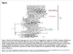
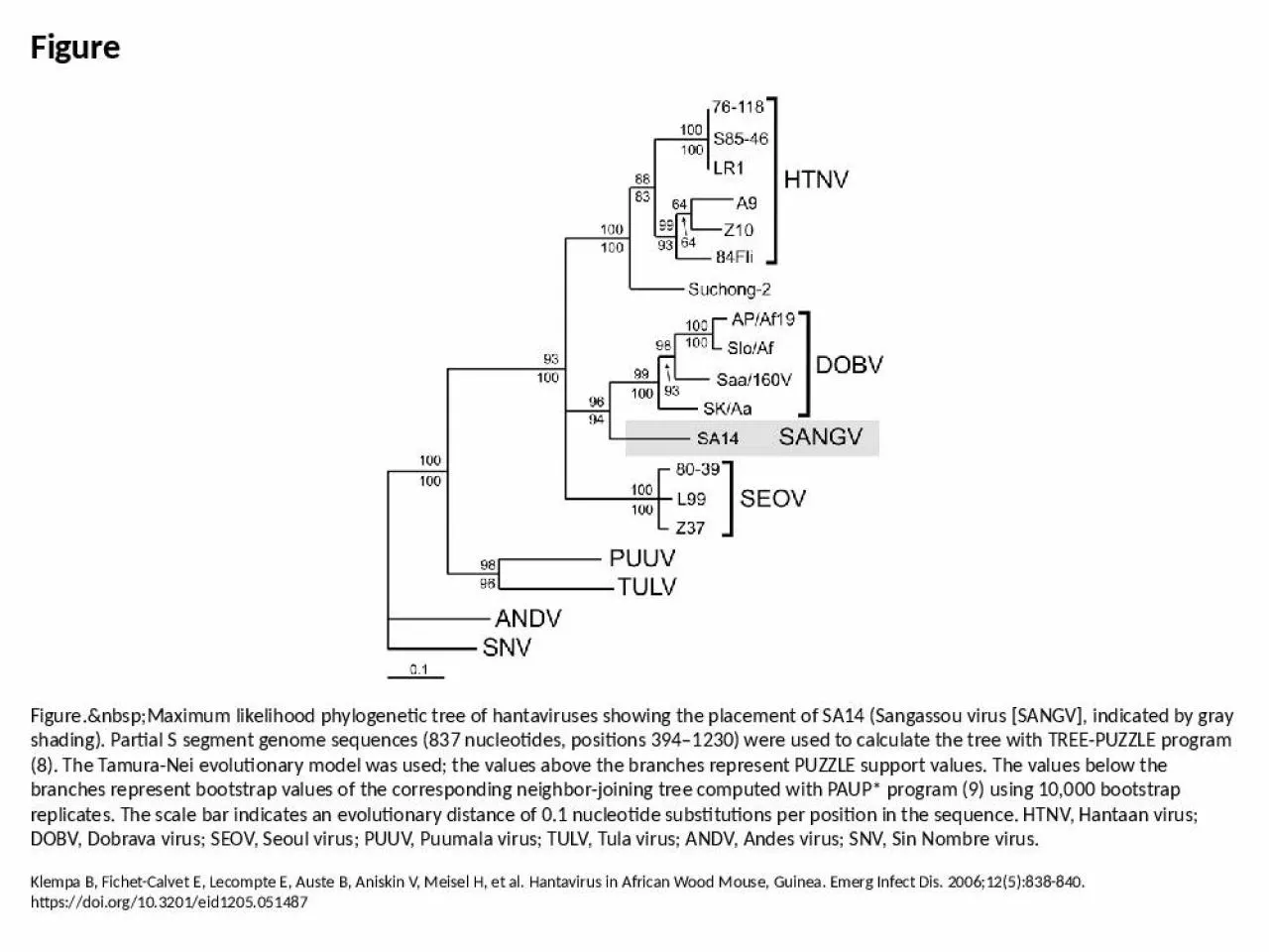
PPT-Figure Figure. Maximum likelihood phylogenetic tree of hantaviruses showing the

Author : davis | Published Date : 2024-06-10

Klempa B FichetCalvet E Lecompte E Auste B Aniskin V Meisel H et al Hantavirus in African Wood Mouse Guinea Emerg Infect Dis 2006125838840 httpsdoiorg103201eid1205051487

Presentation Embed Code

Download Presentation

Download Presentation The PPT/PDF document "Figure Figure. Maximum likeliho..." is the property of its rightful owner. Permission is granted to download and print the materials on this website for personal, non-commercial use only, and to display it on your personal computer provided you do not modify the materials and that you retain all copyright notices contained in the materials. By downloading content from our website, you accept the terms of this agreement.

Figure Figure. Maximum likelihood phylogenetic tree of hantaviruses showing the: Transcript


Klempa B FichetCalvet E Lecompte E Auste B Aniskin V Meisel H et al Hantavirus in African Wood Mouse Guinea Emerg Infect Dis 2006125838840 httpsdoiorg103201eid1205051487. Sobolev I, Sharshov K, Dubovitskiy N, Kurskaya O, Alekseev A, Leonov S, et al. Highly Pathogenic Avian Influenza A(H5N8) Virus Clade 2.3.4.4b, Western Siberia, Russia, 2020. Emerg Infect Dis. 2021;27(8):2224-2227. https://doi.org/10.3201/eid2708.204969. Le Gal F, Gault E, Ripault M, Serpaggi J, Trinchet J, Gordien E, et al. Eighth Major Clade for Hepatitis Delta Virus. Emerg Infect Dis. 2006;12(9):1447-1450. https://doi.org/10.3201/eid1209.060112. Huong VT, Yoshimatsu K, Luan VD, Van Tuan L, Nhi L, Arikawa J, et al. Hemorrhagic Fever with Renal Syndrome, Vietnam. Emerg Infect Dis. 2010;16(2):363-365. https://doi.org/10.3201/eid1602.091204. Watanabe S, Maeda K, Suzuki K, Ueda N, Iha K, Taniguchi S, et al. Novel Betaherpesvirus in Bats. Emerg Infect Dis. 2010;16(6):986-988. https://doi.org/10.3201/eid1606.091567. Boivin G, De Serres G, Côté S, Gilca R, Abed Y, Rochette L, et al. Human Metapneumovirus Infections in Hospitalized Children. Emerg Infect Dis. 2003;9(6):634-640. https://doi.org/10.3201/eid0906.030017. Monroe MC, Morzunov SP, Johnson AM, Bowen MD, Artsob H, Yates T, et al. Genetic Diversity and Distribution of Peromyscus-Borne Hantaviruses in North America. Emerg Infect Dis. 1999;5(1):75-86. https://doi.org/10.3201/eid0501.990109. Bellini WJ, Rota PA. Genetic Diversity of Wild-Type Measles Viruses: Implications for Global Measles Elimination Programs. Emerg Infect Dis. 1998;4(1):29-34. https://doi.org/10.3201/eid0401.980105. Brown MA, Troyer JL, Pecon-Slattery J, Roelke ME, O’Brien SJ. Genetics and Pathogenesis of Feline Infectious Peritonitis Virus. Emerg Infect Dis. 2009;15(9):1445-1452. https://doi.org/10.3201/eid1509.081573. Takada N, Fujita H, Kawabata H, Takajo I, Sakata A, Takano A, et al. Spotted Fever Group Rickettsia sp. Closely Related to R. japonica, Thailand. Emerg Infect Dis. 2009;15(4):610-611. https://doi.org/10.3201/eid1504.071271. Keawcharoen J, Oraveerakul K, Kuiken T, Fouchier R, Amonsin A, Payungporn S, et al. Avian Influenza H5N1 in Tigers and Leopards. Emerg Infect Dis. 2004;10(12):2189-2191. https://doi.org/10.3201/eid1012.040759. Salzberg SL, Kingsford C, Cattoli G, Spiro DJ, Janies DA, Aly MM, et al. Genome Analysis Linking Recent European and African Influenza (H5N1) Viruses. Emerg Infect Dis. 2007;13(5):713. https://doi.org/10.3201/eid1305.070013. Elsayed S, Zhang K. Human Infection Caused by Clostridium hathewayi. Emerg Infect Dis. 2004;10(11):1950-1952. https://doi.org/10.3201/eid1011.040006. Markey PG, Davis JS, Harnett GB, Williams SH, Speers DJ. Meningitis and a Febrile Vomiting illness Caused by Echovirus Type 4, Northern Territory, Australia. Emerg Infect Dis. 2010;16(1):63-68. https://doi.org/10.3201/eid1601.081519. Ternovoi VA, Kurzhukov GP, Sokolov YV, Ivanov GY, Ivanisenko VA, Loktev AV, et al. Tick-Borne Encephalitis with Hemorrhagic Syndrome, Novosibirsk Region, Russia, 1999. Emerg Infect Dis. 2003;9(6):743-746. https://doi.org/10.3201/eid0906.030007.

Download Document

Here is the link to download the presentation.
"Figure Figure. Maximum likelihood phylogenetic tree of hantaviruses showing the"The content belongs to its owner. You may download and print it for personal use, without modification, and keep all copyright notices. By downloading, you agree to these terms.

Related Documents