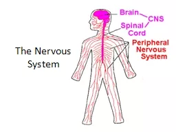
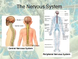
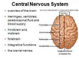
meninges ventricles cerebrospinal fluid and blood supply hindbrain and midbrain forebrain integrative functions the cranial nerves Copyright The McGrawHill Companies Inc Permission required for reproduction or display ID: 775206
Download Presentation The PPT/PDF document " Central Nervous System overview of the ..." is the property of its rightful owner. Permission is granted to download and print the materials on this web site for personal, non-commercial use only, and to display it on your personal computer provided you do not modify the materials and that you retain all copyright notices contained in the materials. By downloading content from our website, you accept the terms of this agreement.
Slide1
Central Nervous System
overview of the brainmeninges, ventricles, cerebrospinal fluid and blood supplyhindbrain and midbrainforebrainintegrative functionsthe cranial nerves
Copyright © The McGraw-Hill Companies, Inc. Permission required for reproduction or display.
Frontal lobe
Occipital lobe
Central sulcus
Longitudinal fissure
Parietal lobe
(a) Superior view
Cerebral
hemispheres
Slide2Directional Terms and Landmarks
rostral - toward the foreheadcaudal - toward the spinal cordbrain weighs about 1600 g (3.5 lb) in men, and 1450 g in womenthree major portions of the brain - cerebrum, cerebellum, brainstemcerebrum is 83% of brain volume; cerebral hemispheres, gyri and sulci, longitudinal fissure, corpus callosumcerebellum contains 50% of the neurons; second largest brain region, located in posterior cranial fossabrainstem the portion of the brain that remains if the cerebrum and cerebellum are removed; diencephalon, midbrain, pons, and medulla oblongata
Brainstem
Cerebellum
Cerebrum
Spinal cord
Rostral
Caudal
Central sulcus
Lateral sulcus
Gyri
(b) Lateral view
Temporal lobe
Copyright © The McGraw-Hill Companies, Inc. Permission required for reproduction or display.
Slide3Cerebrum
longitudinal fissure – deep groove that separates cerebral hemispheresgyri - thick foldssulci - shallow groovescorpus callosum – thick nerve bundle at bottom of longitudinal fissure that connects hemispheres
Copyright © The McGraw-Hill Companies, Inc. Permission required for reproduction or display.
Frontal lobe
Occipital lobe
Central sulcus
Longitudinal fissure
Parietal lobe
(a) Superior view
Cerebral
hemispheres
Slide414-4
Cerebellum
occupies
posterior cranial fossa
marked by
gyri
,
sulci
, and
fissures
about 10% of brain volume
contains over 50% of brain neurons
Slide5Brainstem
brainstem
– what remains of the brain if the cerebrum and cerebellum are removed
major components
diencephalon
midbrain
pons
medulla oblongata
Slide6Median Section of the Brain
Copyright © The McGraw-Hill Companies, Inc. Permission required for reproduction or display.
leaves
Thalamus
Hypothalamus
Frontal lobe
Corpus callosum
Cingulate gyrus
Optic chiasm
Pituitary gland
Mammillary body
Midbrain
Pons
Central sulcus
Parietal lobe
Parieto–occipital sulcus
Occipital lobe
Pineal gland
Habenula
Posterior commissure
Cerebral aqueduct
Fourth ventricle
Cerebellum
(a)
Epithalamus
Anterior
commissure
Temporal lobe
Medulla
oblongata
Slide714-7
Gray and White Matter
gray matter
– the seat of neuron cell bodies, dendrites, and synapses
dull white color when fresh, due to little myelin
forms surface layer,
cortex,
over cerebrum and cerebellum
forms
nuclei
deep within brain
white matter
- bundles of axons
lies
deep to cortical gray matter
, opposite relationship in the spinal cord
pearly white color from
myelin
around nerve fibers
composed of
tracts
, bundles of axons, that connect one part of the brain to another, and to the spinal cord
Slide814-8
Meninges
meninges
– three connective tissue membranes that envelop the brain
lies between the nervous tissue and bone
as in spinal cord, they are the
dura mater
,
arachnoid mater
, and the
pia mater
protect the brain and provide structural framework for its arteries and veins
dura mater
in cranial cavity -
2 layers
outer
periosteal
– equivalent to periosteum of cranial bones
inner
meningeal
– continues into vertebral canal and forms dural sac around spinal cord
cranial dura mater is pressed closely against cranial bones
no epidural space
not attached to bone except: around
foramen magnum
,
sella turcica
, the
crista
galli
, and
sutures of the skull
layers separated by
dural sinuses
– collect blood circulating through brain
folds inward to extend between parts of the brain
falx cerebri
separates the two cerebral hemispheres
tentorium cerebelli
separates cerebrum from cerebellum
falx cerebelli
separates the right and left halves of cerebellum
Slide914-9
Meninges
arachnoid mater and pia mater are similar to those in the spinal cord
arachnoid mater
transparent membrane over brain surface
subarachnoid space
separates it from pia mater below
subdural space
separates it from dura mater above in some places
pia mater
very thin membrane that follows contours of brain, even dipping into sulci
not usually visible without a microscope
Slide1014-10
Meninges of the Brain
Copyright © The McGraw-Hill Companies, Inc. Permission required for reproduction or display.
Subdural space
Skull
Pia mater
Blood vessel
Dura mater:
Periosteal layer
Meningeal layer
Arachnoid mater
Brain:
Gray matter
White matter
Arachnoid villus
Subarachnoid
space
Superior sagittal
sinus
Falx cerebri
(in longitudinal
fissure only)
Figure 14.5
Slide1114-11
Meningitis
meningitis
- inflammation of the meninges
serious disease of infancy & childhood
especially between 3 months and 2 years of age
caused by bacterial and virus invasion of the CNS by way of the nose and throat
pia mater and arachnoid are most often affected
bacterial meningitis
can cause swelling the brain, enlarging the ventricles, and hemorrhage
signs
include high fever, stiff neck, drowsiness, and intense headache and may progress to coma – death within hours of onset
diagnosed by examining the CSF for bacteria
lumbar puncture (spinal tap)
draws fluid from subarachnoid space between two lumbar vertebrae
Slide12Brain Ventricles
Slide13Ventricles of the Brain
Slide1414-14
Ventricles and Cerebrospinal Fluid
ventricles
– four internal chambers within the brain
two
lateral ventricles –
one
in each cerebral hemisphere
interventricular foramen -
a tiny pore that connects to third ventricle
third ventricle
- single narrow medial space beneath corpus callosum
cerebral aqueduct
runs through midbrain and connects third to fourth ventricle
fourth ventricle
– small triangular chamber between pons and cerebellum
connects to
central canal
runs down through spinal cord
choroid plexus
– spongy mass of blood capillaries on the floor of each ventricle
ependyma
– neuroglia that lines the ventricles and covers choroid plexus
produces cerebrospinal fluid
Slide1514-15
Cerebrospinal Fluid (CSF)
cerebrospinal fluid (CSF)
– clear, colorless liquid that fills the ventricles and canals of CNS
bathes its external surface
brain produces and absorbs 500 mL/day
100 – 160 mL normally present at one time
40% formed in subarachnoid space external to brain
30% by the general ependymal lining of the brain ventricles
30% by the choroid plexuses
production begins with the filtration of blood plasma through the capillaries of the brain
ependymal cells modify the filtrate, so CSF has more sodium and chloride than plasma, but less potassium, calcium, glucose, and very little protein
Slide1614-16
Cerebrospinal Fluid (CSF) Circulation
CSF continually flows through and around the CNS
driven by its own pressure, beating of ependymal cilia, and pulsations of the brain produced by each heartbeat
CSF secreted in
lateral ventricles
flows through
intervertebral foramina
into
third ventricle
then down the
cerebral aqueduct
into the
fourth ventricle
third and fourth ventricles add more CSF along the way
small amount of CSF fills the
central canal of the spinal cord
all escapes through three pores
median aperture
and
two lateral apertures
leads into
subarachnoid space
of brain and spinal cord surface
CSF is reabsorbed by
arachnoid villi
cauliflower-shaped extension of the
arachnoid meninx
protrudes through dura mater
into
superior sagittal sinus
CSF penetrates the walls of the villi and mixes with the blood in the sinus
Slide1714-17
Functions of CSF
buoyancy
allows brain to attain considerable size without being impaired by its own weight
if it rested heavily on floor of cranium, the pressure would kill the nervous tissue
protection
protects the brain from striking the cranium when the head is jolted
shaken child syndrome
and
concussions
do occur from severe jolting
chemical stability
flow of CSF rinses away metabolic wastes from nervous tissue and homeostatically regulates its chemical environment
Slide1814-18
Flow of Cerebrospinal Fluid
Copyright © The McGraw-Hill Companies, Inc. Permission required for reproduction or display.
Choroid plexus in fourth
ventricle adds more CSF.
CSF flows out two lateral apertures
and one median aperture.
CSF fills subarachnoid space and
bathes external surfaces of brain
and spinal cord.
At arachnoid villi, CSF is reabsorbed
into venous blood of dural
venous sinuses.
1
2
3
4
5
6
7
7
8
1
2
3
4
5
6
7
8
CSF is secreted by
choroid plexus in
each lateral ventricle.
CSF flows through
Interventricular foramina
into third ventricle.
Choroid plexus in third
ventricle adds more CSF.
CSF flows down cerebral
aqueduct to fourth ventricle.
Arachnoid villus
Superior
sagittal
sinus
Arachnoid mater
Subarachnoid
space
Dura mater
Choroid plexus
Third ventricle
Cerebral
aqueduct
Lateralaper ture
Fourth ventricle
Median aperture
Centralcanal
of spinal cord
Subarachnoid
space of
spinal cord
Figure 14.7
Slide1914-19
Blood Supply to the Brain
brain is only 2% of the adult body weight, and receives 15% of the blood
750 mL/min
neurons have a high demand for ATP, and therefore, oxygen and glucose, so a constant supply of blood is critical to the nervous system
10 second interruption of blood flow may cause loss of consciousness
1 – 2 minute interruption can cause significant impairment of neural function
4 minutes with out blood causes irreversible brain damage
Slide2014-20
Brain Barrier System
blood is also a source of antibodies, macrophages, bacterial toxins, and other harmful agents
brain barrier system
– strictly regulates what substances can get from the bloodstream into the tissue fluid of the brain
two points of entry must be guarded:
blood capillaries throughout the brain tissue
capillaries of the choroid plexus
blood-brain barrier
-
protects blood capillaries throughout brain tissue
consists of tight junctions between endothelial cells that form the capillary walls
astrocytes
reach out and contact capillaries with their perivascular feet
induce the endothelial cells to form tight junctions that completely seal off gaps between them
anything leaving the blood must pass through the cells, and not between them
endothelial cells
can exclude harmful substances from passing to the brain tissue while allowing necessary ones to pass
Slide2114-21
Brain Barrier System
blood-CSF barrier -
protects the brain at the choroid plexus
form tight junctions between the ependymal cells
tight junctions are absent from ependymal cells elsewhere
important to allow exchange between brain tissue and CSF
blood barrier system
is
highly permeable
to water, glucose, and lipid-soluble substances such as oxygen, carbon dioxide, alcohol, caffeine, nicotine, and anesthetics
slightly permeable
to sodium, potassium, chloride, and the waste products urea and creatinine
obstacle for delivering medications such as antibiotics and cancer drugs
trauma and inflammation can damage BBS and allow pathogens to enter brain tissue
circumventricular organs (CVOs)
– places in the third and fourth ventricles where the barrier is absent
blood has direct access to the brain
enables the brain to monitor and respond to fluctuations in blood glucose, pH, osmolarity, and other variables
CVOs afford a route for invasion by the human immunodeficiency virus (HIV)
Slide2214-22
Hindbrain - Medulla Oblongata
embryonic myelencephalon becomes medulla oblongatabegins at foramen magnum of the skullextends for about 3 cm rostrally and ends at a groove between the medulla and ponsslightly wider than spinal cordpyramids – pair of external ridges on anterior surfaceresembles side-by-side baseball batsolive – a prominent bulge lateral to each pyramidposteriorly, gracile and cuneate fasciculi of the spinal cord continue as two pair of ridges on the medullaall nerve fibers connecting the brain to the spinal cord pass through the medullafour pairs of cranial nerves begin or end in medulla - IX, X, XI, XII
Figure 14.2a
leaves
Thalamus
Hypothalamus
Frontal lobe
Corpus callosum
Cingulate gyrus
Optic chiasm
Pituitary gland
Mammillary body
Midbrain
Pons
Central sulcus
Parietal lobe
Parieto–occipital sulcus
Occipital lobe
Pineal gland
Habenula
Posterior commissure
Cerebral aqueduct
Fourth ventricle
Cerebellum
(a)
Epithalamus
Anterior
commissure
Temporal lobe
Medulla
oblongata
Copyright © The McGraw-Hill Companies, Inc. Permission required for reproduction or display.
Slide2314-23
Hindbrain - Medulla Oblongata
cardiac center
adjusts rate and force of heart
vasomotor center
adjusts blood vessel diameter
respiratory centers
control rate and depth of breathing
reflex centers
for coughing, sneezing, gagging, swallowing, vomiting, salivation, sweating, movements of tongue and head
Slide2414-24
Medulla Oblongata
pyramids contain descending fibers called corticospinal tractscarry motor signals to skeletal musclesinferior olivary nucleus – relay center for signals to cerebellumreticular formation - loose network of nuclei extending throughoutthe medulla, pons and midbraincontains cardiac, vasomotor & respiratory centers
Figure 14.9c
Copyright © The McGraw-Hill Companies, Inc. Permission required for reproduction or display.
Hypoglossal nerve
Medial lemniscus
Tectospinal tract
Nucleus
Tract
Gracile nucleus
Cuneate nucleus
Olive
Pyramids of medulla
Corticospinal tract
Trigeminal nerve:
Fourth ventricle
Reticular formation
(c) Medulla oblongata
(a) Midbrain
(c) Medulla
(b) Pons
Posterior spinocerebellar
tract
Nucleus of
vagus nerve
Inferior olivary
nucleus
Nucleus of
hypoglossal nerve
Slide2514-25
Medulla and Pons
Copyright © The McGraw-Hill Companies, Inc. Permission required for reproduction or display.
Diencephalon:
Midbrain:
Thalamus
Optic tract
Cranial nerves:
Oculomotor nerve (III)
Optic nerve (II)
Trochlear nerve (IV)
Trigeminal nerve (V)
Abducens nerve (VI)
Facial nerve (VII)
Vestibulocochlear nerve (VIII)
Glossopharyngeal nerve (IX)
Vagus nerve (X)
Accessory nerve (XI)
Hypoglossal nerve (XII)
Spinal nerves
Infundibulum
Mammillary body
Cerebral peduncle
Pyramid
Anterior median fissure
Pyramidal decussation
Spinal cord
(a) Anterior view
Pons
Medulla oblongata:
Regions of the brainstem
Midbrain
Diencephalon
Pons
Medulla oblongata
Figure 14.8a
Slide2614-26
Posterolateral View of Brainstem
Copyright © The McGraw-Hill Companies, Inc. Permission required for reproduction or display.
Diencephalon:
Midbrain:
Thalamus
Pineal gland
Superior colliculus
Inferior colliculus
Spinal cord
Pons
Olive
Cerebral peduncle
Medial geniculate body
Lateral geniculate body
Optic tract
Fourth ventricle
Cuneate fasciculus
Gracile fasciculus
(b) Posterolateral view
Regions of the brainstem
Midbrain
Diencephalon
Pons
Medulla oblongata
Medulla
oblongata
Superior cerebellar
peduncle
Middle cerebellar
peduncle
Inferior cerebellar
peduncle
Figure 14.8b
Slide2714-27
Pons
Figure 14.2a
Copyright © The McGraw-Hill Companies, Inc. Permission required for reproduction or display.
leaves
Thalamus
Hypothalamus
Frontal lobe
Corpus callosum
Cingulate gyrus
Optic chiasm
Pituitary gland
Mammillary body
Midbrain
Pons
Central sulcus
Parietal lobe
Parieto–occipital sulcus
Occipital lobe
Pineal gland
Habenula
Posterior commissure
Cerebral aqueduct
Fourth ventricle
Cerebellum
(a)
Epithalamus
Anterior
commissure
Temporal lobe
Medulla
oblongata
metencephalon
- develops into the pons and cerebellum
pons
– anterior bulge in brainstem, rostral to medulla
cerebral peduncles
– connect cerebellum to pons and midbrain
Slide2814-28
Pons
ascending sensory tracts
descending motor tracts
pathways in and out of cerebellum
cranial nerves V, VI, VII, and VIII
sensory roles
– hearing, equilibrium, taste, facial sensations
motor roles
– eye movement, facial expressions, chewing, swallowing, urination, and secretion of saliva and tears
reticular formation
in pons contains additional nuclei concerned with:
sleep, respiration, and posture
Slide2914-29
Midbrain
mesencephalon becomes one mature brain structure, the
midbrain
short segment of brainstem that connects the hindbrain to the forebrain
contains
cerebral aqueduct
contains continuations of the
medial lemniscus
and
reticular formation
contains the motor nuclei of
two cranial nerves
that control eye movements – CN III (oculomotor) and CN IV (trochlear)
tectum
– roof-like part of the midbrain posterior to cerebral aqueduct
exhibits four bulges, the
corpora quadrigemina
upper pair, the
superior colliculi
function in visual attention, tracking moving objects, and some reflexes
lower pair, the
inferior colliculi
receives signals from the inner ear
relays them to other parts of the brain, especially the thalamus
cerebral peduncles
– two stalks that anchor the cerebrum to the brainstem anterior to the cerebral aqueduct
Slide3014-30
Midbrain
cerebral peduncles
each consists of
three main components
tegmentum, substantia nigra, and cerebral crus
tegmentum
dominated by the
red nucleus
pink color due to high density of blood vessels
connections go to and from cerebellum
collaborates with cerebellum for fine motor control
substantia nigra
dark gray to black nucleus
pigmented with melanin
motor center that relays inhibitory signals to thalamus & basal nuclei preventing unwanted body movement
degeneration of neurons leads to tremors of Parkinson disease
cerebral crus
bundle of nerve fibers that connect the cerebrum to the pons
carries
corticospinal tracts
Slide3114-31
Midbrain -- Cross Section
Copyright © The McGraw-Hill Companies, Inc. Permission required for reproduction or display.
leaves
Tegmentum
Cerebral peduncle:
Cerebral crus
Tectum
Superior colliculus
Cerebral aqueduct
Medial geniculate nucleus
Reticular formation
Central gray matter
Oculomotor nucleus
Medial lemniscus
Red nucleus
Substantia nigra
Oculomotor nerve (III)
Posterior
Anterior
(a) Midbrain
(a) Midbrain
(c) Medulla
(b) Pons
Figure 14.9a
Slide3214-32
Cerebellum
the largest part of the hindbrain and the second largest part of the brain as a wholeconsists of right and left cerebellar hemispheres connected by vermiscortex of gray matter with folds (folia) and four deep nuclei in each hemispherecontains more than half of all brain neurons, about 100 billiongranule cells and Purkinje cells synapse on deep nucleiwhite matter branching pattern is called arbor vitae
Copyright © The McGraw-Hill Companies, Inc. Permission required for reproduction or display.
leaves
(b) Superior view
Folia
Anterior
Posterior
Anterior lobe
Vermis
Posterior lobe
Cerebellar
hemisphere
Figure 14.11b
Slide3314-33
Cerebellum
cerebellar peduncles – three pairs of stalks that connect the cerebellum to the brainsteminferior peduncles – connected to medulla oblongatamost spinal input enters the cerebellum through inferior pedunclemiddle peduncles – connected to the pons most input from the rest of the brain enters by way of middle pedunclesuperior peduncles – connected to the midbraincarries cerebellar outputconsist of thick bundles of nerve fibers that carry signals to and from the cerebellum
Copyright © The McGraw-Hill Companies, Inc. Permission required for reproduction or display.
Superior colliculus
Posterior commissure
Pineal gland
Inferior colliculus
Mammillary body
Midbrain
Cerebral aqueduct
Oculomotor nerve
Pons
Fourth ventricle
Medulla oblongata
Gray matter
(a) Median section
White matter
(arbor vitae)
Figure 14.11a
Slide3414-34
Cerebellar Functions
monitors muscle contractions and aids in motor coordination
evaluation of sensory input
comparing textures without looking at them
spatial perception and comprehension of different views of 3D objects belonging to the same object
timekeeping center
predicting movement of objects
helps predict how much the eyes must move in order to compensate for head movements and remain fixed on an object
hearing
distinguish pitch and similar sounding words
planning and scheduling tasks
lesions may result in emotional overreactions and trouble with impulse control
Slide3514-35
The Forebrain
Diencephalon
Mesencephalon
Telencephalon
Forebrain
Pons
Cerebellum
Metencephalon
Spinal cord
Hindbrain
(c) Fully developed
Midbrain
Myelencephalon
(medulla oblongata)
Copyright © The McGraw-Hill Companies, Inc. Permission required for reproduction or display.
forebrain consists of :
the
diencephalon
encloses the third ventricle
most rostral part of the brainstem
has three major embryonic derivatives
thalamus
hypothalamus
epithalamus
the
telencephalon
develops chiefly into the
cerebrum
Figure 14.4c
Slide3614-36
Diencephalon: Thalamus
thalamus – ovoid mass on each side of the brain perched at the superior end of the brainstem beneath the cerebral hemispheresconstitutes about four-fifths of the diencephalontwo thalami are joined medially by a narrow intermediate masscomposed of at least 23 nuclei – we will consider five major functional groupsthe “gateway to the cerebral cortex” – nearly all input to the cerebrum passes by way of synapses in the thalamic nuclei, filters information on its way to cerebral cortexplays key role in motor control by relaying signals from cerebellum to cerebrum and providing feedback loops between the cerebral cortex and the basal nucleiinvolved in the memory and emotional functions of the limbic system – a complex of structures that include some cerebral cortex of the temporal and frontal lobes and some of the anterior thalamic nuclei
Copyright © The McGraw-Hill Companies, Inc. Permission required for reproduction or display.
leaves
(a) Thalamus
Anterior group
Medial group
Ventral group
Lateral group
Posterior group
Lateral geniculate nucleus
Medial geniculate nucleus
Thalamic Nuclei
Part of limbic system;
memory and emotion
Emotional output to prefrontal
cortex; awareness of emotions
Somesthetic output to
postcentral gyrus; signals
from cerebellum and basalnuclei to motor areas of cortex
Somesthetic output toassociation areas of cortex;contributes to emotional functionof limbic system
Relay of visual signals tooccipital lobe (via lateralgeniculate nucleus) and auditorysignals to temporal lobe (viamedial geniculate nucleus)
Figure 14.12a
Slide37hypothalamus – forms part of the walls and floor of the third ventricleextends anteriorly to optic chiasm and posteriorly to the paired mammillary bodieseach mammillary body contains three or four mammillary nucleirelay signals from the limbic system to the thalamusinfundibulum – a stalk that attaches the pituitary gland to the hypothalamusmajor control center of autonomic nervous system and endocrine systemplays essential roll in homeostatic regulation of all body systems
Diencephalon: Hypothalamus
Slide38functions of hypothalamic nucleihormone secretioncontrols anterior pituitaryregulates growth, metabolism, reproduction ,and stress responsesautonomic effectsmajor integrating center for the autonomic nervous systeminfluences heart rate, blood pressure, gastrointestinal secretions and motility, and othersthermoregulationhypothalamic thermostat monitors body temperatureactivates heat-loss center when temp is too highactivates heat-promoting center when temp is too lowfood and water intake hunger and satiety centers monitor blood glucose and amino acid levelsproduce sensations of hunger and satietythirst center monitors osmolarity of the bloodrhythm of sleep and wakingcontrols 24 hour circadian rhythm of activitymemory-mammillary nuclei receive signals from hippocampusemotional behavior anger, aggression, fear, pleasure, and contentment
Diencephalon: Hypothalamus
Slide39Diencephalon: Epithalamus
epithalamus
– very small mass of tissue composed of:
pineal gland
– endocrine gland
habenula
– relay from the limbic system to the midbrain
thin roof over the third ventricle
Slide40Telencephalon: Cerebrum
cerebrum
– largest and most conspicuous part of the human brain
seat of sensory perception, memory, thought, judgment, and voluntary motor actions
Slide4114-41
Cerebrum - Gross Anatomy
two cerebral hemispheres divided by longitudinal fissureconnected by white fibrous tract the corpus callosumgyri and sulci – increases amount of cortex in the cranial cavitygyri increases surface area for information processing capabilitysome sulci divide each hemisphere into five lobes named for the cranial bones that overly them
Frontal lobe
Occipital lobe
Central sulcus
Longitudinal fissure
Parietal lobe
(a) Superior view
Cerebral
hemispheres
Figure 14.1a,b
Brainstem
Cerebellum
Cerebrum
Spinal cord
Rostral
Caudal
Central sulcus
Lateral sulcus
Gyri
(b) Lateral view
Temporal lobe
Copyright © The McGraw-Hill Companies, Inc. Permission required for reproduction or display.
Slide4214-42
frontal lobevoluntary motor functions motivation, foresight, planning, memory, mood, emotion, social judgment, and aggressionparietal lobereceives and integrates general sensory information, taste and some visual processingoccipital lobeprimary visual center of braintemporal lobeareas for hearing, smell, learning, memory, and some aspects of vision and emotioninsula (hidden by other regions)understanding spoken language, taste and sensory information from visceral receptors
Functions of Cerebrum - Lobes
Figure 14.13
Copyright © The McGraw-Hill Companies, Inc. Permission required for reproduction or display.
Postcentral gyrus
Occipital lobe
Temporal lobe
Lateral sulcus
Frontal lobe
Parietal lobe
Insula
Rostral
Caudal
Central
sulcus
Precentral
gyrus
Slide4314-43
Cerebral White Matter
Copyright © The McGraw-Hill Companies, Inc. Permission required for reproduction or display.
Association tracts
Frontal lobe
Corpus callosum
Temporal lobe
(a) Sagittal section
Projection tracts
Parietal lobe
Occipital lobe
Longitudinal fissure
Corpus callosum
Basal nuclei
Cerebral peduncle
Projection tracts
Decussation in pyramids
Commissuralta tracts
Lateral ventricle
Thalamus
Third ventricle
Mammillary body
pons
Pyramid
Medulla oblongata
(b) Frontal section
Figure 14.14
Slide4414-44
The Cerebral White Matter
most of the volume of cerebrum is white matter
glia and
myelinated nerve fibers
transmitting signals from one region of the cerebrum to another and between cerebrum and lower brain centers
three types of tracts
projection tracts
extends vertically between higher and lower brain and spinal cord centers
carries information between cerebrum and rest of the body
commissural tracts
cross from one cerebral hemisphere through bridges called
commissures
most pass through
corpus callosum
anterior
and
posterior commissures
enables the two sides of the cerebrum to communicate with each other
association tracts
connect different regions within the same cerebral hemisphere
long association fibers
– connect different lobes of a hemisphere
to each other
short association fibers
– connect different gyri within a single lobe
Slide4514-45
Cerebral Cortex
neural integration is carried out in the gray matter of the cerebrumcerebral gray matter found in three placescerebral cortexbasal nucleilimbic systemcerebral cortex – layer covering the surface of the hemispheresonly 2 – 3 mm thickcortex constitutes about 40% of the mass of the braincontains 14 – 16 billion neurons
Copyright © The McGraw-Hill Companies, Inc. Permission required for reproduction or display.
I
II
III
IV
V
VI
Cortical surface
Stellate cells
Small pyramidal
cells
Large pyramidal
cells
White
matter
Figure 14.15
Slide4614-46
Cerebral Cortex
contains two principal types of neuronsstellate cellshave spheroid somas with dendrites projecting in all directionsreceive sensory input and process information on a local levelpyramidal cellstall, and conical, with apex toward the brain surfacea thick dendrite with many branches with small, knobby dendritic spinesinclude the output neurons of the cerebrumonly neurons that leave the cortex and connect with other parts of the CNSneocortex – six layered tissue that constitutes about 90% of the human cerebral cortexrelatively recent in evolutionary origin
I
II
III
IV
V
VI
Cortical surface
Stellate cells
Small pyramidal
cells
Large pyramidal
cells
White
matter
Figure 14.15
Slide4714-47
Limbic System
limbic system – important center of emotion and learningmost anatomically prominent components are:cingulate gyrus – arches over the top of the corpus callosum in the frontal and parietal lobeshippocampus – in the medial temporal lobe - memoryamygdala – immediately rostral to the hippocampus - emotionlimbic system components are connected through a complex loop of fiber tracts allowing for somewhat circular patterns of feedbacklimbic system structures have centers for both gratification and aversiongratification – sensations of pleasure or rewardaversion –sensations of fear or sorrow
Copyright © The McGraw-Hill Companies, Inc. Permission required for reproduction or display.
Basal nuclei
Amygdala
Temporal lobe
Fornix
Hippocampus
Medial
prefrontal
cortex
Corpus
callosum
Cingulate
gyrus
Orbitofrontal
cortex
Thalamic
nuclei
Mammillary
body
Figure 14.17
Slide4814-48
Higher Brain Functions
higher brain functions - sleep, memory, cognition, emotion, sensation, motor control, and language
involve interactions between cerebral cortex and basal nuclei, brainstem and cerebellum
functions of the brain do not have easily defined anatomical boundaries
integrative functions of the brain focuses mainly on the cerebrum, but involves combined action of multiple brain levels
Slide4914-49
The Electroencephalogram
electroencephalogram (EEG)
– monitors surface electrical activity of the brain waves
useful for studying normal brain functions as sleep and consciousness
in diagnosis of degenerative brain diseases, metabolic abnormalities, brain tumors, etc.
brain waves
– rhythmic voltage changes resulting from synchronized postsynaptic potentials at the superficial layer of the cerebral cortex
4 types distinguished by amplitude (mV) and frequency (Hz)
persistent absence of brain waves is common clinical and legal criterion of brain death
Slide5014-50
Brain Waves
alpha waves 8 – 13 Hz
awake and resting with eyes closed and mind wandering
suppressed when eyes open or performing a mental task
beta waves 14 – 30 Hz
eyes open and performing mental tasks
accentuated during mental activity and sensory stimulation
theta waves 4 – 7 Hz
drowsy or sleeping adults
if awake and under emotional stress
delta waves high amplitude, less than 3.5 Hz
deep sleep in adults
Slide5114-51
Sleep
sleep occurs in cycles called
circadian rhythms
events that reoccur at intervals of about 24 hours
sleep
- temporary state of unconsciousness from which one can awaken when stimulated
characterized by
stereotyped posture
lying down with eyes closed
sleep paralysis
- inhibition of muscular activity
resembles unconsciousness but can be aroused by sensory stimulation
coma
or
hibernation
– states of prolonged unconsciousness where individuals cannot be aroused from those states by sensory stimulation
restorative effect
brain glycogen and ATP levels increase in non-REM sleep
memories strengthened in REM sleep
synaptic connections reinforced
Slide5214-52
Four Stages of Sleep
Stage 1
feel drowsy, close our eyes, begin to relax
often feel drifting sensation, easily awakened if stimulated
alpha waves dominate EEG
Stage 2
pass into light sleep
EEG declines in frequency but increases in amplitude
exhibits
sleep spindles
– high spikes resulting from interactions between neurons
of the thalamus and cerebral cortex
Stage 3
moderate to deep sleep
about 20 minutes after stage 1
theta and delta waves appear
muscles relax and vital signs (body temperature, blood pressure, heart and respiratory rate) fall
Stage 4
called
slow-wave-sleep
(SWS)
– EEG dominated by low-frequency, high amplitude delta waves
muscles now very relaxed, vital signs at their lowest, and we become more difficult to awaken
Slide53Rhythm of Sleep
rhythm of sleep
controlled by a complex interaction between the cerebral cortex, thalamus, hypothalamus, and reticular formation
arousal induced in the upper reticular formation, near junction of
pons
and midbrain
sleep induced by nuclei below
pons
, and in
ventrolateral
preoptic
nucleus in the hypothalamus
sleep has a restorative effect, and sleep deprivation can be fatal to experimental animals
bed rest alone does not have the restorative effect of sleep…why must we lose consciousness?
sleep may be the time to replenish such energy sources as glycogen and ATP
REM sleep may consolidate and strengthen memories by reinforcing some synapses, and eliminating others
Slide54Sleep Stages
Slide5514-55
Cognition
cognition
– the range of mental processes by which we acquire and use knowledge
such as sensory perception, thought, reasoning, judgment, memory, imagination, and intuition
association areas of cerebral cortex
has above functions
constitutes about 75% of all brain tissue
studies of patients with brain lesions, cancer, stroke, and trauma yield information on cognition
parietal lobe association area
– perceiving stimuli
contralateral neglect syndrome
– unaware of objects on opposite side of their body
temporal lobe association area
– identifying stimuli
agnosia
– inability to recognize, identify, and name familiar objects
prosopagnosia
– person cannot remember familiar faces
frontal lobe association area
– planning our responses and personality – inability to execute appropriate behavior
Slide5614-56
Memory
information management requires
learning
– acquiring new information
memory
– information storage and retrieval
forgetting
– eliminating trivial information; as important as remembering
amnesia
– defects in
declarative memory
– inability to describe past events
procedural memory
– ability to tie your shoes
anterograde amnesia
– unable to store new information
retrograde amnesia
– cannot recall things they knew before the injury
hippocampus
– important memory-forming center
does not store memories
organizes sensory and cognitive information into a unified long-term memory
memory consolidation
– the process of “teaching the cerebral cortex” until a long-term memory is established
long-term memories are stored in various areas of the
cerebral cortex
vocabulary and memory of familiar faces stored in superior
temporal lobe
memories of one’s plans and social roles stored in the
prefrontal cortex
cerebellum
– helps learn motor skills
amygdala
- emotional memory
Slide5714-57
Emotion
emotional feelings and memories are interactions between prefrontal cortex and diencephalon
prefrontal cortex
-
seat of judgment, intent, and control over expression of emotions
feelings
come from hypothalamus and amygdala
nuclei generate feelings of fear or love
amygdala
receives input from sensory systems
role in food intake, sexual behavior, and drawing attention to novel stimuli
one output
goes to
hypothalamus
influencing somatic and visceral motor systems
heart races, raises blood pressure, makes hair stand on end, induce vomiting
other output
to
prefrontal cortex
important in controlling expression of emotions
ability to express love, control anger, or overcome fear
behavior
shaped by learned associations between stimuli, our responses to them, and the reward or punishment that results
Slide5814-58
Sensation
primary sensory cortex - sites where sensory input is first received and one becomes conscious of the stimulusassociation areas nearby to sensory areas that process and interpret that sensory informationprimary visual cortex is bordered by visual association area which interprets and makes cognitive sense of visual stimulimultimodal association areas – receive input from multiple senses and integrate this into an overall perception of our surroundings
Anterior
Posterior
(a)
Precentral
gyrus
Central
sulcus
Postcentral
gyrus
Parietal
lobe
Frontal
lobe
Occipital
lobe
Copyright © The McGraw-Hill Companies, Inc. Permission required for reproduction or display.
Figure 14.22a
Slide5914-59
Functional Regions of Cerebral Cortex
Wernicke area
Broca area
Primary motor
cortex
Motor association
area
Prefrontal
cortex
Olfactory
association
area
Primary somesthetic
cortex
Somesthetic
association area
Primary gustatory
cortex
Visual association
area
Primary
visual cortex
Primary
auditory cortex
Auditory
association area
Figure 14.21
Copyright © The McGraw-Hill Companies, Inc. Permission required for reproduction or display.
Slide6014-60
Motor Control
the intention to contract a muscle begins in
motor association (premotor) area
of frontal lobes
where we plan our behavior
where neurons compile a program for degree and sequence of muscle contraction required for an action
program transmitted to neurons of the
precentral gyrus (primary motor area)
most posterior gyrus of the frontal lobe
neurons send signals to the brainstem and spinal cord
ultimately resulting in muscle contraction
precentral gyrus
also exhibits
somatotopy
neurons for toe movement are deep in the longitudinal fissure of the medial side of the gyrus
the summit of the gyrus controls the trunk, shoulder, and arm
the inferolateral region controls the facial muscles
motor homunculus
has a distorted look because the amount of cortex devoted to a given body region is proportional to the number of muscles and motor units in that body region
Slide6114-61
Motor Control
pyramidal cells of the precentral gyrus are called
upper motor neurons
their fibers project caudally
about 19 million fibers ending in nuclei of the brainstem
about 1 million forming the corticospinal tracts
most fibers decussate in lower medulla oblongata
form lateral corticospinal tracts on each side of the spinal cord
in the brainstem or spinal cord, the fibers from upper motor neurons synapse with
lower motor neurons
whose axons innervate the skeletal muscles
basal nuclei
and
cerebellum
are also important in muscle control
Slide6214-62
Motor Control
basal nuclei
determines the onset and cessation of intentional movements
repetitive hip and shoulder movements in walking
highly practiced, learned behaviors that one carries out with little thought
writing, typing, driving a car
lies in a feedback circuit from the cerebrum to the basal nuclei to the thalamus and back to the cerebrum
dyskinesias
– movement disorders caused by lesions in the basal nuclei
cerebellum
highly important in motor coordination
aids in learning motor skills
maintains muscle tone and posture
smoothes muscle contraction
coordinates eye and body movements
coordinates the motions of different joints with each other
ataxia – clumsy, awkward gait
Slide6314-63
Input and Output to Cerebellum
Copyright © The McGraw-Hill Companies, Inc. Permission required for reproduction or display.
Cerebrum
Cerebrum
Motor cortex
Cerebellum
Cerebellum
Brainstem
Brainstem
Inner ear
Eye
Reticular formation
Muscle and joint proprioceptors
(a) Input to cerebellum
(b) Output from cerebellum
Spinocerebellar
tracts of spinal cord
Reticulospinal
and vestibulospinal
tracts of spinal cord
Limb and posturalmuscles
Figure 14.24
Slide6414-64
Language
language include several abilities:
reading
,
writing
,
speaking
, and
understanding words
assigned to different regions of the cerebral cortex
Wernicke area
permits recognition of spoken and written language and creates plan of speech
when we intend to speak, Wernicke area formulates phases according to learned rules of grammar
transmits plan of speech to Broca area
Broca area
generates motor program for the muscles of the larynx, tongue, cheeks and lips
transmits program to primary motor cortex for commands to the lower motor neurons that supply relevant muscles
Affective language area
lesions produce
aprosody
- flat emotionless speech
Slide6514-65
Language Centers
Copyright © The McGraw-Hill Companies, Inc. Permission required for reproduction or display.
leaves
Precentral gyrus
Anterior
Posterior
Speech center of
primary motor cortex
Primary auditory
cortex
(in lateral sulcus)
Postcentral
gyrus
Angular
gyrus
Primary
visual cortex
Wernicke
area
Broca
area
Figure 14.25
Slide6614-66
Cerebral Lateralization
Copyright © The McGraw-Hill Companies, Inc. Permission required for reproduction or display.
leaves
Olfaction, left nasal cavity
Memory for shapes
Left hand motor control
Musical ability
Intuitive, nonverbal thought
Vision, left field
Vision, right field
Speech
Verbal memory
Olfaction, right nasal cavity
Left hemisphere
Right hemisphere
Posterior
Anterior
(Limited language
comprehension, mute)
Feeling shapes with
left hand
Hearing nonvocal sounds
(left ear advantage)
Superior recognition of
faces and spatial
relationships
Right hand
motor control
Feeling shapes
with right hand
Hearing vocal sounds
(right ear advantage)
Rational, symbolic
thought
Superior language
comprehension
Figure 14.26
Slide6714-67
Cerebral Lateralization
cerebral lateralization
– the difference in the structure and function of the cerebral hemispheres
left hemisphere
-
categorical hemisphere
specialized for spoken and written language
sequential and analytical reasoning (math and science)
breaks information into fragments and analyzes it in a linear way
right hemisphere
-
representational hemisphere
perceives information in a more integrated holistic way
seat of imagination and insight
musical and artistic skill
perception of patterns and spatial relationships
comparison of sights, sounds, smells, and taste
highly correlated with
handedness
left hemisphere is the categorical one in 96% of right-handed people
right hemisphere in 4%
left handed people – right hemisphere is categorical in 15% and left in 70%
lateralization develops with age
males exhibit more lateralization than females and suffer more functional loss when one hemisphere is damaged
Slide6814-68
Cranial Nerves
the brain must communicate with the rest of the body
most of the input and output travels by way of the spinal cord
12 pairs of cranial nerves
arise from the base of the brain
exit the cranium through
foramina
lead to muscles and sense organs located mainly in the
head and neck
Slide6914-69
Cranial Nerve Pathways
most
motor fibers
of the cranial nerves begin in nuclei of brainstem and lead to glands and muscles
sensory fibers
begin in receptors located mainly in head and neck and lead mainly to the brainstem
sensory fibers for
proprioception
may travel to brain in a different nerve from motor nerve
most cranial nerves carry fibers between brainstem and
ipsilateral
receptors and effectors
lesion in left brainstem causes sensory or motor deficit on same side
exceptions are: optic nerve where half the fibers decussate, and
trochlear
nerve where all efferent fibers lead to a muscle of the
contralateral
eye
Slide7014-70
Cranial Nerve Classification
some cranial nerves are classified as
motor
, some
sensory
, others
mixed
sensory (
I
,
II
, and
VIII
)
motor (
III
,
IV
,
VI
,
XI
, and
XII
)
stimulate muscle but also contain fibers of proprioception
mixed (
V
,
VII
,
IX
,
X
)
sensory functions may be quite unrelated to their motor function
facial nerve (VII) has sensory role in taste and motor role in facial expression
Slide7114-71
Cranial Nerves
leaves
Cranial nerves:
Optic nerve (II)
Trochlear nerve (IV)
Trigeminal nerve (V)
Abducens nerve (VI)
Facial nerve (VII)
Vestibulocochlear nerve (VIII)
Glossopharyngeal nerve (IX)
Accessory nerve (XI)
Hypoglossal nerve (XII)
Vagus nerve (X)
Oculomotor nerve (III)
Frontal lobe
Frontal lobe
Cerebellum
Cerebellum
Olfactory tract
Temporal lobe
Infundibulum
Pons
Medulla
Optic chiasm
Optic chiasm
Olfactory tract
Temporal lobe
Pons
Spinal cord
Spinal cord
(a)
(b)
Olfactory bulb
(from olfactory nerve, I)
Longitudinal
fissure
Medulla
oblongata
b: © The McGraw-Hill Companies, Inc./Rebecca Gray, photographer/Don Kincaid, dissections
Copyright © The McGraw-Hill Companies, Inc. Permission required for reproduction or display.
Figure 14.27a-b
Slide7214-72
I Olfactory Nerve
sense of smelldamage causes impaired sense of smell
Copyright © The McGraw-Hill Companies, Inc. Permission required for reproduction or display.
Olfactory bulb
Olfactory tract
Nasal mucosa
Cribriform plate of
ethmoid bone
Fascicles of
olfactory nerve (I)
Figure 14.28
Slide7314-73
II Optic Nerve
provides visiondamage causes blindness in part or all of the visual field
Figure 14.29
Copyright © The McGraw-Hill Companies, Inc. Permission required for reproduction or display.
Eyeball
Optic nerve (II)
Optic chiasm
Optic tract
Pituitary gland
Slide7414-74
III Oculomotor Nerve
controls muscles that turn the eyeball up, down, and medially, as well as controlling the iris, lens, and upper eyeliddamage causes drooping eyelid, dilated pupil, double vision, difficulty focusing and inability to move eye in certain directions
Copyright © The McGraw-Hill Companies, Inc. Permission required for reproduction or display.
Oculomotor nerve (III):
Superior orbital fissure
Superior branch
Inferior branch
Ciliary ganglion
Figure 14.30
Slide7514-75
IV Trochlear Nerve
eye movement (superior oblique muscle)damage causes double vision and inability to rotate eye inferolaterally
Copyright © The McGraw-Hill Companies, Inc. Permission required for reproduction or display.
Trochlear nerve (IV)
Superior orbital fissure
Superior oblique muscle
Figure 14.31
Slide7614-76
V Trigeminal Nerve
largest of the cranial nervesmost important sensory nerve of the faceforks into three divisions:ophthalmic division (V1) – sensorymaxillary division (V2) – sensorymandibular division (V3) - mixed
Figure 14.32
Copyright © The McGraw-Hill Companies, Inc. Permission required for reproduction or display.
Lingual nerve
V
1
V2
V3
Superior orbital fissure
Ophthalmic division (V1)
Trigeminal ganglion
Trigeminal nerve (V)
Maxillary division (V2)
Mandibular division (V3)
Foramen ovale
Foramen rotundum
Temporalis muscle
Medial pterygoid muscle
Masseter muscle
Lateral pterygoid muscle
Infraorbital
nerve
Superior
alveolar nerves
Inferior
alveolar nerve
1
Anterior belly of
digastric muscle
Anterior trunk of V
3
to
chewing muscles
Motor branches of the
mandibular division (V
3
)
Distribution of sensory
fibers of each division
Slide7714-77
VI Abducens Nerve
provides eye movement (lateral rectus m.)damage results in inability to rotate eye laterally and at rest eye rotates medially
Figure 14.33
Copyright © The McGraw-Hill Companies, Inc. Permission required for reproduction or display.
Abducens nerve (VI)
Superior orbital fissure
Lateral rectus muscle
Slide7814-78
VII Facial Nerve
motor – major motor nerve of facial muscles: facial expressions; salivary glands and tear, nasal and palatine glands sensory - taste on anterior 2/3’s of tonguedamage produces sagging facial muscles and disturbed sense of taste (no sweet and salty)
Copyright © The McGraw-Hill Companies, Inc. Permission required for reproduction or display.
Geniculate ganglion
Pterygopalatine ganglion
Lacrimal (tear) gland
Submandibular gland
Stylomastoid foramen
Sublingual gland
Internal acoustic meatus
Facial nerve (VII)
(a)
Parasympathetic fibers
Submandibular ganglion
Motor branch
to muscles of
facial expression
to (b)
Chorda tympani
branch (taste and
salivation)
Figure 14.34a
Slide7914-79
Five Branches of Facial Nerve
clinical test: test anterior 2/3’s of tongue with substances such as sugar, salt, vinegar, and quinine; test response of tear glands to ammonia fumes; test motor functions by asking subject to close eyes, smile, whistle, frown, raise eyebrows, etc.
Figure 14.34 b-c
Temporal
Zygomatic
Buccal
Mandibular
Cervical
(b)
© The McGraw-Hill Companies, Inc./Joe DeGrandis, photographer
(c)
Copyright © The McGraw-Hill Companies, Inc. Permission required for reproduction or display.
Slide8014-80
VIII Vestibulocochlear Nerve
nerve of hearing and equilibriumdamage produces deafness, dizziness, nausea, loss of balance and nystagmus (involuntary rhythms oscillation of the eyes from side to side
Copyright © The McGraw-Hill Companies, Inc. Permission required for reproduction or display.
leaves
Cochlear nerve
Cochlea
Semicircular
ducts
Vestibular ganglia
Vestibular nerve
Vestibulocochlear
nerve (VIII)
Internal
acoustic meatus
Vestibule
Figure 14.35
Slide8114-81
IX Glossopharyngeal Nerve
swallowing, salivation, gagging, control of BP and respirationsensations from posterior 1/3 of tonguedamage results in loss of bitter and sour taste and impaired swallowing
Figure 14.36
Copyright © The McGraw-Hill Companies, Inc. Permission required for reproduction or display.
Glossopharyngeal nerve (IX)
Parotid salivary gland
Jugular foramen
Superior ganglion
Inferior ganglion
Otic ganglion
Carotid sinus
Pharyngeal muscles
Slide8214-82
X Vagus Nerve
most extensive distribution of any cranial nervemajor role in the control of cardiac, pulmonary, digestive, and urinary functionswallowing, speech, regulation of visceradamage causes hoarseness or loss of voice, impaired swallowing and fatal if both are cut
Figure 14.37
Copyright © The McGraw-Hill Companies, Inc. Permission required for reproduction or display.
Heart
Lung
Liver
Spleen
Small intestine
Stomach
Kidney
Carotid sinus
Laryngeal nerve
Pharyngeal nerve
Jugular foramen
Vagus nerve (X)
Colon
(proximal portion)
Slide8314-83
XI Accessory Nerve
swallowing, head, neck and shoulder movementdamage causes impaired head, neck, shoulder movement;head turns towards injured side
Accessory nerve (XI)
Cranial root of XI
Spinal root of XI
Posterior view
Vagus nerve
Sternocleidomastoid
muscle
Trapezius muscle
Jugular
foramen
Foramen
magnum
Spinal nerves
C3 and C4
Figure 14.38
Copyright © The McGraw-Hill Companies, Inc. Permission required for reproduction or display.
Slide8414-84
XII Hypoglossal Nerve
tongue movements for speech, food manipulation and swallowingif both are damaged – can’t protrude tongue if one side is damaged – tongue deviates towards injured side; see ipsilateral atrophy
Hypoglossal canal
Hypoglossal nerve (XII)
Intrinsic muscles
of the tongue
Extrinsic muscles
of the tongue
Figure 14.39
Copyright © The McGraw-Hill Companies, Inc. Permission required for reproduction or display.
Slide8514-85
Cranial Nerve Disorders
Trigeminal neuralgia (tic douloureux)
recurring episodes of intense stabbing pain in trigeminal nerve area (near mouth or nose)
pain triggered by touch, drinking, washing face
treatment may require cutting nerve
Bell palsy
degenerative disorder of facial nerve causes paralysis of facial muscles on one side
may appear abruptly with full recovery within 3 - 5 weeks
Slide8614-86
Images of the Mind
positron emission tomography
(PET) and
MRI
visualize increases in blood flow when brain areas are active
injection of radioactively labeled glucose
busy areas of brain “light up”
functional magnetic resonance imaging (fMRI)
looks at increase in blood flow to an area (additional glucose is needed in active area) – magnetic properties of hemoglobin depend on how much oxygen is bound to it (additional oxygen is there due to additional blood flow)
quick, safe and accurate method to see brain function