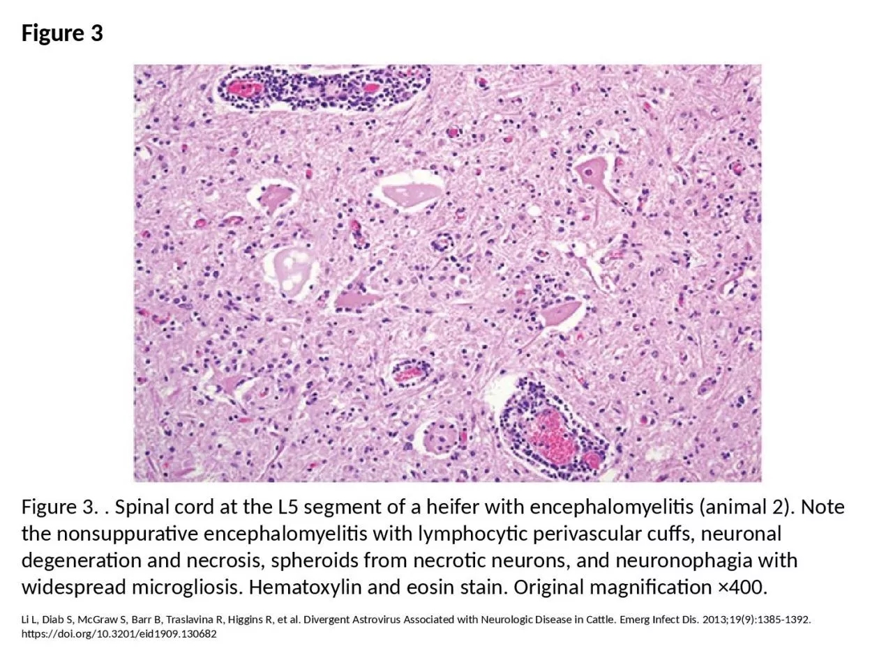
PPT-Figure 3 Figure 3. . Spinal cord at the L5 segment of a heifer with encephalomyelitis
Author : elena | Published Date : 2024-03-13
Li L Diab S McGraw S Barr B Traslavina R Higgins R et al Divergent Astrovirus Associated with Neurologic Disease in Cattle Emerg Infect Dis 201319913851392 httpsdoiorg103201eid1909130682
Presentation Embed Code
Download Presentation
Download Presentation The PPT/PDF document "Figure 3 Figure 3. . Spinal cord at the ..." is the property of its rightful owner. Permission is granted to download and print the materials on this website for personal, non-commercial use only, and to display it on your personal computer provided you do not modify the materials and that you retain all copyright notices contained in the materials. By downloading content from our website, you accept the terms of this agreement.
Figure 3 Figure 3. . Spinal cord at the L5 segment of a heifer with encephalomyelitis: Transcript
Download Rules Of Document
"Figure 3 Figure 3. . Spinal cord at the L5 segment of a heifer with encephalomyelitis"The content belongs to its owner. You may download and print it for personal use, without modification, and keep all copyright notices. By downloading, you agree to these terms.
Related Documents