/
Physiology of the Respiratory system Physiology of the Respiratory system

Physiology of the Respiratory system - PowerPoint Presentation

elina
elina . @elina
Follow
67 views
Uploaded On 2023-07-07

Physiology of the Respiratory system - PPT Presentation

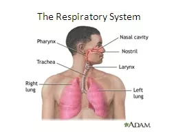
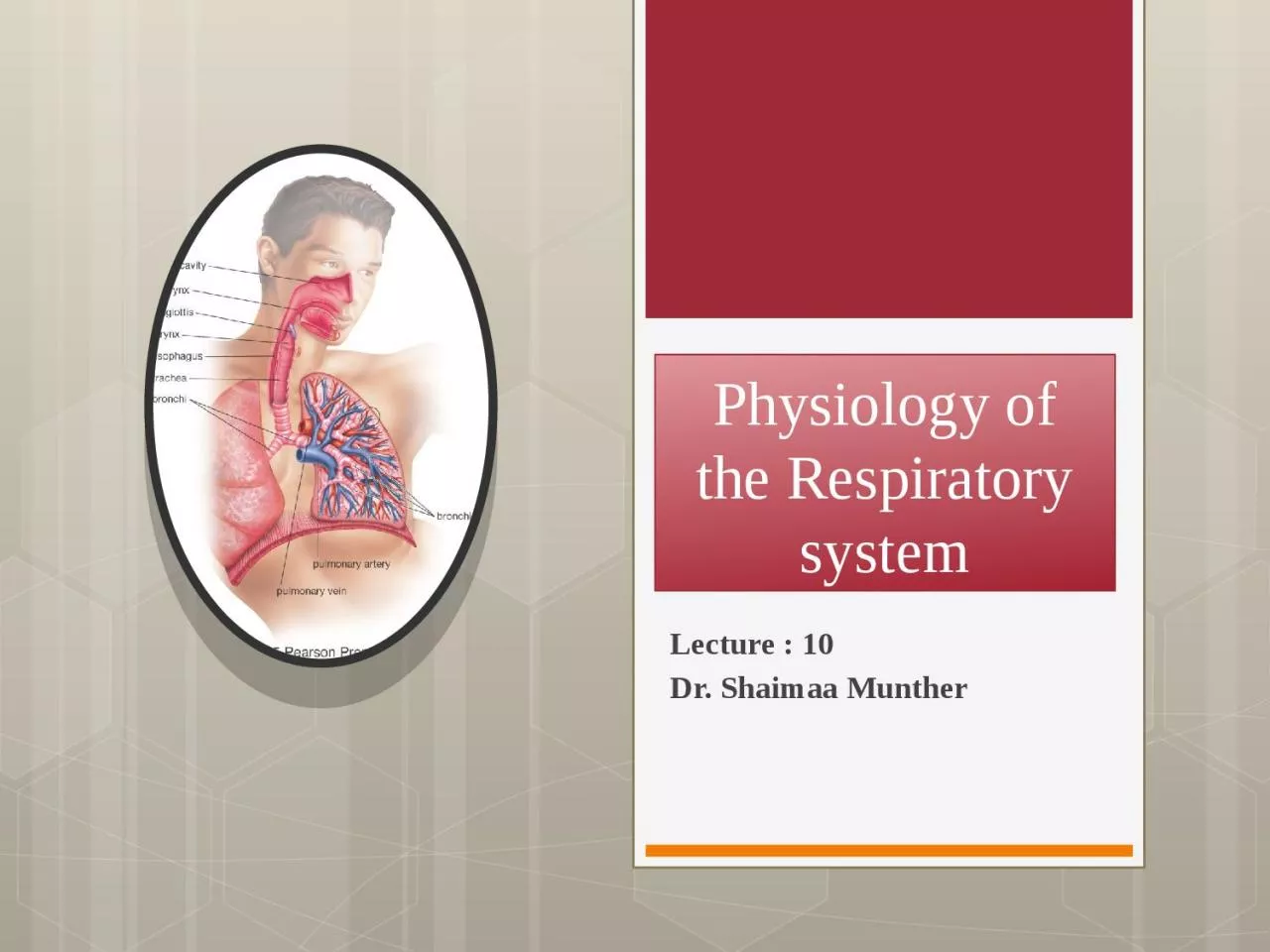
Lecture 10 Dr Shaimaa Munther Physiology of the Respiratory system Overview Respiration includes two processes External respiration the absorption of O2 and removal of CO2 from the body as a whole ID: 1006425

air lungs pulmonary cells lungs air cells pulmonary alveoli alveolar surface surfactant respiratory called blood trachea lung tension amp

Share:

Link:

Embed:

Download Presentation from below link

Download Presentation The PPT/PDF document "Physiology of the Respiratory system" is the property of its rightful owner. Permission is granted to download and print the materials on this web site for personal, non-commercial use only, and to display it on your personal computer provided you do not modify the materials and that you retain all copyright notices contained in the materials. By downloading content from our website, you accept the terms of this agreement.


Presentation Transcript

1. Physiology of the Respiratory systemLecture : 10Dr. Shaimaa Munther

2. Physiology of the Respiratory systemOverview Respiration, includes two processes:External respiration, the absorption of O2 and removal of CO2 from the body as a wholeInternal respiration: the utilization of O2 and production of CO2 by cells and the gaseous exchanges between the cells and their fluid medium.

3.

4. THE RESPIRATORY SYSTEMThe respiratory system is made up of a gas-exchanging organ (The lungs ) and a “pump” that ventilates the lungs.The pump consists of: The chest wall &the respiratory muscles, which increase and decrease the size of the thoracic cavityThe areas in the brain that control the muscles and the tracts Nerves that connect the brain to the muscles.

5. THE RESPIRATORY SYSTEMAt rest, a normal human breathes 12 to 15 times a minute. About 500 mL of air per breath, or 6 to 8 L/min, is inspired and expired. This air mixes with the gas in the alveoli, and, by simple diffusion, O2 enters the blood in the pulmonary capillaries while CO2 enters the alveoli for excretion through the lung with Traces of other gases, such as methane from the intestines are also found in expired air. Alcohol and acetone are expired when present in appreciable quantities in the body. Indeed over 250 different volatile substances have been identified in human breath.

6. Diffusion

7. Perfusion

8. The air after passing through the nasal passages and pharynx, warmed and takes up water vapor.The inspired air passes down the trachea and through the bronchioles, and alveolar ducts to the alveoli, where gas exchange occurs .AIR PASSAGES

9. The Trachea (windpipe) The Trachea is located in the neck in front of the esophagus , it is a hollow tube supported by rings of cartilage Air enters the trachea from the pharynx.In order for air to enter or leave the trachea and lungs, however, it must pass through a valve like opening called the glottis between the vocal folds. The ventricular and vocal folds are part of the larynx, or voice box AIR PASSAGES

10. The projection at the front of the throat, commonly called the “Adam’sapple,” is formed by the largest cartilage of the larynxBetween the trachea and the alveolar sacs, the airways divide into bronchi & bronchioles, and terminal bronchioles, alveolar ducts, and alveoli. These multiple divisions greatly increase the total cross-sectional area of the airways.The alveoli are surrounded by pulmonary capillaries .In most areas, air and blood are separated only by the alveolar epithelium and the capillary endothelium, Humans have 300 million alveoli, and the total area of the alveolar walls in contact with capillaries in both lungs is about 70 m2.AIR PASSAGES

11.

12.

13. The alveoli are lined by two types of epithelial cells: Type I cells : Are flat cells with large cytoplasmic extensions and are the primary lining cells of the alveoli, covering approximately 95% of the alveolar epithelial surface area.Type II cells (granular pneumocytes) : Are thicker and contain numerous lamellar inclusion bodies. A primary function of these cells is to secrete surfactant; however, they are also important in alveolar repair as well as other cellular physiology. Although these cells make up approximately 5% of the surface area, they represent approximately 60% of the epithelial cells in the alveoliThe Alveolar Cells

14. The alveoli also contain other specialized cells, including: Pulmonary alveolar macrophages (PAMs, or AMs)LymphocytesPlasma cellsNeuroendocrine cellsMast cells.The mast cells contain heparin, various lipids, histamine, and various proteases that participate in allergic reactionsThe Alveolar Cells

15.

16.

17.

18. THE BRONCHI & THEIR INNERVATIONThe trachea and bronchi have cartilage in their walls but relatively little smooth muscle. They are lined by a ciliated epitheliumthat contains mucous and serous glands. Cilia are presentas far as the respiratory bronchiolesThe walls of the bronchi and bronchioles are innervated by the autonomic nervous system. Muscarinic receptors are abundant, and cholinergic discharge causes bronchoconstriction on cholinergic endings, by acetylcholine release. The adrenergic β2 receptors mediate bronchodilation, it increase bronchial secretion, while α1 adrenergicreceptors inhibit secretion.

19. ANATOMY OF BLOOD FLOW IN THE LUNGBoth the pulmonary circulation and the bronchial circulation contribute to blood flow in the lung. In the pulmonary circulation, almost all the blood in the body passes via the pulmonary artery to the pulmonary capillary bed, where it is oxygenated and returned to the left atrium via the pulmonary veins .The bronchial circulation nourishes the trachea down to the terminal bronchioles and also supplies the pleura and lymph nodes. It should be noted that lymphatic channels are more abundant in the lungs than in any other organ

20. MECHANICS OF RESPIRATIONINSPIRATION & EXPIRATIONThe lungs and the chest wall are elastic structures. Normally no more than a thin layer of fluid is present between the lungs and the chest wall (intrapleural space) , which make the lungs slide easily on the chest wall.The pressure in the “space” between the lungs and chest wall called (intrapleural pressure)

21. Ventilation CyclePulmonary ventilation consists of two phases: inspiration and expiration. Inspiration (inhalation) and expiration (exhalation) are accomplished by alternately increasing and decreasing the volumes of the thorax and lungs .

22. Physical Properties of the Lungs A-Compliance: In order for inspiration to occur, the lungs must be able to expand when stretched; they must have high compliance.The lungs are very distensible (stretchable), they are, in fact, about a hundred times more distensible than a toy balloon.Lung compliance can be defined as the change in lung volume per change in trans pulmonary pressure, expressed symbolically as Δ V /Δ P.A pathological condition called pulmonary fibrosis, decreases lung compliance.

23. B-Elasticity : The tendency to get smaller is also aided by surface tension forces within the alveoli.The term elasticity refers to the tendency of a structure to return to its initial size after being distended. Because of their high content of elastin proteins, the lungs are very elastic and resist distension. Physical Properties of the Lungs

24. C-Surface TensionThe forces that act to resist distension include elastic resistance and the surface tension that is exerted by fluid in the alveoli. ''The thin film of fluid normally present in the alveolus has a surface tension, produced because water molecules at the surface are attracted more to other water molecules than to air.Alveolar fluid contains a substance that reduces surface tension. This substance is called surfactant .Physical Properties of the Lungs

25. surfactant is substance that secreted into the alveoli by type II alveolar cells and consists of phospholipids—primarily phosphatidylcholine and phosphatidylglycerolTogether with hydrophobic surfactant proteins , Surfactant becomes interspersed between water molecules at the water–air interface; this reduces the hydrogen bonds between water molecules at the surface and thereby reduces the surface tension. Surfactant prevents alveoli from collapsing during expiration by reducing the surface tension , thus the alveoli remain open and a residual volume of air remains in the lungs. SURFACTANT

26. Surfactant begins to be produced in late fetal life. For this reason, premature babies are sometimes born with lungs that lack sufficient surfactant and their alveoli are collapsed as a result. This condition is called respiratory distress syndrome RDS). A full-term pregnancy lasts 37 to 42 weeks. RDS occurs in about 60% of babies born at less than 28 weeks 30% of babies born at 28 to 34 weeks, and less than 5% of babies born after 34 weeks of gestation. The risk of RDS can be assessed by analysis of amniotic fluid (surrounding the fetus), and mothers can be given an exogenous corticosteroids to accelerate the maturation of their fetus’s lungs. SURFACTANT

27. LUNG VOLUMESTidal volume : is the amount of air that moves into the lungs with each inspiration or the amount that moves out with each expiration. The air left in the lungs after a maximal expiratory effort is the residual volume

28. LUNG VOLUMESThe space in the conducting zone of the airways occupied by gas that does not exchange with blood in the pulmonary vessels is the respiratory dead space.The forced vital capacity (FVC ) the largest amount of air that can be expired after a maximal inspiratory effort, is frequently measured clinically as an index of pulmonary function. It gives useful information about the strength of the respiratory muscles and other aspects of pulmonary function.The fraction of the vital capacity expired during the first second of a forced expiration is referred to as the forced expiratory volume in one second (FEV1) The FEV1 to FVC ratio (FEV1/FVC) is a useful tool in the diagnosis of airway disease . 

29.

30. In addition to their functions in gas exchange, the lungs have a number of metabolic functions :They manufacture surfactant for local use, They also contain a fibrinolytic system that lyses clots in the pulmonary vessels.They release a variety of substances that enter the systemic arterial blood (Table 35–5), and they remove other substances from the systemic venous blood that reach them via the pulmonary artery.The lungs also activate a hormone called angiotensin I , that converted to angiotensin II, causing vasoconstriction & enhancing aldosterone secretion from the adrenal gland. METABOLIC &ENDOCRINE FUNCTIONS OF THE LUNGS

31.

32. Pulmonary Function TestsPulmonary function may be assessed clinically by means of a technique known as spirometry. In this procedure, a subject breathes in a closed system in which air is trapped within a light plastic bell floating in water. The bell moves up when the subject exhales and down when the subject inhales. The movements of the bell cause corresponding movements of a pen, which traces a record of the breathing called a spirogram ( fig. 16.15 ). More sophisticated computerized devices are now more commonly employ

33.

34.