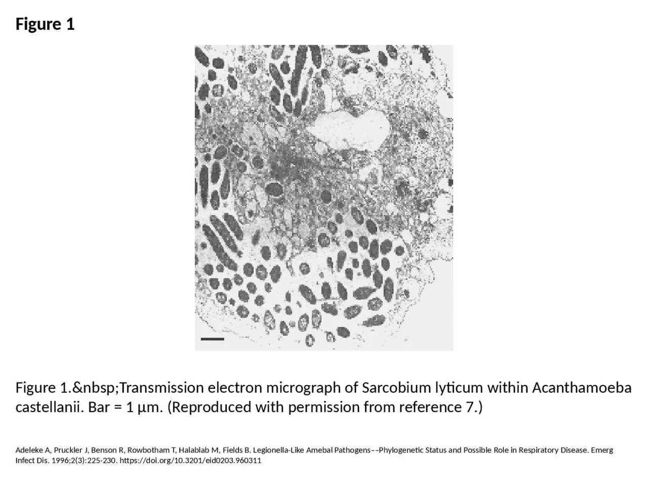
PPT-Figure 1 Figure 1. Transmission electron micrograph of Sarcobium lyticum within

Author : elise | Published Date : 2023-07-19

Adeleke A Pruckler J Benson R Rowbotham T Halablab M Fields B LegionellaLike Amebal PathogensPhylogenetic Status and Possible Role in Respiratory Disease Emerg Infect

Presentation Embed Code

Download Presentation

Download Presentation The PPT/PDF document "Figure 1 Figure 1. Transmission..." is the property of its rightful owner. Permission is granted to download and print the materials on this website for personal, non-commercial use only, and to display it on your personal computer provided you do not modify the materials and that you retain all copyright notices contained in the materials. By downloading content from our website, you accept the terms of this agreement.

Figure 1 Figure 1. Transmission electron micrograph of Sarcobium lyticum within: Transcript


Adeleke A Pruckler J Benson R Rowbotham T Halablab M Fields B LegionellaLike Amebal PathogensPhylogenetic Status and Possible Role in Respiratory Disease Emerg Infect Dis 199623225230 httpsdoiorg103201eid0203960311. Figure C54. SEM micrograph of compressed mature yellow-poplar (4mm), TRT3. Figure C55. SEM micrograph of uncompressed mature yellow-poplar (4mm), TRT3. Figure C56. SEM micrograph of uncompressed matur Figure C28. SEM micrograph of uncompressed mature yellow-poplar (4mm), TRT1. Figure C29. SEM micrograph of compressed mature yellow-poplar (4mm), TRT1. Figure C30. SEM micrograph of compressed mature Figure C80. SEM micrograph of compressed mature southern pine, TRT1. Figure C81. SEM micrograph of compressed mature southern pine, TRT1, showing multiplefractures in earlywood tracheids. Figure C82. Goosney DL, Knoechel DG, Finlay BB. Enteropathogenic E. coli, Salmonella, and Shigella: Masters of Host Cell Cytoskeletal Exploitation. Emerg Infect Dis. 1999;5(2):216-223. https://doi.org/10.3201/eid0502.990205. Dey R, Mount H, Ensminger AW, Tyrrell GJ, Ward LP, Ashbolt NJ. Isolation of Legionella pneumophila by Co-culture with Local Ameba, Canada. Emerg Infect Dis. 2019;25(11):2104-2107. https://doi.org/10.3201/eid2511.190522. Holmes AH, Govan J, Goldstein R. Agricultural Use of Burkholderia (Pseudomonas) cepacia: A Threat to Human Health?. Emerg Infect Dis. 1998;4(2):221-227. https://doi.org/10.3201/eid0402.980209. Donlan RM. Biofilms: Microbial Life on Surfaces. Emerg Infect Dis. 2002;8(9):881-890. https://doi.org/10.3201/eid0809.020063. Sonntag M, Mühldorfer K, Speck S, Wibbelt G, Kurth A. New Adenovirus in Bats, Germany. Emerg Infect Dis. 2009;15(12):2052-2055. https://doi.org/10.3201/eid1512.090646. Goosney DL, Knoechel DG, Finlay BB. Enteropathogenic E. coli, Salmonella, and Shigella: Masters of Host Cell Cytoskeletal Exploitation. Emerg Infect Dis. 1999;5(2):216-223. https://doi.org/10.3201/eid0502.990205. Foucault C, Brouqui P, Raoult D. Bartonella quintana Characteristics and Clinical Management. Emerg Infect Dis. 2006;12(2):217-223. https://doi.org/10.3201/eid1202.050874. Donlan RM. Biofilms and Device-Associated Infections. Emerg Infect Dis. 2001;7(2):277-281. https://doi.org/10.3201/eid0702.700277. Baseman JB, Tully JG. Mycoplasmas: Sophisticated, Reemerging, and Burdened by Their Notoriety. Emerg Infect Dis. 1997;3(1):21-32. https://doi.org/10.3201/eid0301.970103. Jabra-Rizk MA, Falkler WA, Meiller TF. Fungal Biofilms and Drug Resistance. Emerg Infect Dis. 2004;10(1):14-19. https://doi.org/10.3201/eid1001.030119. Kim MJ, Bancroft E, Lehnkering E, Donlan RM, Mascola L. Alcaligenes xylosoxidans Bloodstream Infections in Outpatient Oncology Office. Emerg Infect Dis. 2008;14(7):1046-1052. https://doi.org/10.3201/eid1407.070894.

Download Document

Here is the link to download the presentation.
"Figure 1 Figure 1. Transmission electron micrograph of Sarcobium lyticum within"The content belongs to its owner. You may download and print it for personal use, without modification, and keep all copyright notices. By downloading, you agree to these terms.

Related Documents